Clinical Measurements
ICG Impedance Cardiography Non-invasive hemodynamic measurements
Impedance Cardiography The technology behind ICG Impedance cardiography (ICG) is a safe, non-invasive method to measure a patient’s hemodynamic status. The ICG waveform is generated by thoracic electrical bioimpedance (TEB) technology, which measures the level of change in impedance in the thoracic fluid. Four small sensors send and receive a low amplitude electrical current through the thorax to detect the level of change in resistance in the thoracic fluid. With each cardiac cycle, fluid levels change, which affects the impedance to the electrical signal transmitted by the sensors.
• Detects B, C, and X points on the ICG waveform, timed with the vECG • Adjusts for patient gender, height, weight, and age • Accepts or rejects each beat
Advanced ICG algorithm Philips ICG uses a sophisticated algorithm to determine the level of change in impedance, to generate the ICG wave, and to calculate or derive hemodynamic parameters. This advanced algorithm: • Removes pacemaker spikes and respiratory artifact • Stores an adaptive mean curve as representative of typical curve shape • Holds three heart beats of current patient data, as typical for that patient
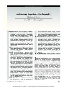
O B C X
Mitral Valve Opens,Ventricular Filling Aortic Valve Opens Peak Systolic Flow Aortic Valve Closes
SV VET P B-C Slope
Stroke Volume Ventricular Ejection Time Atrial systole Acceleration cardiac index
ICG sensor placement for impedance cardiography • Two sensors placed above the clavicle on each side of the neck, level with the sternal notch • Two sensors placed on either side of the thorax, level with the xyphoid process and in line with the midaxillary line • Sensors measure the change in impedance in the thoracic fluid • Studs on the sensor are spaced 5 cm apart for proper signal emission and detection • Electrical signals follow the path of least resistance, traveling through the fluidfilled aorta • The outer studs continuously send and receive the electrical signal; the inner electrodes constantly detect and measure the impedance
• The resulting ICG waveform is used to calculate or derive 12 hemodynamic parameters
ICG Impedance Non-invasive, Philips Impedance Cardiography (ICG) continuously measures hemodynamic parameters without the associated risks of traditional invasive methods. ICG is based on enhanced thoracic electrical bioimpedance technology, which allows a simple, quick, and easy method for clinicians to perform hemodynamic assessment or continuous monitoring of a patient’s hemodynamic status. It measures 12 different hemodynamic parameters in addition to non-invasive blood pressure, which can facilitate more rapid decision making in differentiating causes of dyspnea and hyper/hypotension.
Philips ICG measurement is easy to use and ideal for hemodynamic evaluation of adult patients in the emergency department, step-down units, special procedure areas, and other areas where non-invasive continuous hemodynamic monitoring is required. It is also appropriate for select ICU patients. Impedance cardiography is designed for assessment and management of congestive heart failure, hypertension, and pacemaker patients to name a few.
12 hemodynamic parameters for comprehensive assessment, including: • Thoracic Fluid Content (TFC) • Accelerated Cardiac Index (ACI) • Stroke Volume (SV) • Cardiac Output (CO) • Systemic Vascular Resistance (SVR)
VueLink connection to Philips IntelliVue, CMS, and V24 and V26 patient monitors.
Cardiography safe, and timely measurements Signal Quality Indicator for validation of ICG waveforms at a glance. Signal Quality Indicator (SQI) visually supports the operator by showing the quality of the beats used for calculations, the percentage of accepted beats, signal amplitude (to know if the signal is adequate for monitoring), and timing with events of the cardiac cycle that is necessary for the algorithm to perform.
Overlapping waveforms show the accepted beats in the last 30 seconds, with the most recently accepted beat in magenta.
Key events of the cardiac cycle are indicated by three magenta vertical lines: aortic valve opens (B) peak systolic flow (C) aortic valve closes (X)
C
Percentage of accepted beats in the last 30 seconds. Total Fluid Content (TFC) is the total conductivity of the thorax representing parallel conductivity of three compartments: intravascular, intra-alveolar, and interstitial.
B X
Waveform amplitude indicated by the lighted bar: Green: acceptable Yellow: reduced accuracy possible Red: inadequate signal for monitoring
Portable, weighing only 2.2 kg (4.9 lbs.). Sharp 12cm (4.96˝) color display allows for easy viewing of information. Advanced patient-specific algorithm accounts for gender, age, and body surface area in calculating hemodynamic values. Signal Quality Indicator gives a real-time assessment of the quality of the ICG signal, indicating the percentage of accepted beats,TFC, signal amplitude, and timing with events of the cardiac cycle. Two-hour battery for emergency and transport support. Navigation wheel operates much like a computer mouse. Built-in recorder/printer for recording waveforms and documenting trends.
All parameters screen displays the complete set of hemodynamic parameters (CO, CI, SVR, SVRI, SV, SI,TFC,ACI, LCWI, PEP).
Tabular trends display shows 12 hours of hemodynamic parameter tabular trends at one-minute or 15-minute intervals.
Graphical trend display shows hemodynamic parameters in a rolling window. 30-minute, two-hour, or four-hour trends allow retrospective review of response to therapy.
IntelliVue monitor with ICG parameters displayed alongside other physiological measurements, including heart rate, blood pressure, pulse oximetry, cardiac output, and system vascular resistance – integrated via the Philips VueLink module.
ICG comes ready for use with accessories included • ICG cable • 10 sets of ICG sensors • Non-invasive blood pressure tubing and one adult, latex-free cuff • Two rolls of recorder paper • Self-paced training guide Flexible mounting options • Rollstand • Wall mount A simple ICG sensor-placement diagram and color-coded lead set illustrate how to conveniently apply the ICG sensors.
The ICG measurement monitor can be conveniently mounted on a roll stand or a tilt/swivel wall mount.
862146
ICG hemodynamic measurement
989803141821 Rollstand
Using ICG for the appropriate patient population The ICG measurement is designed for assessment of most adult patients – height 122-229cm (4´-7´6˝) and weight 30 -159 kg (67-350 lb.) – but may demonstrate reduced accuracy when patients present with the following conditions or anomalies: • Aortic valve regurgitation • Minute ventilation sensor function pacemakers • Connection to a cardiopulmonary bypass machine • Sustained arrhythmias • Connection to an intra-aortic balloon pump or chest tubes • Connection to a respiratory ventilator • Congenital heart defects • Pericardial effusion • Severe hypertension (MAP > 130 mm Hg) • Septic shock • Severe anemia
To jumpstart your ICG program, a discount starter kit of sensors is available from Philips Medical Supplies.
989803141961 Wall mount
Philips Medical Systems is part of Royal Philips Electronics www.medical.philips.com
[email protected]
© Koninklijke Philips Electronics N.V. 2004 Philips Medical Systems 3000 Minuteman Road Andover, MA 01810-1085 (800) 934-7372
All rights are reserved. Reproduction in whole or in part is prohibited without the prior written consent of the copyright holder.
Philips Medical Systems Nederland B.V. reserves the right to make changes in specifications and/or discontinue any product at any time without notice or obligation and will not be liable for any consequences resulting from the use of this publication.
4522 981 88271/862 * OCT 2004