Brain (1997), 120, 1067–1076
The neuroendocrine axis in patients with multiple sclerosis Terence Wei1 and Stafford L. Lightman2 1Department
of Clinical Neurology, Institute of Neurology, London and 2Department of Medicine, Bristol Royal Infirmary, Bristol, UK
Correspondence to: Professor S. Lightman, Department of Medicine, Bristol Royal Infirmary, Marlborough Street, Bristol BS2 8HW, UK
Summary We investigated the basal and dynamic regulation of the hypothalamo–pituitary–adrenal (HPA), hypothalamo– pituitary–thyroid (HPT) and hypothalamo–pituitary–gonadal axes and prolactin secretion in 52 patients with clinically definite multiple sclerosis. These patients also had gadolinium enhanced brain MRI scans and were divided into relapsing– remitting, secondary progressive and primary progressive subgroups. These subgroups were compared with healthy controls and a group of patients with other neurological diseases. The cortisol diurnal rhythm was preserved in all groups of patients. The time-integrated cortisol response to human corticotropin-releasing hormone (CRH) stimulation was lower in the patients with secondary progressive multiple
sclerosis, compared with patients with primary progressive multiple sclerosis and healthy subjects. The time-integrated β-endorphin response to CRH was greater in the patients with relapsing–remitting multiple sclerosis compared with the others. Feedback regulation assessed by dexamethasone suppression was normal. Serum testosterone was low in 24% of male multiple sclerosis patients and oestradiol was low in 25% of pre-menopausal female multiple sclerosis patients, whereas prolactin and the HPT function were normal. Correlations with C-reactive protein (CRP) and MRI suggest that activation of the HPA axis in multiple sclerosis patients is secondary to an active inflammatory stimulus.
Keywords: hypothalamo–pituitary–adrenal axis; diurnal rhythm; multiple sclerosis Abbreviations: ACTH 5 corticotropin; β-END 5 β-endorphin; CRH 5 corticotropin-releasing hormone; CRP 5 C-reactive protein; EAE 5 experimental allergic encephalomyelitis; FSH 5 follicle-stimulating hormone; GnRH 5 gonadotropinreleasing hormone; HPA 5 hypothalamo–pituitary–adrenal; HPG 5 hypothalamo–pituitary–gonadal; HPT 5 hypothalamo– pituitary–thyroid; LH 5 luteinising hormone; TRH 5 thyrotropin-releasing hormone; TSH 5 thyroid-stimulating hormone
Introduction Multiple sclerosis is an immune-mediated inflammatory demyelinating disease of uncertain aetiology. During the time of active demyelination in the rat model of experimental allergic encephalomyelitis (EAE), there is activation of the hypothalamo–pituitary–adrenal (HPA) axis which is vital for recovery from the disease (MacPhee et al., 1989). HPA activity has also been found to be increased in other inflammatory diseases and may indeed act as a protective mechanism against an excessive immune response. In rats with adjuvant-induced arthritis the HPA axis is chronically activated and diurnal rhythms of corticotropin (ACTH) and corticosterone are lost (Sarlis et al., 1992). Patients with rheumatoid arthritis also have an activated HPA axis (Neeck et al., 1990) with an impaired cortisol rhythm and a poor response to stress (Neeck et al., 1990; Chikanza et al., 1992). © Oxford University Press 1997
This impairment of HPA response to stress in patients with rheumatoid arthritis is particularly interesting since it parallels findings in experimental animals in both EAE and in arthritis. Mason et al. (1990) found a lower HPA response to stress in the Lewis rat than in the Piebald Viral Glaxo rat and went on to show that not only was the Lewis rat more susceptible to EAE, but if EAE-resistant Piebald Viral Glaxo rats were adrenalectomized and given corticosterone replacement that mimicked the levels in the Lewis rat, they developed EAE like Lewis rats. EAE in Lewis rats can be suppressed by exogenously administered glucocorticoid and exacerbated by the potent anti-glucocorticoid RU38486 (Bolton and Flower, 1989). Sternberg et al. (1989a) also showed a susceptibility to arthritis and a reduced response to both inflammation and stress in the Lewis rat. Furthermore, these rats had a reduced
1068
T. Wei et al.
hypothalamic activation of corticotropin-releasing hormone (CRH) messenger ribonucleic acid (Sternberg et al., 1989b) and secretion of CRH (Calogero et al., 1992) in response to stress when compared with the arthritis-resistant Fischer rat. This HPA hyporesponsiveness in Lewis rats appears to be associated with a generalized susceptibility to inflammatory diseases (Karalis et al., 1995). These data raise the possibility that HPA hyporesponsiveness in man might also play a role in increasing susceptibility to inflammatory disease. There is already some evidence that this might be the case in rheumatoid arthritis (Chikanza et al., 1992) and it is clearly important to assess whether this might also be the case for multiple sclerosis. Previous studies on the hormonal status in limited numbers of patients with multiple sclerosis have produced fragmentary and conflicting results (Wender and Gutowski, 1959; Teasdale et al., 1967; Millac et al., 1969; Klapps et al., 1992). Data from a small series of multiple sclerosis patients has implicated a selective central impairment of the HPA axis (Michelson et al., 1994) and other studies have shown variable adrenal sensitivity to ACTH (Fog, 1951; Alexander et al., 1971; Snyder et al., 1981). Insulin-induced hypoglycaemia has been used with variable results (Teasdale et al., 1967; Millac et al., 1969), but this is an extremely potent non-specific stimulus with little chance of showing subtle differences between the multiple sclerosis subgroups. Other hormones may also be involved in multiple sclerosis. Thus prolactin, which has been implicated as an immunomodulatory hormone (Jara et al., 1991), has been found to be raised (Grinsted et al., 1989; Kira et al., 1991) or normal (Reder and Lowy, 1993) and the hypothalamo–pituitary–gonadal (HPG) axis which has long been known to be suppressed during stress (Selye, 1936) also deserves attention. The only way to clarify the neuroendocrine consequences or possible aetiological relevance in multiple sclerosis was to carry out a full prospective study in an adequate number of multiple sclerosis patients. Since there may be different aetiological factors (Olerup et al., 1989) and inflammatory activity (Thompson et al., 1991; Revesz et al., 1994) in different subgroups of patients this must also be taken into account in classifying the patients into appropriate diagnostic classes and in correlating with indices of inflammatory disease activity. We now report our findings in 52 patients together with those in healthy controls and patients with other neurological disorders.
and the NMR Research Unit, Institute of Neurology, London. Patients who had hormone-profile studies were investigated as inpatients and recruited during their hospital stay for diagnosis and treatment; others were investigated as inpatients or out-patients. Normal control subjects were recruited from the hospital staff and their friends and were studied as outpatients. Multiple sclerosis patients are subdivided into three subgroups: (i) relapsing–remitting multiple sclerosis, (ii) secondary progressive multiple sclerosis and (iii) primary progressive multiple sclerosis (Thompson et al., 1991). Most patients were in clinical relapse (within four weeks). The characteristics of the subjects are detailed below. Contra-indications to inclusion were the presence of major depressive illness, anxiety neurosis, current sepsis, major cognitive impairment, epilepsy, pituitary adenoma, hypophysectomy and other inflammatory diseases. No patient under study was on current steroid or other immunomodulatory therapy. The study had prior approval by the National Hospital’s medical ethics committee and the subjects’ informed and signed consent.
Hormone-study protocols Blood was sampled via an i.v. indwelling Venflon catheter into Vacutainer tubes. All samples were stored at 220°C until analysis.
Basal hormone and C-reactive protein (CRP) concentrations Basal serum free T3, free T4, thyroid-stimulating hormone (TSH), testosterone (males), oestradiol (females), luteinizing hormone (LH), follicle-stimulating hormone (FSH) and plasma CRP were determined from morning samples taken between 08.00 and 09.30 hours.
Baseline cortisol and prolactin profiles Samples of blood were taken throughout the day (06.30, then 2-hourly 08.00–24.00 and, in addition, half-hourly 08.00–10.00 and 20.00–22.00) for serum cortisol and prolactin measurements.
Stimulation and suppression tests Methods Patients Fifty-two patients with clinically definite multiple sclerosis (Poser et al., 1983), 10 normal control subjects and five patients with other neurological diseases (benign intracranial hypertension, headache, non-organic gait disorder, motor neuron disease and stiff-man’s syndrome) were recruited from the National Hospital for Neurology and Neurosurgery
Human CRH (CRH Ferring, Kiel, Germany) was injected i.v. (100 µg over 1 min) in the morning (Oppermann, 1986), blood was taken at 20-min intervals for 60 min. The combined thyrotropin-releasing hormone (TRH), gonadotropin-releasing hormone (GnRH) and synthetic ACTH tests (Hall et al., 1980) followed the CRH test. Injection of each peptide was given sequentially in quick succession: TRH (TRH-Cambridge®, Newcastle, UK) 200 µg i.v., GnRH (HRF® Monmouth, Guildford, UK) 100 µg i.v. and 1–24ACTH (Synacthen®, Ciba, Horsham, UK) 250 µg
Neuroendocrine axis in multiple sclerosis i.v.; blood was taken at 20-min intervals for 60 min. For the dexamethasone-suppression test the patient took dexamethasone 1 mg orally at 23:00 hours the previous night and blood was taken at 09.00 hours the following morning (Hall et al., 1980).
Assay methods
Plasma ACTH and β-endorphin (β-END) were assayed by in-house radioimmunoassay (Jessop et al., 1994). The ACTH antiserum did not recognize β-END and the β-END antiserum did not cross-react with ACTH. Serum testosterone was measured by in-house radioimmunoassay. Other serum hormones were assayed using enzyme-linked immunosorbant assay in an automated analyser. Concentrations of CRP in plasma of ,2 mg/l was measured by an in-house enzymelinked immunosorbant assay and CRP .2 mg/l by a commercial radial immunodiffusion assay (Giovannoni et al., 1996). Between-batch coefficients of variation were: ACTH and β-END, ,15%; cortisol 3.7%; prolactin 8.8%; free T4 2.5%; free T3 3.5%; TSH 3.4%; LH 6.5%; FSH 6.6%; oestradiol 12.5%; testosterone 7.8% and CRP 7.9%.
MRI Brain images were obtained in a 1.5 Tesla superconducting system (General Electric, Milwaukee, USA). Duo-echo T2weighted (TR3500, TEef90/100) and proton density-weighted (TR2900, TEef18/20) axial brain fast spin echo images were obtained using head coil before gadolinium (Thorpe et al., 1994) and T1-weighted spin echo images (TR640, TE14) after gadolinium (5-mm thick, contiguous slices, 2563256 matrix and one excitation). Gadolinium (Magnevist®, Schering, Burgess Hill, UK) was given at 0.1 mmol/kg as an i.v. bolus.
Curve-fitting method For cortisol profiles, data were fitted to cosine curves (Nelson et al., 1979). The general equation for the curve is: f(t) 5 C0 1 C cos(ωt – φ), in which: f(t) represents cortisol concentration at time t, C0 is the mesor mean, C is the amplitude, ω is the angular frequency, φ is the acrophase. Constraints were: C ø C0; 40 ø φ ø 135; 0 ø C0; ωt 5 360; 10 ø ω ø 15 and (φ/9) ø ω ø (φ/4).
1069
Table 1 Male multiple sclerosis patients with low serum testosterone Age
Subgroup
Testosterone (nmol/l)
LH (U/l)
FSH (U/l)
32 33 36 38 48 40
2PMS 2PMS 2PMS 2PMS 2PMS 1PMS
7.8 1.7 8.1 6.9 8.7 7.0
3.3 6.3 1.9 2.2 2.7 4.6
13 6.7 3.2 7.3 6.3 4.0
See footnote of Table 2 for abbreviations.
used to investigate aspects of hormonal data with one another and the loge of CRP concentrations. P , 0.05 was regarded as statistically significant.
Results Basal thyroid hormones Data were obtained from 52 multiple sclerosis patients in four groups (relapsing–remitting multiple sclerosis : secondary progressive multiple sclerosis : primary progressive multiple sclerosis 5 19 : 21 : 12), also from five patients with other neurological diseases and 10 normal control subjects. The mean ages (range) in these five groups were 39.8 (25–68), 43.2 (24–70), 46.4 (32–57), 42.4 (19–60) and 33.1 (24–53) years, respectively. There were no significant differences between the groups for free T3, free T4 or TSH (data not shown).
Basal testosterone, LH and FSH in males Data for testosterone, LH and FSH were obtained from 25 male multiple sclerosis patients (relapsing–remitting multiple sclerosis : secondary progressive multiple sclerosis : primary progressive multiple sclerosis 5 7 : 10 : 8) and six normal control subjects. Their mean ages (range) were 36.6 (25–49), 40.4 (29–56), 44.9 (32–57) and 34.7 (26–53) years, respectively. There was no significant difference in the mean testosterone concentrations between groups (data not shown). Of all 25 of the male multiple sclerosis patients, six (24.0%, mean age 37.8 years, range: 32–48 years) had serum testosterone below the lower limit of normal (Table 1) and did not have appropriately elevated LH or FSH.
Statistics Data are presented as the mean (6 SE) and hormone concentrations were rounded to one decimal place unless otherwise stated. Time-integrated hormone responses were calculated as the area under curve by integration of fitted curves (cortisol profiles) or a trapezium method. CRP data were analysed a loge transformation. Data were compared by Student’s t test or single-factor analysis of variance, as appropriate. Statistically significant data were examined post hoc by the Newman–Keuls test. Correlation analysis was
Basal oestradiol, LH and FSH in females Data for oestradiol were available in 26 female multiple sclerosis patients (relapsing–remitting multiple sclerosis : secondary progressive multiple sclerosis : primary progressive multiple sclerosis 5 14 : 9 : 3) and 4 normal control subjects. Their mean ages (range) were 41.0 (25–68), 45.7 (24–70), 47.3 (35–51) and 30.8 (24–51) years, respectively. Four out of 16 (25%) pre-menopausal patients had low oestradiol concentrations (, 100 pmol/l). Six patients were post-
1070
T. Wei et al.
Table 2 Characteristics of the patient groups for profile study
Age (years), mean (range) EDSS, mean (range) Males : females Gd-MRI (enhanced) Fatigue
RRMS (n 5 9)
2PMS (n 5 8)
1PMS (n 5 4)
OND (n 5 4)
43 (25–68) 3.1 (1–7) 2:7 5 (3) 3
49 (32–70) 6.7 (3–9) 5:3 2 (0) 1
49 (41–57) 7.4 (6.5–8) 3:1 3 (0) 1
48 (37–60) NA 3:1 NA NA
RRMS 5 relapsing–remitting multiple sclerosis; 1PMS 5 primary progressive multiple sclerosis; 2PMS 5 secondary progressive multiple sclerosis; OND 5 other neurological diseases; EDSS 5 Kurtzke’s expanded disability-status scale; Gd-MRI (enhanced) 5 number of patients who had MRI with gadolinium (and number who enhanced); NA 5 not applicable. Age and EDSS are given as mean (and range), rounded to the nearest integer and by one decimal place, respectively.
menopausal and not on hormone replacement therapy; all had low oestradiol concentrations consistent with postmenopausal status. Five of them had normal post-menopausal levels of LH and FSH but one patient with relapsing– remitting multiple sclerosis had an inappropriately low serum FSH of 12.7 U/l.
Cortisol profiles Details of patient groups are summarized in Table 2. The mean profile characteristics of cortisol are shown in Fig. 1A. Although there were individual variations in the shape of the curves, a diurnal rhythm was present in each case. The mean area under the curve for the whole profile period did not differ between the groups. The mean cortisol curve amplitude (6SE) was just significantly different between groups (relapsing–remitting multiple sclerosis, 163.7 6 13.5; secondary progressive multiple sclerosis, 141.8 6 7.6; primary progressive multiple sclerosis, 124.1 6 6.7; other neurological diseases, 184.6 6 18.8 nmol/l, P 5 0.048), but on post hoc testing there was only a trend for the mean cortisol curve amplitude from both primary progressive multiple sclerosis and secondary progressive multiple sclerosis groups to be significantly lower than that in the group with other neurological diseases (P , 0.06, P , 0.07, respectively).
Prolactin profiles The mean total area under the prolactin curve did not differ significantly between groups (Fig. 1B).
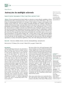
CRH test The patterns of cortisol responses were similar in the patient and normal control groups (Table 3). The mean total area under the curve of cortisol over 60 min differed significantly between groups (P 5 0.016; Fig. 2A), with that of the secondary progressive multiple sclerosis being significantly lower than those of the primary progressive multiple sclerosis and normal control groups (P , 0.05 in both, Table 3). Data were available for ACTH in multiple sclerosis patients only; the mean basal ACTH concentrations and the total areas
under the curve were not significantly different between groups (data not shown). The basal β-END concentrations were not significantly different between groups (Table 3). The mean total area under the curve for β-END was significantly different between groups (P 5 0.015, Fig. 2B); in the relapsing–remitting multiple sclerosis group it was significantly greater than that in the primary progressive multiple sclerosis group (P , 0.05), secondary progressive multiple sclerosis (P , 0.01) and normal control (P , 0.05) groups.
TRH test There were no significant differences in the TSH and prolactin responses between groups (see group details in Table 4; data otherwise not shown).
Synthetic ACTH test All the patients and control subjects (Table 4) had final serum cortisol concentrations of .550 nmol/l. There was a trend for a difference in the mean baseline cortisol concentrations: which were the lowest in the relapsing–remitting multiple sclerosis group (P 5 0.07). The mean total and the net increase in area under the curve did not differ between groups (Fig. 2C).
GnRH test Only two male multiple sclerosis patients (both relapsing– remitting multiple sclerosis) responded abnormally: one patient had a delayed but adequate response; another patient had an exaggerated response to GnRH (Mortimer, 1977). All except one of 15 female patients responded normally.
Dexamethasone-suppression test This was performed in 10 multiple sclerosis patients (relapsing–remitting multiple sclerosis : secondary progressive multiple sclerosis : primary progressive multiple sclerosis 5 2 : 6 : 2). All patients had cortisol suppression post-dexamethasone. Seven patients had MRI with gadolin-
Neuroendocrine axis in multiple sclerosis
1071
Fig. 1 (A) Cortisol profiles and (B) prolactin profiles in multiple sclerosis and other neurological disease (OND) groups. For other abbreviations see Table 2 footnote. These are actual data, not fitted curves.
ium but had only two enhanced T1 lesions: they both had undetectable post-dexamethasone cortisol.
Correlations between CRP and hormonal data The loge of CRP concentrations ([CRP]) correlated with two parameters of the baseline cortisol profiles: C0 [r(17) 5 0.46, P , 0.05, Fig. 3A] and the total area under the curve (r 5 0.46, P , 0.05, not shown). The loge ([CRP]) was negatively correlated with the net increase in area under the curve in the cortisol response to CRH [r(21) 5 20.48, P , 0.03, Fig. 3B].
Correlations between MRI and hormone data in multiple sclerosis patients The mean total area under the curve of the ACTH response to CRH in multiple sclerosis patients who enhanced (11092
6 613.8 pg·min/ml) was significantly greater than those who did not (7184.6 6 538.7 pg·min/ml, P , 0.001). The mean total area under the curve for the cortisol response to synthetic ACTH in patients who enhanced was significantly lower (34133.3 6 1236.9 nmol·min/l) than in those who did not (42600 6 2012.2 nmol·min/l, P 5 0.015).
Correlation between hormone data and fatigue No significant difference in hormone measures was found between patients with and without fatigue (data not shown).
Discussion This study was designed to provide definitive evidence for any change (or lack of change) in neuroendocrine activity in well-defined subgroups of patients with multiple sclerosis,
1072
T. Wei et al.
Table 3 CRH test: group characteristics, and cortisol and β-END responses RRMS (n 5 9) Group characteristics Age (years), mean (range) EDSS, mean (range) Males : females Gd-MRI (enhanced) Fatigue Cortisol (mean 6 SE) Basal (nmol/l) Total AUC (nmol·min/l)1 Net increase in AUC (nmol·min/ml) β-END (mean 6 SE) Basal (pg/ml) Total AUC (pg·min/ml)3 Net increase in AUC (pg·min/ml)
2PMS (n 5 11)
1PMS (n 5 5) 45 (40–56) 6.3 (3.5–8) 2:3 5 (0) 1
NC (n 5 9)
38 (26–53) 3.4 (1–6.5) 4:5 7 (3) 2
42 (24–59) 6.4 (3–9) 5:6 6 (2) 2
33 (23–53)
364.4 6 43.5 27647.8 6 1560.1 5781.1 6 1848.1
336.1 6 29.8 25255.5 6 1395.22 5090 6 926.5
471.2 6 78.7 33088 6 3122.4 4816 6 2142.6
471.4 6 43.3 32022.2 6 1870.5 3735.6 6 1592.2
52.1 6 14.9 6693.3 6 1700.3 3566.7 6 1529.8
28.4 6 6.9 2890 6 483.24 1188.2 6 137.3
24.4 6 2.0 2570 6 556.55 1106 6 621.3
28.9 6 6.2 2435.2 6 596.65 702.7 6 343.4
NA 5:4 NA NA
Age and EDSS are given as mean (and range), rounded to the nearest integer and by one decimal place, respectively. AUC 5 area under the curve; NC 5 normal control subjects. For all other abbreviations see Table 2 footnote. 1P 5 0.016 between groups; 2P , 0.05 comparing 2PMS with 1PMS and NC groups; 3P 5 0.015 between groups; 4P , 0.01, RRMS versus 2PMS group; 5P , 0.05, RRMS versus 1PMS and NC groups.
Fig. 2 (A) CRH test and cortisol response, (B) CRH test and β-END response and (C) synthetic ACTH test and cortisol response, in multiple sclerosis and normal control (NC) groups. For other abbreviations see Table 2 footnote.
Neuroendocrine axis in multiple sclerosis
1073
Table 4 Group characteristics for the TRH, GnRH and synthetic ACTH tests
Age (years), mean (range) EDSS mean (range) Males : females Gd-MRI (enhanced) Fatigue
RRMS (n 5 10)
2PMS (n 5 11)
1PMS (n 5 6)
NC (n 5 9)
37 (26–53) 3.5 (1–6.5) 4:6 7 (3) 2
44.3 (24–59) 6.6 (3–9) 5:6 6 (3) 2
45.7 (35–56) 6.5 (3.5–8) 3:3 6 (0) 1
33 (23–53) NA 5:4 NA NA
NC 5 normal control subjects. For all other abbreviations see Table 2 footnote. Age and EDSS are given as mean (and range), rounded to the nearest integer and by one decimal place, respectively.
Fig. 3 Relationships between (A) mesor mean cortisol level (Co) in cortisol profiles, and (B) cortisol response (net increase in AUC) in the CRH test, and C-reactive protein (ln CRP) in multiple sclerosis patients. A regression line is shown in each. The upper limit of the normal concentration of CRP is 2.08 ng/ml (ln 2.08 5 0.73).
with additional information from gadolinium enhanced MRI scans and CRP. In particular, we sought to discover whether there was any evidence for altered HPA activity, as has been demonstrated in EAE susceptible rats. Diurnal cortisol rhythms and time-integrated cortisol production were preserved in patients with multiple sclerosis. Correlation of rhythm parameters with CRP, which is a marker for the acute phase of the systemic response and which is modestly raised during relapses of multiple sclerosis, infection and MRI activity (Giovannoni et al., 1996), reveals upregulation of the HPA axis in response to stressors in multiple sclerosis. A possible common mediator in upregulating CRP and the HPA axis is interleukin-6, which is elevated in multiple sclerosis (Frei et al., 1991; Rovaris et al., 1996). In rheumatoid arthritis patients, the cortisol response to CRH has also been shown to correlate with changes in CRP levels (Gudbjo¨rnsson et al., 1996). The extent of activation of the HPA axis in multiple sclerosis patients (Fig. 3A) is up to 2.5-fold that in normal controls, which is modest in contrast to the six-fold increase in EAE. This appears at first surprising. However, many patients did not have gandolinium enhancement on MRI and their CRP levels were mostly normal or slightly elevated. Detection of inflammatory mediators in the CNS or the periphery in multiple sclerosis has been inconsistent; they have been found to be transiently expressed or expressed at low levels
(Giovannoni et al., 1996; Rovaris et al., 1996). Acute EAE is associated with a marked degree of non-specific stress (Levine et al., 1980). The magnitude of HPA activation in our patients is comparable to patients with rheumatoid arthritis (Neeck et al., 1990) and those with non-inflammatory arthropathy having major surgery (Chikanza, et al., 1992). The magnitude of HPA activation was in agreement with post-mortem immunocytochemical data, which revealed upregulation of immunoreactive-CRH neurons (Erkut et al., 1995; Purba et al., 1995). In contrast, this was not found in patients with other diseases (Erkut et al., 1995) and argues against non-specific HPA activation during illness in general. These CRH neurons co-expressed arginine vasopressin, which has been implicated in the study of Michelson et al. (1994) but its precise role remains uncertain. Multiple sclerosis patients who showed gadolinium enhancement on MRI, which serves as another index of inflammation (Katz et al., 1993), had a significantly higher ACTH response to CRH, but a significantly lower cortisol response to synthetic ACTH than those who did not. The total time-integrated hormonal responses correlated with basal concentrations (data not shown). Therefore, the HPA axis is centrally upregulated in the presence of active inflammation. Despite this, these patients had low cortisol response at the adrenal level; this implies a state of reduced adrenal reserve or adrenal insensitivity. Adrenal hypo-
1074
T. Wei et al.
responsiveness has also been found in patients with rheumatoid arthritis (Gudbjo¨rnsson et al., 1996) and fatigue syndromes (Sternberg, 1993). The possible mechanisms have been discussed by Grasser et al. (1996) who found some multiple sclerosis adrenals were hyporesponsive while most were hyperresponsive. Adrenal size was unknown in our patients but in a post-mortem study was found to be increased (Reder et al., 1994), consistent with chronic hypersecretion of ACTH, which would be expected to result in increased adrenal reserve. The adrenal response to ACTH during therapy has been shown to be variable in multiple sclerosis (Fog, 1951; Alexander et al., 1971; Snyder et al., 1981). Thus, the evidence indicates a spectrum of adrenal responsiveness in multiple sclerosis . The increased β-END response to CRH in the relapsing– remitting multiple sclerosis group suggested central upregulation of CRH neurons although this was mainly due to three patients with huge responses: the apparent discrepancy with measured ACTH response may partly be due to their different plasma half-lives. The negative correlation between the loge([CRP]) and the incremental cortisol response (the net increase in area under the curve) to CRH also implies that basal HPA activity is upregulated, since we found that the net increase in area under the curve negatively correlated with the basal pre-stimulatory cortisol concentrations (data not shown). The mean total time-integrated cortisol response to CRH in the secondary progressive multiple sclerosis group was significantly lower than those of the primary progressive multiple sclerosis and normal control groups: the former also tended to have the lowest mean basal pre-stimulatory cortisol concentrations. In contrast, patients with active rheumatoid arthritis had normal cortisol response to ovine CRH but all of them were on treatment (Chikanza et al., 1992). Thus, there is reduction in the cumulative HPA response to stress in secondary progressive multiple sclerosis patients, but the degree of impairment is less than that in rodent models (Sternberg et al., 1989a; Mason et al., 1990). This is likely to be a consequence of the disease, or related to altered HPA axis regulation from chronic activation. This group of patients might have had more courses of steroid therapy in the distant past, but any suppression of the HPA axis recovers rapidly (Wenning et al., 1994). Our data might have been confounded by the presence of fatigue or depression in multiple sclerosis patients. Fatigue syndromes have been reported to be associated with hyporesponsiveness of the HPA axis (Sternberg, 1993). We had not primarily set out to study this and the number of patients with fatigue was small. Our patients were not clinically depressed although no formal psychiatric evaluation was carried out and alterations of the HPA activity in multiple sclerosis are thought not to be due to depression (Michelson et al., 1994). Our patients had intact feedback regulation, in contrast to nearly half of those of Reder et al. (1987) but these authors used different diagnostic criteria. On the whole, the
hypothalamo–pituitary–thyroid (HPT) and HPG axes in our patients were intact; only a minority had low testosterone/ oestradiol and an even smaller number had a delayed gonadotropin response to GnRH. Although the HPG axis can be suppressed by HPA activation, cytokines and chronic illness, these would not adequately explain the hypogonadotrophic hypogonadism in these isolated patients. As the GnRH test was normal in the vast majority, a hypothalamic rather than pituitary defect was likely; a small part of the hypothalamus might have been selectively affected by multiple sclerosis plaque formation resulting in altered GnRH release. In assessing how closely our data on the HPA axis agree with those in EAE, the relationship between multiple sclerosis and EAE must be borne in mind. In EAE, the hormonal measurements were made during the first attack (MacPhee et al., 1989), but have not been repeated in animals with chronic relapsing EAE, which arguably resembles the disease course and process in multiple sclerosis more closely than acute EAE. Here, the patients were studied subsequent to their first demyelinating episode. Nonetheless, activation of the HPA axis is found in both multiple sclerosis and EAE. Activation of the HPA axis in multiple sclerosis relapses is potentially important for subsequent recovery, according to the known anti-inflammatory actions of steroids. The Lewis rat model of generally increased susceptibility to inflammatory diseases is remarkable but probably an extreme model, as these inbred rats have an HPA response at the lower range of normal of that of outbred Sprague–Dawley rats (Karalis et al., 1995). More importantly, although a robust HPA response is crucial for recovery from EAE, it is clearly not responsible for the subsequent refractory state (MacPhee et al., 1989). The role of the HPA axis in the initial pathogenesis of multiple sclerosis appears at best to be minor. The Lewis rat is susceptible to EAE because of consistently impaired HPA response to stress. Most multiple sclerosis patients have a normal stress response. We have argued that the modest degree of HPA activation in multiple sclerosis is not necessarily inappropriate. Some rat strains are resistant to EAE even if adrenalectomized (Mason et al., 1990). Clearly, non-hormonal factors are implicated. The hypothesis that hyporesponsiveness of the HPA axis in humans predisposes to multiple sclerosis cannot, at present, be directly tested before disease develops. Nonetheless, by analogy to animal models, the finding of intact HPA function in most of our patients implies that HPA impairment is unlikely to make a substantial contribution to its initial pathogenesis. Also, the normal HPA responses in most patients do not support an important role for HPA impairment in disease perpetuation or progression in general, but it may be a contributory factor in a small proportion of patients with hyporesponsive adrenals.
Acknowledgements We wish to thank Mr G. Carter, Charing Cross Hospital, for serum hormone assays; Dr G. Giovannoni of the Institute of
Neuroendocrine axis in multiple sclerosis
1075
Neurology for CRP assays; Professor W. I. McDonald for discussions and reviewing the earlier manuscript; Mr D. MacManus and colleagues, NMR Research Unit, for obtaining MRI; the physicians of the National Hospital for permission to study their patients, and the neuroradiologists of the National Hospital for reporting on the MRI scans. Funding for this research was generously provided by The Multiple Sclerosis Society of Great Britain and Northern Ireland.
M. Fundamentals of clinical endocrinology. 3rd ed. Tunbridge Wells (UK): Pitman Medical, 1980: 752–4.
References Alexander L, Cass LJ. ACTH-induced adrenocortical response patterns in multiple sclerosis and their relation to the clinical effectiveness of ACTH therapy. Confin Neurol 1971; 33: 1–24.
Karalis K, Crofford L, Wilder RL, Chrousos GP. Glucocorticoid and/or glucocorticoid antagonist effects in inflammatory diseasesusceptible Lewis rats and inflammatory disease-resistant Fischer rats. Endocrinology 1995; 136: 3107–12.
Bolton C, Flower RJ. The effects of the anti-glucocorticoid RU 38486 on steroid-mediated suppression of experimental allergic encephalomyelitis (EAE) in the Lewis rat. Life Sci 1989; 45: 97–104.
Katz D, Taubenberger JK, Cannella B, McFarlin DE, Raine CS, McFarland HF. Correlation between magnetic resonance imaging findings and lesion development in chronic, active multiple sclerosis. Ann Neurol 1993; 34: 661–9.
Calogero AE, Sternberg EM, Bagdy G, Smith C, Bernardini R, Aksentijevich S, et al. Neurotransmitter-induced hypothalamicpituitary-adrenal axis responsiveness is defective in inflammatory disease-susceptible Lewis rats: in vivo and in vitro studies suggesting globally defective hypothalamic secretion of corticotropin-releasing hormone. Neuroendocrinology 1992; 55: 600–8. Chikanza IC, Petrou P, Kingsley G, Chrousos G, Panayi GS. Defective hypothalamic response to immune and inflammatory stimuli in patients with rheumatoid arthritis [see comments]. Arthritis Rheum 1992; 35: 1281–8. Comment in: Arthritis Rheum 1993; 36: 1334–5. Erkut ZA, Hofman MA, Ravid R, Swaab DF. Increased activity of hypothalamic corticotropin-releasing hormone neurons in multiple sclerosis. J Neuroimmunol 1995; 62: 27–33. Fog T. ACTH in therapy of multiple sclerosis. Nord Med 1951; 46: 1742–8. Frei K, Fredrikson S, Fontana A, Link H. Interleukin-6 is elevated in plasma in multiple sclerosis. J Neuroimmunol 1991; 31: 147–53. Giovannoni G, Thorpe JW, Kidd D, Kendall BE, Moseley IF, Thompson AJ, et al. Soluble E-selectin in multiple sclerosis: raised concentrations in patients with primary progressive disease. J Neurol Neurosurg Psychiatry 1996; 60: 20–6. Grasser A, Mo¨ller A, Backmund H, Yassouridis A, Holsboer F. Heterogeneity of hypothalamic-pituitary-adrenal system response to a combined dexamethasone-CRH test in multiple sclerosis. Exp Clin Endocrinol 1996; 104: 31–7. Grinsted L, Heltberg A, Hagen C, Djursing H. Serum sex hormone and gonadotropin concentrations in premenopausal women with multiple sclerosis. J Intern Med 1989; 226: 241–4. ¨ berg K, Wide L, Ha¨llgren R. Gudbjo¨rnsson B, Skogseid B, O Intact adrenocorticotropic hormone secretion but impaired cortisol response in patients with active rheumatoid arthritis. Effect of glucocorticoids [see comments]. J Rheumatol 1996; 23: 596–602. Comment in: J Rheumatol 1996; 23: 577–81. Hall R, Anderson J, Smart GA, Besser M. Appendix D. Tests of adrenocortical function. In: Hall R, Anderson J, Smart GA, Besser
Jara LJ, Lavalle C, Fraga A, Go´mez-Sanchez C, Silveira LH, Martinez-Osuna P, et al. Prolactin, immunoregulation, and autoimmune diseases. [Review]. Semin Arthritis Rheum 1991; 20: 273–84. Jessop DS, Lightman SL, Chowdrey HS. Effects of a chronic inflammatory stress on levels of pro-opiomelanocortin-derived peptides in the rat spleen and thymus. J Neuroimmunol 1994; 49: 197–203.
Kira J, Harada M, Yamaguchi Y, Shida N, Goto I. Hyperprolactinemia in multiple sclerosis. J Neurol Sci 1991; 102: 61–6. Klapps P, Seyfert S, Fischer T, Scherbaum WA. Endocrine function in multiple sclerosis. Acta Neurol Scand 1992; 85: 353–7. Levine S, Sowinski R, Steinetz B. Effects of experimental allergic encephalomyelitis on thymus and adrenal: relation to remission and relapse. Proc Soc Exp Biol Med 1980; 165: 218–24. MacPhee IA, Antoni FA, Mason DW. Spontaneous recovery of rats from experimental allergic encephalomyelitis is dependent on regulation of the immune system by endogenous adrenal corticosteroids. J Exp Med 1989; 169: 431–45. Mason D, MacPhee I, Antoni F. The role of the neuroendocrine system in determining genetic susceptibility to experimental allergic encephalomyelitis in the rat. Immunology 1990; 70: 1–5. Michelson D, Stone L, Galliven E, Magiakou MA, Chrousos GP, Sternberg EM, et al. Multiple sclerosis is associated with alterations in hypothalamic-pituitary-adrenal axis function. J Clin Endocrinol Metab 1994; 79: 848–53. Millac P, Book DB, Chase K. Endocrine function in multiple sclerosis. J Neurol Neurosurg Psychiatry 1969; 32: 414–8. Mortimer CH. Gonadotropin-releasing hormone. In: Martini L, Besser GM, editors. Clinical Neuroendocrinology. New York: Academic Press, 1977: 213–36. Neeck G, Federlin K, Graef V, Rusch D, Schmidt KL. Adrenal secretion of cortisol in patients with rheumatoid arthritis. J Rheumatol 1990; 17: 24–9. Nelson W, Tong YL, Lee JK, Halberg F. Methods for cosinorrhythmometry. Chronobiologia 1979; 6: 305–23. Olerup O, Hillert J, Fredrikson S, Olsson T, Kam-Hansen S, Mo¨ller E, et al. Primarily chronic progressive and relapsing/remitting multiple sclerosis: two immunogenetically distinct disease entities. Proc Natl Acad Sci USA 1989; 86: 7113–7. Oppermann D. Safety of human and ovine corticotropin-releasing hormone [letter]. Lancet 1986; 2: 1031–2.
1076
T. Wei et al.
Poser CM, Paty DW, Scheinberg L, McDonald WI, Davis FA, Ebers GC, et al. New diagnostic criteria for multiple sclerosis: guidelines for research protocols. Ann Neurol 1983; 13: 227–31. Purba JS, Raadsheer FC, Hofman MA, Ravid R, Polman CH, Kamphorst W, et al. Increased number of corticotropin-releasing hormone expressing neurons in the hypothalamic paraventricular nucleus of patients with multiple sclerosis. Neuroendocrinology 1995; 62: 62–70. Reder AT, Lowy MT. Serum prolactin levels in active multiple sclerosis and during cyclosporin treatment. J Neurol Sci 1993; 117: 192–6. Reder AT, Lowy MT, Meltzer HY, Antel JP. Dexamethasone suppression test abnormalities in multiple sclerosis: relation to ACTH therapy. Neurology 1987; 37: 849–53. Reder AT, Makowiec RL, Lowy MT. Adrenal size is increased in multiple sclerosis. Arch Neurol 1994; 51: 151–4. Revesz T, Kidd D, Thompson AJ, Barnard RO, McDonald WI. A comparison of the pathology of primary and secondary progressive multiple sclerosis. Brain 1994; 117: 759–65. Rovaris M, Barnes D, Woodrofe N, du Boulay GH, Thorpe JW, Thompson AJ, et al. Patterns of disease activity in multiple sclerosis patients: a study with quantitative gadolinium-enhanced brain MRI and cytokine measurement in different clinical subgroups. J Neurol 1996; 243: 536–42. Sarlis NJ, Chowdrey HS, Stephanou A, Lightman SL. Chronic activation of the hypothalamo-pituitary-adrenal axis and loss of circadian rhythm during adjuvant-induced arthritis in the rat. Endocrinology 1992; 130: 1775–9. Selye H. A syndrome produced by diverse nocuous agents. Nature (London) 1936; 138: 32. Snyder BD, Lakatua DJ, Doe RP. ACTH-induced cortisol production in multiple sclerosis. Ann Neurol 1981; 10: 388–9. Sternberg EM, Hill JM, Chrousos GP, Kamilaris T, Listwak SJ,
Gold PW, et al. Inflammatory mediator-induced hypothalamicpituitary-adrenal axis activation is defective in streptococcal cell wall arthritis-susceptible Lewis rats. Proc Natl Acad Sci USA 1989a; 86: 2374–8. Sternberg EM, Young WS III, Bernardini R, Calogero AE, Chrousos GP, Gold PW, et al. A central nervous system defect in biosynthesis of corticotropin-releasing hormone is associated with susceptibility to streptococcal cell wall-induced arthritis in Lewis rats. Proc Natl Acad Sci USA 1989b; 86: 4771–5. Sternberg EM. Hyperimmune fatigue syndromes: diseases of the stress response? [editorial] [corrected] [published erratum appears in J Rheumatol 1993; 20: 925] [comment]. J Rheumatol 1993; 20: 418–21. Comment on: J Rheumatol 1993; 20: 469–74. Teasdale GM, Smith PA, Wilkinson R, Latner AL, Miller H. Endocrine activity in multiple sclerosis. Lancet 1967; 1: 64–8. Thompson AJ, Kermode AG, Wicks D, MacManus DG, Kendall BE, Kingsley DP, et al. Major differences in the dynamics of primary and secondary progressive multiple sclerosis. Ann Neurol 1991; 29: 53–62. Thorpe JW, Halpin SF, MacManus DG, Barker GJ, Kendall BE, Miller DH. A comparison between fast and conventional spin-echo in the detection of multiple sclerosis lesions. Neuroradiology 1994; 36: 388–92. Wender M, Gutowski J. Zur Frage der Funktion der Nebennierenrinde bei der Multiplen Sklerose. Wien Klin Wochenschr 1959; 71: 648–50. Wenning GK, Wietho¨lter H, Schnauder G, Mu¨ller PH, Kanduth S, Renn W. Recovery of the hypothalamic-pituitary-adrenal axis from suppression by short-term, high-dose intravenous prednisolone therapy in patients with multiple sclerosis. Acta Neurol Scand 1994; 89: 270–3.
Received July 17, 1996. Revised January 25, 1997. Accepted January 28, 1997