ORIGINAL ARTICLES Extracranial Doppler sonographic criteria of chronic cerebrospinal venous insufficiency in the patients with multiple sclerosis M. SIMKA 1, J. KOSTECKI 2, M. ZANIEWSKI 2, 3, E. MAJEWSKI 2, 3, M. HARTEL 4 1Department of Angiology, Private Healthcare Institution SANA, Pszczyna, Poland 2Department of General and Vascular Surgery, Regional Specialistic Hospital, Tychy, 3Medical University of Silesia, Katowice, Poland 4Magnetic Resonance Imaging Department VOXEL, Medical University Hospital No
Aim. The aim of this open-label study was to assess extracranial Doppler criteria of chronic cerebrospinal venous insufficiency in multiple sclerosis patients. Methods. Seventy patients were assessed: 49 with relapsing-remitting, 5 with primary progressive and 16 with secondary progressive multiple sclerosis. The patients were aged 15-58 years and they suffered from multiple sclerosis for 0.5-40 years. Sonographic signs of abnormal venous outflow were detected in 64 patients (91.4%). Results. We found at least two of four extracranial criteria in 63 patients (90.0%), confirming that multiple sclerosis is stronghly associated with chronic cerebrospinal venous insufficiency. Additional transcranial investigations may increase the rate of patients found positive in our survey. Reflux in internal jugular and/or vertebral veins was present in 31 cases (42.8%), stenosis of internal jugular veins in 61 cases (87.1%), not detectable flow in internal jugular and/or vertebral veins in 37 cases (52.9%) and negative difference in cross-sectional area of the internal jugular vein assessed in the supine vs. sitting position in 28 cases (40.0%). Flow abnormalities in the vertebral veins were found in 8 patients (11.4%). Pathologic structures (membranaceous or netlike septa, or inverted valves) in the junction of internal jugular vein with brachiocephalic vein were found in 41 patients (58.6%), in 15 patients (21.4%) on one side only and in 26 patients (37.1%) bilaterally. Conclusion. Multiple sclerosis is highly correlated with chronic cerebrospinal venous insufficiency. These abnormalities in the extracranial veins draining the central nervous system can exist in various combinations. The most common pathology in our patients was the presence of an inverted valve or another pathologic structure (like membranaceous or netlike septum) in the area of junction of the IJV with the brachiocephalic vein. [Int Angiol 2010;29:109-14] Key words: Color Ultrasonography, Doppler, color - Multiple sclerosis - Venous insufficiency. Received on November 6, 2009; resubmitted on December 3, 2009; accepted for publication on December 30, 2009. Vol. 29, No. 2
Poland 1, Zabrze, Poland
Multiple sclerosis (MS) is a chronic disease of the central nervous system. This disease was commonly regarded as an autoimmune disorder, although some phenomena associated with MS were difficult to explain on the basis of autoimmunity.1 The discovery of the so-called chronic cerebrospinal venous insufficiency (CCSVI), which comprises of stenoses and occlusions in the extracranial veins draining the central nervous system: the azygous and the internal jugular veins (IJVs) has shed new light on potential cause of MS.2-4 It has been recently hypothesized that CCSVI might lead to clinically overt MS, either through activating autoimmune reaction by cerebrovascular endothelium in the settings of refluxing blood flow,5 or through noxious activity of iron deposits,6-9 which can be stored in brain parenchyma as a result of breakdown of the blood-brain barrier. It should be emphasized, however, that these hypotheses ought to be proven, both in clinical, as well as experimental settings. Importantly, significant improvement of clinical symptoms of MS (especially: chronic fatigue, heat intolerance and impaired cognitive functions) after endovascular correction of these venous obstacles 10, 11 favors the idea of direct association of CCSVI with MS. Different venous abnormalities have been found in patients with CCSVI: stenoses, complete occlusions, distortions and intravenously localized pathologic structures (membranes, webs and inverted valves). All these obstacles that profoundly disturb venous outflow
INTERNATIONAL ANGIOLOGY
109
Figure 2.—Significant stenosis of the internal jugular vein (venous contour is marked with arrows).
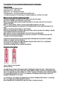
Figure 1.—Pathologic valve (arrow) in the lower portion of the internal jugular vein.
from the brain and spinal cord are found in very different combinations, regarding the type and the localization.3, 4, 12, 13 At the moment, the only published study on a larger group of MS patients, which describes anatomic characteristics of venous lesions comes from Italy.4 However, in this study a selected group of MS patients was assessed, with a relatively high percentage of primary progressive cases. This paper presents the findings in a less selected population of MS patients coming from 5 European countries (Poland, Slovakia, Germany, Switzerland and Netherlands). The aim of this study was to measure in an openlabel series the prevalence of extracranial venous flow anomalies according to the criteria elaborated by Italian researches.2, 4 Materials and methods Seventy patients were assessed (48 women and 22 men) with confirmed diagnosis of MS (according to McDonald criteria).14 There were 49 patients 110
with relapsing-remitting, 5 with primary progressive and 16 with secondary progressive clinical type of MS (according to Lublin et al. criteria 15). The patients were aged 15-58 years; median patients’ age was 38.5 years. They suffered from MS for 0.5-40 years; median duration of the disease was 7 years. All examined MS patients were Caucasians and they were citizens of the following European countries: Poland – 45 patients, Slovakia — 14 patients, Germany — 8 patients, Switzerland — 2 patients and Netherlands — 1 patient. This open-label study was the initial step of the ongoing randomized controlled clinical trial on surgical and endovascular treatments for CCSVI. The entire study has been approved by the ethics committee of the local medical university (Silesian Medical University in Katowice, Poland) and the patients have given their consent to have sonographic examination of veins in the neck. The patients were examined with color Doppler sonography: the IJVs and the vertebral veins (VVs) were assessed. Imaging of the veins was conducted on color Doppler ultrasound machines: GE Logiqe with 7.5-12 MHz linear probe and ATL Apogee 800 with 7.5 MHz linear probe. Examinations were done in the lying and the sitting positions in order to reveal four Zamboni’s extracranial criteria of CCSVI: 1) reflux in the internal jugular and/or vertebral veins over 0.88 s; 2) at least 50% stenosis of the proximal internal jugular vein/veins; 3) no detectable flow in the internal jugular and/or vertebral veins; 4) no position-dependent change in diameter of the internal jugular vein/veins.4 Because of the lack of special skills and sophisticated software needed on the ultrasonographic system used, we were unable to assess unequivo-
INTERNATIONAL ANGIOLOGY
April 2010
TABLE I.—Extracranial Doppler sonographic criteria of CCSVI in the present vs. Italian Study. Criteria of CCSVI
Number of patients assessed Reflux in IJV and/or VV Stenosis in IJV Not detectable flow in IJV and/or VV Negative ∆CSA* in the IJV Reflux in intracranial veins Conclusive analysis: ≥2 positive criteria At least one positive parameter
Present study
Italian study*
70 31/70 (42.8%) 61/70 (87.1%) 37/70 (52.9%) 28/70 (40.0%) Not assessed 63/70 (90.0%) 64/70 (91.4%)
109 76/109 (69.7%) 30/109 (27.5%) 35/109 (32.1%) 61/109 (56.0%) 55/109 (50.5%) 109/109 (100%) 109/109 (100%)
*Difference in cross-sectional area of the internal jugular vein assessed in the supine and sitting position.
cally the fifth Zamboni’s criterion 4 (reflux in the deep cerebral veins over 0.5 s), we relied only on the above-described four parameters for assessing the extracranial veins. The IJV, as well as the VVs, have been assessed using a high-frequency linear probe, similarly to the examination of carotid arteries. The probe applied minimal pressure to the skin, in order to prevent undesired compression of the examined vein. The search for venous lesions: stenoses, pathologic valves, etc. (Figures 1, 2) was primarily performed in the supine position, since in this hemodynamic condition the veins were physiologically dilated and therefore it was easier to find pathologies. However, all those veins were also examined in the sitting position, in order to look closer at hemodynamics,16-18 refluxes that were present only in this body posture and for changes in diameter of the IJVs.4, 12 Statistical analysis Distributions of venous lesions in the subgroups of the patients (clinical type of MS, sex, duration of the disease and citizenship) were assessed statistically using the χ2 and the Kolmogorov-Smirnov tests. Significance of these tests was set at P