Autoimmunity, February 2006; 39(1): 3 –8
Molecular mimicry in multiple sclerosis MIREIA SOSPEDRA1 & ROLAND MARTIN1,2 1
Unitat de Neuroimmunologia Clinica, Hospital Universitari Vall d’Hebron, Pg. Vall d’Hebron, 119-129, Barcelona 08035, Spain, and 2Institucio Catalana de Recerca i Estudis Avanc¸ats (ICREA), Barcelona, Spain
Abstract Two main etiological components are considered important in human autoimmune diseases including multiple sclerosis (MS), first the immunogenetic background and second environmental factors. Among the latter, infectious organisms are probably the most relevant, and epidemiological studies in MS firmly support that viral infections often precede disease exacerbations or the onset of MS. Infectious agents can contribute to disease development or phenotypic expression in different ways. Our focus will be directed on molecular mimicry, i.e. antigenic similarity between structural epitopes or peptide sequences from infectious organisms with those found in self proteins of the host. The intriguing concept of molecular mimicry has evolved substantially since its introduction over 20 years ago. We will summarize the most important developments and discuss puzzling questions, which remain open despite many claims that molecular mimicry is involved in the development of human autoimmune disease after infections or vaccinations.
Keywords: Multiple sclerosis, antigen-presenting cells, toll-like receptors, T cells
Introduction Infectious agents may lead to the onset or exacerbation of autoimmune diseases by different mechanisms. These include: (a) direct damage of a target tissue such as axons, myelin or oligodendrocytes in MS and subsequent immune responses to the released autoantigens or (b) indirect damage via activated immune cells that secrete proinflammatory cytokines and radicals, alteration of the stimulatory requirements of immune cells such as increased expression of costimulatory molecules and expression of molecular structures that activate either antigen-presenting cells (APCs) via toll-like receptors (TLRs) or T cells via superantigens. Both types of damage can lead to bystander activation of innate and adaptive immune mechanisms, but also to specific immune responses via molecular mimicry between infectious agent and self protein. Only molecular mimicry will be addressed here, and we will focus on antigen-specific T cells due to space limitations. Molecular mimicry in the context of antibodymediated immune responses refers primarily to the recognition of structural determinants on
macromolecules such as lipids, proteins or sugar moieties [1– 5]. Molecular mimicry and cross-recognition by T cells usually refers to the response to short peptide fragments that are generated from foreign or self proteins by the proteolytic machinery of APCs and then loaded onto to self major histocompatibility complex (MHC; human leukocyte antigens or HLA in humans) molecules. Since T cells do not recognize peptide in free form, but only as a complex with an MHC molecule, the distinction between structural or sequence mimicry is artificial, as will become clear below. Since the term molecular mimicry first appeared in the immunological literature over two decades ago [1 – 5], many studies have tried to establish its biological relevance in the context of the induction of autoimmune diseases or adverse events following vaccinations [6 – 8]. While there is no doubt that the ability for cross-recognition of antigens by both T cells and antibodies are essential properties of our adaptive immune system that assure protection from infections, it has become clear that molecular similarities between foreign and self proteins are usually not sufficient to lead to pathologic consequences.
Correspondence: R. Martin, Institucio Catalana de Recerca i Estudis Avanc¸ats (ICREA), Barcelona, Spain. E-mail:
[email protected] ISSN 0891-6934 print/ISSN 1607-842X online q 2006 Taylor & Francis DOI: 10.1080/08916930500484922
4
M. Sospedra & R. Martin
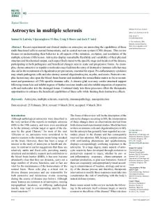
Figure 1. The evolution of the molecular mimicry concept in the context of T cell recognition is schematically depicted. (a) Sequence homology; (b) sharing of conserved TCR- and MHC- contact motifs between peptides. Upward arrows are towards the TCR, downward areas towards the MHC molecule; (c) no aa needs to be shared and molecular mimicry/T cell cross-reactivity can still occur as long as the additive stimulatory contribution of each aa in the peptides surpass a threshold that is necessary to stimulate the T cell clones. Upward arrows are towards the TCR, downward areas towards the MHC molecule; (d) structural mimicry. A single MBP (85–99)-specific TCR recognizes the MBP peptide in the context of DR2b (DRB1*1501) and a structurally similar complex of an EBV peptide in the context of DR2a (DRB5*0101).
Evolution of the concept of molecular mimicry of T cell responses Parallel to the growing knowledge as to how T cells recognize antigen, the molecular mimicry concept has changed considerably over time. While it was initially held that antigen recognition is very specific, and the analogy was often invoked that T cells fit to an antigen like a specific key into a lock, this view has given way to one that considers T cell recognition as degenerate or highly cross-reactive. To illustrate the evolution of these concepts, we will briefly recapitulate some important steps, but focus on examples that have been made in the fields of MS research. Soon after the realization that T cells recognize short peptides rather than conformational epitopes [9], investigators searched for homologous stretches of amino acids (aa) in proteins of foreign agents and those derived from an interesting self protein. To give one example, an essential study at this stage by Fujinami and Oldstone described that myelin basic protein (MBP) aa 66 – 75 shares six identical aa with
hepatitis B virus (HBV) polymerase peptide 589 –598 (Figure 1(a)), and injection of the HBV peptide into rabbits resulted in an encephalitis due to a cross-reactive immune reaction against the myelin peptide [3]. The reasoning that molecular mimicry only occurs if the same stretch of aa is shared by two proteins implied that cross-recognition is a rare event, since the likelihood of reoccurrence of for example a nonamer peptide in a randomly composed pool of proteins would be 1/209. Based on theoretical considerations that an animal’s/human’s T cell repertoire is orders of magnitude smaller than the potential realm of antigens that the immune system may encounter and hence potentially not protective [10], but also based on experimental observations that single T cell clones (TCCs) recognized more than one antigen and the progress in understanding the structural interactions between TCR and MHC/peptide complex [11,12], it was realized that cross-recognition does not require sequence homology. According to these considerations, Wucherpfennig and Strominger devised a search
Molecular mimicry in multiple sclerosis algorithm that was based on the assumption that molecular mimics need to share certain aa in distinct positions and with the proper spacing to assure binding to the respective HLA molecule that serves as restriction element for a TCC and other aa that contact the TCR (Figure 1(b)) [13]. When searching for such shared aa motifs in viral and bacterial databases that are recognized by MBP (85 –99)specific TCC, they identified seven viral- and one bacterial peptides that were recognized by three TCC. Based on these data, it was clear that molecular mimicry is probably a much more frequent event than originally assumed. As a next step, we speculated the overall contact surface of TCR and MHC/peptide complex is largely formed by direct interactions of the TCR with residues of the MHC molecule, while the peptide contributes relatively less, though importantly. Based on findings that every aa in a peptide can be exchanged as long as the primary TCR contact aa is kept in place [14], we further assumed that the primary TCR contact can probably also be replaced as long as the overall contribution to MHC/peptide recognition is kept above a threshold, i.e. critical for activating the TCC [15,16]. Using systematic aa exchanges of MBP (83– 99) and testing their relative stimulatory influence on a TCC, Hemmer et al. could thus show that a peptide that shares no aa with MBP (83 –99) can nevertheless be a molecular mimic (Figure 1(c)) [15]. While this case may be relatively rare and also difficult to demonstrate in most systems, it illustrated that the propensity for cross-reactivity of T cells is very broad and a very frequent event. Recently, these observations were extended beyond cross-reactivity at the level of the peptide. Lang et al. demonstrated in vitro, in vivo and in part also at the structural level that an MBP (85 –99)-specific TCC that is restricted by DRB1*1501, one of the MSassociated HLA-DR molecules in the DR15 haplotype, cross-reacts with MHC/peptide complexes formed by an Epstein –Barr virus (EBV) peptide and DRB5*0101, the second MS-associated DR allele in the DR15 haplotype (Figure 1(d)) [17]. The latter study broke an important barrier, i.e. that a given TCC uses only one restriction element, although prior work on alloreactivity had clearly suggested that crossrecognition and molecular mimicry must extend beyond variations of the antigenic peptide. As a further extension of these findings, we have recently identified in an unbiased fashion using combinatorial peptide libraries in the positional scanning format, that a single TCC that was isolated from the cerebrospinal fluid during MS exacerbation recognizes sets of identical foreign and self peptides in the context of all HLA-DR and even—DQ molecules that are co-expressed in the MS-associated DR15 haplotype [18]. Further research on this subject will have to address to which extent variations of the antigenic peptide
5
and/or the use of single or multiple restriction elements contribute to molecular mimicry. It has, however, already become clear that molecular mimicry and T cell cross-reactivity are frequent and physiologically essential to assure a T cell repertoire that is protective and not fraught with major holes. Evidence supporting a role of molecular mimicry in MS According to the above argument regarding the frequency of molecular mimicry and the physiological role of T cell cross-reactivity, one logical conclusion is that many claims that specific peptides from infectious organisms, vaccines or molecular mimics from autoantigens are relevant to the etiology of MS must be viewed with caution [19]. Establishing a possible causal relationship between an infectious pathogen and a T cell response against an autoantigen in MS requires a number of pieces of evidence that are rarely assembled and are in part impossible to provide. The search for a putative foreign agent that might be involved in MS can be started from several directions, from a reasonable candidate agent, a putative target autoantigen or disease-relevant T cells. The three main paths are summarized in Table I, and while these are not mutually exclusive, it is clear that the chain of evidence is not easy to establish. If one started with path I, epidemiological studies support that MS exacerbations are often preceded by common viral infections such as those caused by respiratory viruses [20]. Among the most recent candidate agents, the herpes viruses, human herpes virus type 6 (HHV6) [21 – 23] and EBV [24 – 28], appear most interesting. Both cause latent and life-long infections, have tropism for immune cells (EBV) and/or CNS (HHV6), can be periodically reactivated and restimulate immune cells, are highly prevalent in the population, and increased B- and/or T cell responses against these viruses have been demonstrated in MS patients. Searches for molecular mimics have identified homologous sequences or structural mimicry with MBP for both viruses [13,17,23,27]. However, while the reasoning is plausible that molecular mimicry between these common agents and a myelin component might be involved in MS, its role is far from established. Furthermore, creating suitable animal models for herpes viruses is impeded by their often exquisite tropism for human cells. The largest number of human studies has followed path II, based on the assumption that MBP is one relevant target autoantigen in MS. Mimicry searches for MBP-specific TCC that had with few exceptions [27] been established from the peripheral blood of MS patients with moderate to high concentrations of antigen yielded a number of interesting candidate molecular mimics, among them HHV6, EBV, papillomavirus and others [11,13,23,29]. Support for a
6
M. Sospedra & R. Martin Table I. Investigational steps to establish a role for molecular mimicry in MS.
Steps 1 2
3
Path I Identify disease-associated foreign agent (virus/bacterium); Examine T cell response against foreign agent in patients vs controls;
Identify disease-associated autoantigen Examine T cell response against autoantigen in patients versus controls;
If a disease-relevant foreign-agent-specific T cell response exists, search for autoantigens that are cross-recognized
If a disease-relevant autoantigen-specific T cell response exists, search for foreign agents that are cross-recognized
Following step 3, all paths follow the same steps 4 –6 4
5
6
Path II
Path III Identify disease-associated T cell population Use assumption as to which foreign agent or autoantigen might be recognized or (better) use unbiased search strategy*; Establish relevance of indentified agent/autoantigen by examining T cell response in several patients, ideally in a disease-relevant tissue compartment
Demonstrate infection of patient during or shortly after disease activity and/or release of autoantigen Animal model based on putative organism or autoantigen or T cell population or TCR specific for foreign agent Molecular mimicry relevant in animal model, that is, disease induction possible with autoantigen or foreign antigen, respectively?
Additional evidence: restriction of T cell response by disease-associated HLA-class II or -class I molecules? Association of certain foreign agents and/or molecular mimics with specific phenotypic subtypes or courses of the disease? * Examples are approaches that are either based (a) on all antigens or agents that are expressed in a certain tissue, e.g. by using expression libraries that reflect in an unbiased way the antigenic repertoire in a tissue that is affected, or alternatively or (b) on combinatorial peptide chemistry, e.g. combinatorial peptide libraries in the positional scanning format that reflect all possible antigens.
pathogenic role stems from animal models that showed that transgenic mice expressing MBP-specific TCR and the human HLA-DR molecule are susceptible to EAE or even develop spontaneous disease [30,31], however, evidence that infection with a pathogen that contains a molecular mimic for MBP causes an MS-like disease in animals is still scarce [32 – 34]. We have recently followed path III and focused on T cells that were isolated from a disease-relevant tissue (CSF) and clonally expanded during MS exacerbation in this compartment. We then searched for peptides that are recognized by these TCC in an unbiased way using positional scanning combinatorial peptide libraries and bioinformatics approaches that allowed us to scan the entire peptide repertoire that these clones might respond to. While so far limited to a single patient, three clonally expanded TCC that are restricted by MS-associated HLA-DR molecules, recognized evolutionary conserved protein domains in multiple ubiquitous pathogens and autoantigens [35]. The latter protein domains are characterized by a high content of arginine residues, which is typical for functional domains in nuclear localization signals and proteins binding to nucleic acids, thus providing a bridge how T cell recognition of domains that are common and shared between infectious organisms and self proteins can lead to autoreactivity. These observations are intriguing, but
will need to be expanded in more patients and larger numbers of TCC. In summary, there are a number of interesting leads that molecular mimicry might play a role in MS, however, each of these requires further follow-up. Circumstances leading to “pathologic” molecular mimicry It is not sufficient to demonstrate the pathologic relevance of molecular mimicry at the level of T cell recognition in vitro, the structural level, or even less by sequence comparisons. As stipulated in Table I, additional information is required to approach plausibility and possible relevance. The fact that cross-reactivity is so frequent and an inherent property of the adaptive immune system also implies that many safeguards are in place that prevent pathologic consequences of cross-recognition. Autoreactive T cells with high affinity TCR for self-MHC/self-peptide complexes are usually eliminated during maturation by clonal deletion in the thymus. Peripheral tolerance mechanisms such as anergy, activation-induced cell death and regulatory T cells represent further checkpoints in the case of autoreactive T cells that escape deletion due to lack of thymic expression of some autoantigens, or due to the low affinity of their TCRs. Our current understanding of the circumstances that are required to start a pathologic
Molecular mimicry in multiple sclerosis autoimmune response are incomplete, particularly in humans. However, most likely a combination of factors contributes. These include the expression of a susceptibility-conferring genetic background, particularly certain HLA-class II genes. Further factors are an infectious context and strong activation of both innate and adaptive immune systems, conditions that bias the immune response towards a proinflammatory T cell phenotype such as high antigen dose and the expression of certain cytokines, tropism of the inciting infectious organism for the target tissue, and local tissue destruction and/or immune activation in the target tissue. These interactions are supported by elegant experiments conducted by Miller et al. [32,33,36], who engineered a bacterial mimic peptide (from Haemophilus influenzae) of the immunodominant myelin proteolipid protein (PLP) peptide (139– 151) into a non-pathogenic strain of Theiler’s murine encephalitis virus (TMEV). They document that tolerance to PLP (139 – 151) cannot be broken by immunization with the mimic peptide even if injected in complete Freund’s adjuvant, while infection with the engineered TMEV strain leads to rapid-onset demyelinating disease via activation of a PLP (139 – 151)-specific Th1 response [33]. These data not only provide strong evidence for infection-induced molecular mimicry with pathologic consequences, but also demonstrate that a mimic peptide can be processed in a way that results in strong activation of proinflammatory cells recognizing the self-peptide.
Conclusions Molecular mimicry remains an elegant concept to explain how cross-reactive T cell responses can elicit autoimmune diseases, but it is also important to understand how a limited T cell repertoire can protect us against a much larger number of potential pathogens. As is highlighted in this article, molecular mimicry is a very frequent and in most instances physiologic event. When considering it as an etiologic factor for autoimmune diseases, a number of criteria should be met in order to provide plausibility of such an interaction. Future studies should try to address as many of these criteria as possible when proposing a role of molecular mimicry in an autoimmune disease.
References [1] Fujinami RS, Oldstone MBA, Wroblewska Z, Frankel ME, Koprowski H. Molecular mimicry in virus infection: Crossreaction of measles virus phosphoprotein or of herpes simplex virus protein with human intermediate filaments. Proc Natl Acad Sci USA 1983;80:2346 –2350. [2] Abromson-Leeman SR, Cantor H. Specificity of T cell clones for antigen and autologous major histocompatibility complex products determines specificity for foreign major histocompatibility complex products. J Exp Med 1983;158:428–437.
7
[3] Fujinami RS, Oldstone MBA. Amino acid homology between the encephalitogenic site of myelin basic protein and virus: Mechanism for autoimmunity. Science 1985;230:1043– 1045. [4] Dyrberg T, Oldstone MBA. Peptides as probes to study molecular mimicry and virus-induced autoimmunity. Curr Top Microbiol Immunol 1986;130:25–37. [5] Schwimmbeck PL, Yu DT, Oldstone MBA. Autoantibodies to HLA B27 in the sera of HLA B27 patients with ankylosing spondylitis and Reiter’s syndrome. Molecular mimicry with Klebsiella pneumoniae as potential mechanism of autoimmune disease. J Exp Med 1987;166:173–181. [6] Benoist C, Mathis D. Autoimmunity provoked by infection: How good is the case for T cell epitope mimicry? Nat Immunol 2001;9:797 –801. [7] Regner M, Lambert PH. Autoimmunity through infection or immunization? Nat Immunol 2001;2:185– 188. [8] Wraith DC, Goldman M, Lambert PH. Vaccination and autoimmune disease: What is the evidence? Lancet 2003; 362:1659–1666. [9] Townsend AR, Rothbard J, Gotch FM, Bahadur G, Wraith D, McMichael AJ. The epitopes of influenza nucleoprotein recognized by cytotoxic T lymphocytes can be defined with short synthetic peptides. Cell 1986;44:959–968. [10] Mason D. A very high level of crossreactivity is an essential feature of the T-cell receptor. Immunol Today 1998; 19:395–404. [11] Hemmer B, Fleckenstein B, Vergelli M, Jung G, McFarland ¨ ller K-H. Identification of high HF, Martin R, WiesmY potency microbial and self ligands for a human autoreactive class II-restricted T cell clone. J Exp Med 1997;185: 1651–1659. [12] Garboczi DN, Ghosh P, Utz U, Fan QR, Biddison WE, Wiley DC. Structure of the complex between human T-cell receptor, viral peptide and HLA-A2. Nature 1996;384:134 –141. [13] Wucherpfennig KW, Strominger JL. Molecular mimicry in T cell-mediated autoimmunity: Viral peptides activate human T cell clones specific for myelin basic protein. Cell 1995; 80:695–705. [14] Evavold BD, Sloan-Lancaster J, Wilson KJ, Rothbard JB, Allen PM. Specific T cell recognition of minimally homologous peptides: Evidence for multiple endogenous ligands. Immunity 1995;6:655–663. [15] Hemmer B, Vergelli M, Gran B, Ling N, Conlon P, Pinilla C, Houghten R, McFarland HF, Martin R. Predictable TCR antigen recognition based on peptide scans leads to the identification of agonist ligands with no sequence homology. J Immunol 1998;160:3631–3636. [16] Hemmer B, Pinilla C, Gran B, Vergelli M, Ling N, Conlon P, McFarland HF, Houghten R, Martin R. Contribution of individual amino acids within MHC molecule or antigenic peptide to TCR ligand potency. J Immunol 2000; 164:861– 871. [17] Lang HL, Jacobsen H, Ikemizu S, Andersson C, Harlos K, Madsen L, Hjorth P, Sondergaard L, Svejgaard A, Wucherpfennig K, Stuart DI, Bell JI, Jones EY, Fugger L. A functional and structural basis for TCR cross-reactivity in multiple sclerosis. Nat Immunol 2002;3:940–943. [18] Sospedra M, Muraro PA, Stefanova I, Zhao Y, Chung K, Li Y, Giulianotti M, Simon R, Mariuzza R, Pinilla C, Martin R. Redundancy in antigen presenting function of the HLA-DR and -DQ molecules in the multiple sclerosis-associated HLA-DR15 haplotype. J Immunol 2005; in press. [19] Fourneau JM, Bach JM, van Endert PM, Bach JF. The elusive case for a role of mimicry in autoimmune diseases. Mol Immunol 2004;40:1095 –1102. [20] Kriesel JD, Sibley WA. The case for rhinoviruses in the pathogenesis of multiple sclerosis. Mult Scler 2005;11:1–4. [21] Challoner PB, Smith KT, Parker JD, MacLeod DL, Coulter SN, Rose TM, Schultz ER, Bennett JL, Garber RL, Chang M,
8
[22]
[23]
[24]
[25]
[26]
[27]
[28]
M. Sospedra & R. Martin Schad PA, Stewart PM, Nowinski RC, Brown JP, Burmer GC. Plaque-associated expression of human herpesvirus 6 in multiple sclerosis. Proc Natl Acad Sci USA 1995;92:7440 –7444. Soldan SS, Berti R, Salem N, Secchiero P, Flamand L, Calabresi PA, Brennan MB, Maloni HW, McFarland HF, Lin HC, Patnaik M, Jacobson S. Association of human herpes virus 6 (HHV-6) with multiple sclerosis: Increased IgM response to HHV-6 early antigen and detection of serum HHV-6 DNA. Nat Med 1997;3:1394–1397. Tejada-Simon MV, Zang YC, Hong J, Rivera VM, Zhang JZ. Cross-reactivity with myelin basic protein and human herpesvirus-6 in multiple sclerosis. Ann Neurol 2003; 53:189–197. Wandinger K, Jabs W, Siekhaus A, Bubel S, Trillenberg P, Wagner H, Wessel K, Kirchner H, Hennig H. Association between clinical disease activity and Epstein– Barr virus reactivation in MS. Neurology 2000;55:178–184. Ascherio A, Munger KL, Lennette ET, Spiegelman D, Hernan MA, Olek MJ, Hankinson SE, Hunter DJ. Epstein-Barr virus antibodies and risk of multiple sclerosis: A prospective study. JAMA 2001;286:3083–3088. Cepok S, Zhou D, Srivastava R, Nessler S, Stei S, Bussow K, Sommer N, Hemmer B. Identification of Epstein–Barr virus proteins as putative targets of the immune response in multiple sclerosis. J Clin Investig 2005;115:1352–1360. Holmoy T, Kvale EO, Vartdal F. Cerebrospinal fluid CD4+T cells from a multiple sclerosis patient cross-recognize Epstein– Barr virus and myelin basic protein. J Neurovirol 2004; 10:278–283. Levin LI, Munger KL, Rubertone MV, Peck CA, Lennette ET, Spiegelman D, Ascherio A. Temporal relationship between elevation of Epstein–Barr virus antibody titers and initial onset of neurological symptoms in multiple sclerosis. JAMA 2005;293:2496–2500.
[29] Ufret-Vincenty RL, Quigley L, Tresser N, Pak SH, Gado A, Hausmann S, Wucherpfennig KW, Brocke S. In vivo survival of viral antigen-specific T cells that induce experimental autoimmune encephalomyelitis. J Exp Med 1988;188: 1725–1738. [30] Madsen LS, Andersson EC, Jansson L, Krogsgaard M, Andersen CB, Engberg J, Strominger JL, Svejgaard A, Hjorth JP, Holmdahl R, Wucherpfennig KW, Fugger L. A humanized model for multiple sclerosis using HLA-DR2 and a human T-cell receptor. Nat Genet 1999;23:343–347. [31] Quandt JA, Baig M, Yao K, Kawamura K, Huh J, Ludwin SK, Bian HJ, Bryant M, Quigley L, Nagy ZA, McFarland HF, Muraro PA, Martin R, Ito K. Unique clinical and pathological features in HLA-DRB1*0401-restricted MBP 111-129specific humanized TCR transgenic mice. J Exp Med 2004; 200:223– 234. [32] Croxford JL, Anger HA, Miller SD. Viral delivery of an epitope from Haemophilus influenzae induces central nervous system autoimmune disease by molecular mimicry. J Immunol 2005; 174:907– 917. [33] Croxford JL, Olson JK, Anger HA, Miller SD. Initiation and exacerbation of autoimmune demyelination of the central nervous system via virus-induced molecular mimicry: Implications for the pathogenesis of multiple sclerosis. J Virol 2005; 79:8581– 8590. [34] Fujinami RS. Molecular mimicry that primes for autoimmunity which is triggered by infection. Mol Psychiatry 2002; 2(7):S32–S33. [35] Sospedra M, Zhao Y, Zur Hausen H, Muraro PA, Hamashin C, de Villiers E-M, Pinilla C, Martin R. Recognition of conserved amino acid motifs of common viruses and its role in autoimmunity. PLoS Pathogens 2005; in press. [36] Olson JK, Croxford JL, Calenoff MA, Dal Canto MC, Miller SD. A virus-induced molecular mimicry model of multiple sclerosis. J Clin Investig 2001;108:311 –318.