Chapter 11
After a fracture, the primary goal of healing is to regenerate mineralized tissue in the fracture area and restore mechanical strength to the bone. In addition, reconstituting normal soft tissue gliding and movement about the fracture site is important.
u
Answer: E LaStayo, Winters, Hardy, pp. 81-82
2. The phases of fracture healing include which of the following? A. B. C. D.
Inflammation, repair, and remodeling phase Inflammation, repair, and revascularization phase Repair, revascularization, and remodeling phase Repair, regeneration, and remodeling phase
The phases of fracture healing include inflammation, repair, and remodeling. In the inflammation phase, the
Answer: A
u
A. Regenerate mineralized tissue in the fracture area B. Restore mechanical strength to the bone C. Reconstitute normal soft tissue gliding and movement about the fracture site D. A and C only E. All of the above
hematoma develops and provides stability at the site and tissue regeneration is initiated. In the repair phase, the callus is formed to stabilize the fracture even more and granulation tissue replaces the hematoma. In the remodeling phase, there is resorption and deposition of bone to restore the strength of the bone.
LaStayo, Winters, Hardy, pp. 82-84
3. True or False: Secondary healing occurs without a hematoma.
Primary fracture healing occurs without a hematoma. Primary healing is characterized by simultaneous union of fracture ends. It occurs with a mechanically stable environment with motionless fixation such as with plates and screws. In contrast, the process of secondary healing is characterized by three discrete yet overlapping stages: inflammation, repair, and remodeling. The inflammatory stage begins immediately after fracture, initiating cellular and vascular responses to injury that promote fracture stabilization through early hematoma clotting. Secondary healing occurs with fractures managed by closed reduction or with semirigid fixation methods (e.g., Kirschner wires, external fixators, intramedullary pins). Answer: False
u
1. The goal of fracture healing is to do which of the following?
LaStayo, Winters, Hardy, pp. 82-83
Refer to Fig. 11-1
201
11 FRACTURES
Fractures
202
CHAPTER 11 ■ Fractures
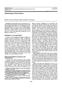
Fig. 11-1 ■ Secondary fracture healing. The three phases of fracture healing with relative time duration are pictured. In the inflammatory phase, the fracture hematoma clots and serves as the initial support. Inflammatory cells invade the hematoma to remove necrosed bone and debris. During the reparative phase, new capillaries are formed, thus providing nutrition for the formation of callus by fibroblasts, chondroblasts, and osteoblasts. The initial soft callus is converted into hard callus, then mineralized into bone during the remodeling phase. Return of medullary and periosteal blood flow also occurs in this final phase. Remodeling of the bone in response to stress to its normal preinjury configuration can take years. (From Brand RA: Fracture healing. In Albright JA, Brand RA: The scientific basis of orthopaedics, New York, 1979, McGraw-Hill.)
CHAPTER 11 ■ Fractures
203
4. True or False: Stable internal fixation allows the therapist to initiate rehabilitation sooner than closed reduction.
Stable internal fixation provides the rigidity necessary to allow initiation of range of motion (ROM) to the involved extremity earlier than closed reduction. Internal fixation allows the increased stability of bone fragments in proper alignment to allow rapid rehabilitation.
Jabaley, Wegener, p. 96 Krop in Mackin, Callahan, Skirven, et al, p. 377
CLINICAL GEM: Despite stability gained from the procedure, the therapist must respect the soft tissue and the healing process. Fig. 11-2
A. B. C. D. E.
Shaft of the proximal phalanx Shaft of the middle phalanx Metacarpal neck Metacarpal base Distal interphalangeal (DIP) joint
A boxer’s fracture is a fracture involving the metacarpal neck. It usually involves the ring and small fingers and occurs when a clenched metacarpophalangeal (MCP) joint strikes a solid object. Nonunion almost never occurs, but malunion can be a complication. Patients complain of a loss of prominence of the metacarpal head and decreased ROM, and they can palpate the metacarpal head in the palm on occasion. Treatments include closed reduction, closed reduction with percutaneous pin fixation, and/or open reduction (Fig. 11-2).
u
Answer: C Green, pp. 698-700
6. A boxer’s fracture is most commonly seen in which digits? A. B. C. D.
First and second metacarpals Second and third metacarpals Third and fourth metacarpals Fourth and fifth metacarpals
A metacarpal neck fracture, or boxer’s fracture, is most commonly seen in the fourth and fifth metacarpals. This fracture occurs when the clenched fist strikes an object at an oblique angle. The boxer’s fracture often is treated with a cast or ulnar gutter splint for approximately 3 to 3.5 weeks to allow the fracture pain to subside and sufficient healing to occur. Surgical treatment can be performed for cosmetic reasons and to avoid a palmar metacarpal head deformity, which interferes with highdemand grasping activities. Answer: D
u
5. The term “boxer’s fracture” refers to a fracture of which of the following?
■ Radiograph of a minimally displaced fracture of the metacarpal neck (boxer’s fracture). (From Hunter JM, Mackin EJ, Callahan AD: Rehabilitation of the hand: surgery and therapy, ed 4, St Louis, 1995, Mosby.)
Diao, p. 564
Refer to Fig. 11-2
11 FRACTURES
u
Answer: True
CHAPTER 11 ■ Fractures
7. True or False: Stable, internal fixation allows fractures to heal faster.
Stable, internal fixation allows for a precise restoration of parts, but it does not make fractures heal faster. It does, however, help them heal more precisely; it also allows for primary bone healing. During primary bone healing, direct deposition of bone in the fracture site occurs without the intermediate phase of cartilage formation and without the formation of an external callus. The major benefit of stable fixation is that early rehabilitation can be initiated. Stable internal fixation also is a deterrent to the development of a chronically painful, swollen, and stiff hand. Although internal fixation does not necessarily speed fracture healing, it may reduce the time required before the patient can return to productive work or leisure.
The stability of a fracture significantly affects the quantity and quality of callus formation. In general, greater amounts of motion at a fracture site result in a greater amount of callus. It is as if the fracture forms an internal splint. In contrast, very stable fixation with accurate reduction results in very small amounts of callus formation. Immobilization and very stable fixation both have advantages and disadvantages. Answer: True
u
204
McCollister, p. 105
10. The cylindrical shaft of a long bone is which of the following? A. B. C. D.
Metaphysis Epiphysis Diaphysis None of the above
Freeland, Jabaley, Hughes, p. 28
8. Which joint(s) in the hand, after developing stiffness, causes the most serious functional loss? A. B. C. D.
DIP Proximal interphalangeal (PIP) MCP All of the above contribute equally to functional loss
PIP joint stiffness results in the most serious functional loss. The PIP joint is crucial to function, and once it is stiffened, correction is quite difficult. The MCP joint can become stiffened in extension, but typically it can be released. The DIP joint contributes minimally to the flexion arc; therefore loss of motion at this level is not as crucial.
u
Answer: B McCollister, p. 1297
9. True or False: The stability of a fracture significantly affects the quantity and quality of callus formation.
The diaphysis is the cylindrical shaft of a bone. The metaphysis is the growing portion of a bone; this is the part between the diaphysis and the epiphysis. The epiphysis is the ossification center at each extreme end of the long bones. When bone growth is complete, the diaphysis is fused with the epiphysis by bony synostosis. Fusion of the epiphysis with the diaphysis occurs approximately 1 to 2 years earlier in females than in males. In general, male bone growth is complete by age 20 and female bone growth is complete by age 18. A radiologist can determine the bone age of a person by studying the ossification center. Answer: C
u
u
Answer: False
Taber’s cyclopedic medical dictionary Netter, p. 131
Refer to Fig. 11-3
CLINICAL GEM: The medial epiphysis of the clavicle, which is the last epiphysis of the long bones to appear in the body, develops between the ages of approximately 18 and 20. This epiphysis also is the last to close; closure occurs between the ages of approximately 23 and 25.
CHAPTER 11 ■ Fractures
205
12. What is the physician’s treatment of choice for stable hyperextension and dorsal dislocation injuries of the PIP joint? A. B. C. D.
Open reduction internal fixation (ORIF) Order a dynamic splint K-wire DIP joint in full extension Closed reduction
u
Answer: D
■ A long bone in a child. (From Mackin EJ, Callahan AD,
Skirven TM, et al: Rehabilitation of the hand and upper extremity, ed 5, St Louis, 2002, Mosby.)
11. Which of the following fractures is easiest to rehabilitate? A. B. C. D.
Distal phalanx Middle phalanx Proximal phalanx Metacarpal
The distal phalanx is the easiest to rehabilitate because of its anatomic relationship with bone and the surrounding soft tissue. There is only a slight chance of the flexor tendon and the terminal tendon becoming adherent as they insert on the distal phalanx. The primary difficulties noted with rehabilitation of these fractures are pain and hypersensitivity due to the sensory nerve endings surrounding the fingertip. Desensitization of the digit tip is initiated as the site is healed.
u
Answer: A Chinchalkar, Gan, p. 118 Purdy, Wilson in Mackin, Callahan, Skirven, et al, p. 385
13. True or False: Pilon fractures can be treated safely using dynamic traction.
A pilon fracture is a comminuted intraarticular fracture of the base of the middle phalanx (Fig. 11-4, A). It can be treated with ORIF, external fixation, or dynamic traction (Fig. 11-4, B). Schenck has popularized the concept of dynamic traction. Using this method, a K-wire is put through the head of the middle phalanx, and traction is applied to the phalanx with rubber-band traction attached to a hoop mount on a forearm splint. The patient regularly performs passive ROM for both flexion and extension in a specified range. Splinting is continued for 6 to 8 weeks. Studies have shown that the skeletal traction technique can be safer and that it produces results equivalent to those of ORIF. Answer: True
u
Fig. 11-3
Chinchalkar, Gan, p. 121
Baratz, Divelbiss, pp. 541-555 Hunter, Mackin, Callahan, p. 384 Campbell, Wilson in Mackin, Callahan, Skirven, et al, p. 401 Jebson, Kasdan, p. 157 Jacobs, Austin, p. 328 Chinchalkar, Gan, p. 125
11 FRACTURES
The physician’s treatment of choice for stable hyperextension and dorsal dislocation injuries of the PIP joint is a closed reduction. The patient is often placed in a dorsal block splint with buddy taping to the next digit for approximately 4 to 6 weeks to allow early motion to begin. ROM exercises are performed with focus on active blocking exercises, which also assist with decreasing any tightness in the oblique retinacular ligament.
206
CHAPTER 11 ■ Fractures
The most commonly fractured bone in the hand is the distal phalanx, which accounts for 45% to 50% of all hand fractures. The thumb and middle fingers are most commonly involved. Fractures of the distal phalanx often are the result of crushing injuries. Fortunately, fractures of the distal phalanx usually heal without excessive treatment.
u
Answer: A Hunter, Mackin, Callahan, p. 360 Purdy, Wilson in Mackin, Callahan, Skirven, et al, p. 384
16. Which of the following is the most serious complication of a proximal phalanx fracture? Fig. 11-4
14. A 27-year-old male laborer presents with a bullet wound through the MCP joint of his dominant thumb. He works in construction, has three children, and finished the tenth grade in high school. He has intact sensation and intact flexor and extensor tendon function but cannot pinch because of severe posttraumatic arthritis of the MCP joint. The best treatment includes which of the following? A. B. C. D. E.
Arthrodesis of the first MCP joint MCP joint arthroplasty Short opponens splint Radial thumb spica splint Second metatarsal phalangeal (MTP) toe-to-thumb transplant
A. B. C. D.
PIP joint extension contracture MCP joint extension contracture MCP joint flexion contracture PIP joint flexion contracture
After a proximal phalanx fracture, development of a fixed PIP joint flexion contracture is the most serious complication because of the associated functional loss. The most effective way to avoid this complication is to splint the PIP joint in full extension to avoid collateral ligament tightness. A dynamic PIP joint extension splint should be initiated at the first sign of flexion deformity. PIP joint flexion contracture also is a serious complication after middle phalanx fractures. Answer: D
u
■ A, Pilon fracture. B, Dynamic traction. (From Hunter JM, Mackin EJ, Callahan AD: Rehabilitation of the hand: surgery and therapy, ed 4, St Louis, 1995, Mosby.)
Hunter, Mackin, Callahan, p. 367 Purdy, Wilson in Mackin, Callahan, Skirven, et al, p. 389
17. What is considered functional flexion for the MCP, PIP, and DIP joints, respectively? A painful unstable MCP joint in a laborer with a need to return to work is best treated by arthrodesis. Arthrodesis will allow the patient to return to work the quickest with the least amount of rehabilitation.
A. B. C. D.
51 69 61 28
degrees, degrees, degrees, degrees,
39 50 60 42
degrees, degrees, degrees, degrees,
32 21 39 30
degrees degrees degrees degrees
u
Answer: A
15. Which bone in the hand is most commonly fractured? A. B. C. D.
Distal phalanx Middle phalanx Proximal phalanx Metacarpal shaft
Functional flexion averages 61 degrees at the MCP joint level, 60 degrees at the PIP joint level, and 39 degrees at the DIP joint level. Functional motion for flexion of the thumb is 21 degrees at the MCP joint level and 18 degrees at the interphalangeal (IP) joint level. These measures are based on common activities of daily living. They are not used for addressing individual activities or work skills. They do, however, provide a basic guideline for functional performance of the hand.
CHAPTER 11 ■ Fractures
A. B. C. D.
Tuft fracture Shaft fracture Base fracture All the above
Base fractures usually are unstable because of the pull of the flexor and extensor tendons at the fracture site; they also tend to angulate the fracture with a dorsal apex. Closed fractures usually can be managed with a short Alumafoam splint, which holds the distal phalanx in extension. If the fracture is unstable or open, it is best to treat it with K-wire fixation. Tuft fractures often are caused by crush injuries and usually are very painful, but they are inherently stable. However, if the disruption of the nail and pulp occur with an open fracture, the fracture is likely to be unstable. Shaft fractures usually have minimal displacement and are stable; they also may be either longitudinal or transverse.
u
Answer: C Hunter, Mackin, Callahan, pp. 361-362
Refer to Fig. 11-5
Fig. 11-5
■ From Hunter JM, Mackin EJ, Callahan AD: Rehabilitation of the hand: surgery and therapy, ed 4, St Louis, 1995, Mosby.
CLINICAL GEM: The most common problems after distal phalanx fractures, especially crush type injures, are pain and hypersensitivity.
A. Figure 8 splints to maintain PIP in slight flexion B. Volar MCP joint blocking splint to maximize flexor tendon excursion C. Dorsal MCP blocking splints D. A and B only E. A and C only
A swan-neck deformity is hyperextension of the PIP joint and flexion of the DIP joint. When the PIP joint loses flexion because of the swan-neck deformity, a hyperflexion at the MCP joint may occur. This is increased hyperflexion causes decreased gliding of the flexor digitorum profundus (FDP) tendons because of the overload of forces on the intrinsic musculature. To treat this deformity, splint with figure 8 splints to maintain the PIP joint in slight flexion and a volar MCP joint blocking splint to maximize flexor tendon excursion. Answer: D Jacobs, Austin, p. 355 Chinchalkar, Gan, pp. 126-127
20. The functional position splint for a nondisplaced fracture of the proximal phalanx at rest should be which of the following? A. MCPs at 60 to 70 degrees of flexion, PIPs left free B. MCPs at 60 to 70 degrees of flexion, IPs at 0 degrees of extension C. MCPs at 60 to 80 degrees of flexion, IPs at 20 to 30 degrees of flexion D. MCPs at 60 to 80 degrees of flexion, IPs at 40 to 50 degrees of flexion
The functional position of a fractured proximal phalanx in the correct position is imperative for proper healing. The splint should place the MCPs in 60 to 70 degrees of flexion and the IPs at 0 degrees of extension. This position allows the MCP joints to keep the collateral ligaments at their appropriate lengths, thereby preventing MCP extension contractures. Placing the IPs in extension prevents flexion contractures from occurring. This functional position splint allows for fracture healing and also allows for mobilization of the uninvolved joints. Answer: B Jacobs, Austin, pp. 328-329
11 FRACTURES
18. Which of the following distal phalanx fractures is inherently unstable because of the pull of the tendons?
19. If a PIP joint develops a swan-neck deformity and produces an unusual hyperflexion at the MCP, corrective splinting could include which of the following to correct both deformities?
u
Hunter, Mackin, Callahan, p. 1185 Cannon in Mackin, Callahan, Skirven, et al, p. 1071
u
u
Answer: C
207
208
CHAPTER 11 ■ Fractures
21. True or False: Buddy taping is the treatment of choice for a stable nondisplaced or minimally displaced fracture of the PIP.
An excellent treatment for a stable nondisplaced or minimally displaced fracture in a motivated compliant patient is buddy taping to the adjacent digit. Many patients, when instructed in the blocking and gliding exercises, are able to progress well with buddy taping. The active motion exercises compress the fracture site and stimulate callus formation, thus allowing healing to occur.
u
Answer: True Clark, Wilgis, Aiello, p. 323 Freeland, Hardy, Singletary, p. 131
Refer to Fig. 11-6
22. A 35-year-old roofer is diagnosed with a boxer’s fracture of the fifth metacarpal, with 30 degrees of angulation through the fracture. The patient is anxious to return to work. Which of the following would be the best treatment and splint application? A. Closed reduction and splint application with the MCP joint flexed at 60 degrees for 3 weeks B. Closed reduction and splint application with the MCP joint neutral for 3 weeks C. Application of a static extension splint to the fifth digit with immediate active ROM D. Application of a hand-based splint with the MCP joint free for 3 weeks, followed by immediate active ROM
Closed reduction, with either a cast application or use of an ulnar gutter splint positioning the fourth and fifth MCP joints at 60 degrees of flexion, has been a successful treatment of the boxer’s fracture of the fifth metacarpal. The splint typically is worn for 3 to 6 weeks. Active ROM may be initiated as early as 2 weeks. Immediate motion is contraindicated in patients with boxer’s fractures because a loss of reduction may occur. However, immediate ROM may be initiated if the fracture is absolutely stable, as in ORIF.
u
Answer: A Light, Bednar, pp. 303-314 Hunter, Mackin, Callahan, pp. 370-371 Purdy, Wilson in Mackin, Callahan, Skirven, et al, pp. 388-395 Campbell, Wilson in Mackin, Callahan, Skirven, et al, pp. 396-409
CLINICAL GEM: The most common complication after a metacarpal fracture is disproportionate dorsal edema.
23. True or False: A patient with osteoporosis has an accelerated loss of bone mass, which leaves the skeleton weakened and more vulnerable to fracture.
Fig. 11-6
■ Velcro straps can be fabricated in the clinic and used instead of taping. (From Mackin EJ, Callahan AD, Skirven TM, et al: Rehabilitation of the hand and upper extremity, ed 5, St Louis, 2002, Mosby.)
Osteoporosis is insidious in nature. It is a progressive disease that causes an accelerated loss of bone mass, thus leaving the skeleton weak and vulnerable to fracture. Fractures of the proximal humerus, pelvis, distal radius,
CHAPTER 11 ■ Fractures
209
and ribs are present in approximately 20 million osteoporotic individuals in the United States. These fractures cause varying degrees of pain, disability, and loss of independence. Impact activities, such as walking, can increase bone mass before age 35 and maintain bone mass after age 35; therefore it is important that patients with osteoporosis participate in an exercise program that stresses impact exercise.
u
Answer: True McCollister, p. 177
24. If the elbow is dislocated in a posterolateral direction, which structure is ruptured in nearly all cases? Distal biceps insertion Triceps tendon Medial collateral ligament Posterior interosseous ligament
11 FRACTURES
A. B. C. D.
A complete disruption of the medial collateral ligament of the elbow is seen in nearly all cases of posterolateral dislocation.
u
Answer: C Browner, Jupiter, Levine, p. 1144
25. A Galeazzi fracture is which of the following? A. Fracture of the distal radial shaft with subluxation/dislocation of the distal radioulnar (DRU) joint B. Fracture of the radius and ulna at the same level C. Fracture of the ulna shaft with disruption of the radiohumeral joint D. Fracture of the distal ulna with disruption of the DRU joint
Fig. 11-7
■ From Cooney WP, Linscheid RL, Dobyns JH: The wrist: diagnosis and operative treatment, St Louis, 1998, Mosby.
26. A Monteggia lesion is which of the following? A. Fracture of the radius and ulna at the same level B. Fracture of the proximal ulna with dislocation of the radial head C. Radial head fracture with dislocation D. Fracture of the distal radius shaft with disruption of the DRU joint
A Galeazzi fracture is a distal radial shaft fracture with subluxation/dislocation of the DRU joint.
Browner, Jupiter, Levine, p. 1113 Purdy, Wilson in Mackin, Callahan, Skirven, et al, pp. 393-394
Refer to Fig. 11-7
A Monteggia lesion is a fracture of the proximal ulna with dislocation of the radial head (see Fig. 9-10). Answer: B
u
u
Answer: A
Browner, Jupiter, Levine, p. 1117
CHAPTER 11 ■ Fractures
27. True or False: Boxer’s fractures can be treated by the physician with the Jahss maneuver.
A boxer’s fracture is a fracture of the metacarpal neck, primarily of the ring and small digits. Metacarpal neck fractures are the most common of all the metacarpal fractures. The Jahss maneuver is used to reduce a fracture of the metacarpal neck. To perform the maneuver, the metaphalangeal (MP) joint is flexed to 90 degrees, and an upward pressure is exerted while using the proximal phalanx on the metacarpal head. Most boxer’s fractures are treated with closed reduction and splint immobilization for 3 to 4 weeks.
Brach, Goitz, p. 153
30. The most significant limitation for a patient suffering from a fracture of the hamate is which of the following? A. B. C. D.
Loss Loss Loss Loss
of of of of
motion grip strength pinch strength sensation
Jebson, Kasdan, pp. 159-160 McNemar, Howell, Chang, pp. 147-148
28. The central slip and the lateral bands work together to do which of the following? A. B. C. D.
Answer: C
Flex the PIP joint Extend the PIP joint Flex and extend the PIP joint Flex the MCP joint
An intricate part of fracture management is tendon gliding. Exercises are performed to prevent adhesions and regain joint motion. After a proximal phalanx fracture, achieving 0 to 40 degrees of motion in the initial 4 weeks after injury is important. When the PIP joint is flexed, the central tendon initiates extension, and the lateral bands work more at the end of PIP joint extension.
Fractures of the hook of the hamate are the most common of hamate fractures. They occur primarily from direct trauma or crush injuries. Computed tomography (CT) scans are helpful in determining the fracture. Acute nondisplaced fractures are treated in a short arm cast, whereas displaced fractures require surgery with hook excision. Loss of grip strength is the primary limitation that these patients suffer, and this can be addressed with a strengthening program. Hypersensitivity versus loss of sensitivity caused by ulnar nerve irritation is the second most common limitation noted. Answer: B
u
u
Answer: True
The scaphoid is the most commonly fractured carpal bone. A humpback deformity is noted when displacement of the scaphoid with excessive flexion at the fracture site occurs. Surgical intervention is necessary via a dorsal or volar approach to correct this deformity.
u
210
Dell, Dell in Mackin, Callahan, Skirven, et al, p. 1175 Brach, Goitz, pp. 156-157
31. True or False: Angulation is more disabling than malrotation with respect to metacarpal shaft fractures.
Freeland, Hardy, Singletary, pp. 137-138
29. The term humpback deformity refers to which of the following? A. Displacement of the scaphoid and excessive extension at the fracture site B. Fracture of the distal pole of the scaphoid C. Displacement of the scaphoid with excessive flexion at the fracture site D. Fracture of the proximal pole of the scaphoid
Malrotation can be more disabling than angulation because of the tendency for digits to overlap. Some authors have stated that for every degree of malrotation in the metacarpal there are 5 degrees of malrotation at the fingertip. As little as 5 degrees of rotation produces a 1.5-cm overlap in the fingertips on flexion. Answer: False
u
u
Answer: B
Hunter, Mackin, Callahan, p. 368 Purdy, Wilson in Mackin, Callahan, Skirven, et al, pp. 390-391 Smith, p. 69
Refer to Fig. 11-8
211
Fig. 11-8
■ A and B, This patient had sustained a fracture of the metacarpal of the right ring finger, and insufficient attention was paid to obtaining the correct rotational alignment. This resulted in a deformity functionally and cosmetically unsatisfactory to the patient and embarrassing for the surgeon. C, Although the spiral fracture of the fourth metacarpal is not easily seen, the entirely unsatisfactory rotation in the finger can be readily appreciated. This must be corrected. (From Smith P: Lister’s The hand: diagnosis and indications, ed 4, London, 2003, Churchill Livingstone.)
32. A volar MCP joint capsulectomy is appropriate for all but which of the following diagnoses? A. B. C. D.
Intrinsic muscle contractures Dupuytren’s contracture Prolonged immobilization Extension contracture
A capsulectomy is surgical removal of a capsule; a capsulotomy involves cutting into the capsule. Some authors use these terms interchangeably. Volar MCP joint capsulectomies are performed less frequently than dorsal MCP capsulectomies because flexion contrac-
tures at the MCP joint level are less common than extensor contractures. Common diagnoses that require volar capsulectomy include long-standing intrinsic muscle contractures, Volkmann’s contracture, Dupuytren’s contracture, crush injuries, spasticity, prolonged immobilization, soft-tissue contractures along the volar surface of the MCP joints, and burst injuries to the palm. Postoperative therapy—including edema and pain management, ROM exercises, splinting, and therapeutic modalities—should be initiated within 24 hours after surgery. Splinting of choice is with the MCP joints in full extension for 4 to 6 weeks to maintain gains in surgery. Extension contractures are more common and are treated with dorsal MCP joint capsulectomies. Additional common diagnoses that require dorsal MCP joint capsulectomy are metacarpal fractures, proximal phalanx fractures, crush injuries, nerve palsies, Volkmann’s contracture, burns, and Colles’ fracture with secondary stiffness. Answer: D
u
CLINICAL GEM: When assessing malrotation, examine the patient’s nails. If the nail bed is not facing up, malrotation of the digit has occurred.
Hunter, Mackin, Callahan, pp. 1173-1174, 1185 Cannon in Mackin, Callahan, Skirven, et al, p. 1073
11 FRACTURES
CHAPTER 11 ■ Fractures
212
CHAPTER 11 ■ Fractures
33. After a crush injury of the forearm, a patient develops severe forearm pain, exquisite forearm muscle tenderness, and excruciating pain with passive stretching of the fingers and wrist. The most concerning diagnosis is which of the following? A. B. C. D.
Compartment syndrome Reflex sympathetic dystrophy (RSD) Tendonitis Fictitious lymphedema
Compartment syndrome after crush injury commonly presents with pain during passive stretch, tenderness over involved muscle, sensory deficits, and weakness. Compartment syndromes can be caused by traumatic insults and crush injuries and can occur after postischemic reperfusion (refill of blood to an area previously lacking blood).
u
Answer: A Browner, Jupiter, Levine, pp. 289-298 Hunter, Mackin, Callahan, p. 967 Taras, Lemel, Nathan in Mackin, Callahan, Skirven, et al, p. 887
34. The treatment for compartment syndrome is which of the following? A. B. C. D. E.
Pain medication Evaluation of the extremity Sympathetic block Fasciotomy Custom pressure garments
Fasciotomy, on an urgent basis, is indicated for compartment syndrome. Compartment pressure measurement may be beneficial to determine whether release is warranted, but clinical evaluation often provides enough evidence. Normal tissue pressure is between 8 and 10 mm Hg. Critical pressures are noted at levels of 30 to 45 mm Hg.
u
Answer: D Browner, Jupiter, Levine, pp. 285-289, 297-298 Hunter, Mackin, Callahan, p. 967 Taras, Lemel, Nathan in Mackin, Callahan, Skirven, et al, p. 887
Refer to Fig. 11-9
Fig. 11-9 ■ A, This young woman developed compartment syndrome after an accident in which her car rolled over and pinned her forearm. B, Fasciotomy was performed. C, After the swelling receded, the wound was approximated by using vessel loops stapled to the skin edges. (From Mackin EJ, Callahan AD, Skirven TM, et al: Rehabilitation of the hand and upper extremity, ed 5, St Louis, 2002, Mosby.)
CLINICAL GEM: The four Ps for compartment syndrome include pain with passive stretch, paresthesias, pallor (pale), and pulselessness.
CHAPTER 11 ■ Fractures
A. B. C. D.
Central slip Volar plate Transverse retinacular ligament Terminal tendon
reduction is not maintained. Surgery is indicated in irreducible dislocations. Answer: A
u
35. Which structure is most likely to be injured with a dorsal dislocation of the PIP joint?
213
Dorsal dislocations of the PIP joint usually occur because of hyperextension stress injuries, which result in volar plate damage. These often occur with ballhandling sports. There are three grades of dorsal dislocation injuries.
Green, p. 771 Hunter, Mackin, Callahan, p. 383 Smith, Price in Mackin, Callahan, Skirven, et al, pp. 339-343 Levin, Moorman, Heller in Mackin, Callahan, Skirven, et al, pp. 344-356 Nathan, Taras in Mackin, Callahan, Skirven, et al, pp. 359-368 Krop in Mackin, Callahan, Skirven, et al, pp. 371-381 Purdy, Wilson in Mackin, Callahan, Skirven, et al, pp. 382-395 Campbell, Wilson in Mackin, Callahan, Skirven, et al, pp. 396-402
Green, pp. 769-770 Hunter, Mackin, Callahan, p. 383 Smith, Price in Mackin, Callahan, Skirven, et al, p. 339 Purdy, Wilson in Mackin, Callahan, Skirven, et al, pp. 389-399
11 FRACTURES
u
Answer: B
36. You are treating a patient who is referred to you with a grade two PIP joint dorsal dislocation from a football injury. Orders are to splint, evaluate, and treat. Which splint will you apply to this patient?
Most dorsal dislocations—as well as fracture dislocations of the PIP joint—are treated nonoperatively. For a grade-two injury (Fig. 11-10, A), immobilization should be in a dorsal splint (Fig. 11-10, B) or a figure 8 splint (Fig. 11-11) with 20 to 30 degrees of PIP joint flexion for approximately 7 to 14 days. It is important not to immobilize the PIP joint in too much flexion because this will predispose the joint to the development of a flexion contracture. After immobilization, the finger can be taped to an adjacent finger (buddy taping) for additional protection while active exercises are initiated. It is not unusual to have stiffness and swelling for months after this injury. Grade-one injuries can be treated in slight flexion until acute pain subsides. Grade-three injuries are treated conservatively as grade-two injuries, unless
Fig. 11-10 ■ A and B From Hunter JM, Mackin EJ, Callahan AD: Rehabilitation of the hand: surgery and therapy, ed 4, St Louis, 1995, Mosby.
37. True or False: When a patient sustains a stable midshaft metacarpal fracture, it is important to immobilize the MCP joint and the wrist.
Not long ago, immobilization of the joint above and below a fracture was required for treatment of fractures with a closed reduction treatment technique. Currently, whenever possible, we immobilize only the fracture and mobilize the adjacent joints as well as the musculotendinous units. In this case, mobilization of the MCP joint and the wrist is acceptable while protecting the fracture site. This early motion, in addition to maintaining joint function, helps to prevent adhesions between the fracture callus and adjacent tendons. Early motion is an important aspect of fracture care to prevent fracture disease and obtain optimal results. Answer: False
u
A. Dorsal finger splint or figure 8 splint in 20 to 30 degrees of PIP joint flexion B. Dorsal finger or figure 8 splint in 50 degrees of PIP joint flexion C. Dorsal finger splint or figure 8 splint in 0 degrees of PIP joint flexion D. A splint is not indicated for this injury.
Freeland, Jabaley, Hughes, p. 12
214
CHAPTER 11 ■ Fractures
Fig. 11-11
■ A and B, This is a figure 8 splint, which positions the finger with 20 to 30 degrees of flexion while allowing full flexion of the digit. (From Mackin EJ, Callahan AD, Skirven TM, et al: Rehabilitation of the hand and upper extremity, ed 5, St Louis, 2002, Mosby.)
A. B. C. D.
In In In In
24 to 72 hours 7 to 10 days 2 weeks 3 to 4 weeks
For a proximal phalanx fracture that has been internally fixed with absolute stability, the patient should be referred to therapy for ROM within 24 to 72 hours after surgery. Of primary concern for the therapist is managing edema, increasing PIP joint mobility, and avoiding PIP joint flexion contracture. Active and passive ROM exercises are performed regularly. A digital extension splint should be worn in between exercise sessions. If absolute fracture stability has not been achieved, active and passive ROM should not be performed immediately. Absolute fracture stability often is not obtained with ORIF. In these cases, ROM can be initiated as soon as 3 to 7 days if sufficient stabilization is obtained; the doctor will give the therapist insight regarding the patient’s fracture stability.
u
Answer: A Hunter, Mackin, Callahan, p. 366 Purdy, Wilson in Mackin, Callahan, Skirven, et al, p. 388
39. Fractures to the ______________ are the second most fractured carpal bone. A. B. C. D.
Triquetrum Capitate Trapezoid Trapezium
The triquetrium is the second most commonly fractured carpal bone. The most commonly fractured carpal bone is the scaphoid. The trapezoid is the least commonly fractured carpal bone. Fractures of the triquetrium usually occur with other fractures of the carpal bones or with distal radius fracture injury. Treatment usually involves cast immobilization for 6 weeks. Answer: A
u
38. You are treating a patient who is referred to you after a proximal phalanx fracture. The fracture has been fixed internally with mini plates and screws and is considered absolutely stable by the physician. When should ROM begin?
Jebson, Kasdan, p. 151 Clark, Wilgis, Aiello, p. 318 Brach, Goitz, p. 157
40. Fifty percent of hand fractures occur in which sport? A. B. C. D.
Men’s basketball Men’s football Women’s basketball Men’s wrestling
CHAPTER 11 ■ Fractures
Sports-related injuries cause many hand fractures. The injuries range from very simple sprains to fractures. Many are treated with splinting and/or return to play with protective devices. The more serious injuries require surgical intervention and reduction in the individual’s playing time. Men’s football accounts for 50% of all hand injuries in sports-related injuries.
u
Answer: B Singletary, Freeland, Jarrett, p. 171 Wright, p. 49
41. A volarly comminuted displaced, angulated fracture of the distal radius is known as which of the following? A. B. C. D.
Colles’ fracture Smith’s fracture Barton’s fracture Chauffeur’s fracture
215
A. Long arm cast for 4 weeks and then therapy B. Long arm splint for two to 3 weeks and then therapy C. Fragment excision and then long arm cast for 3 weeks D. Fragment excision and then long arm splint for 3 weeks
There are three types of radial head fractures. Type I is treated nonoperatively with a sling or a long arm splint for 1 to 4 days, and motion is begun as pain decreases and the patient is able to tolerate motion. Type II fractures are also treated nonoperatively with a long arm splint for 2 to 3 weeks, especially if displacement occurred. With the elbow at 90 degrees of flexion and the forearm and wrist in neutral, sometimes range of motion is initiated sooner. Type III is sometimes treated with fragment excision, followed with a long arm cast or splint for up to 3 weeks with the elbow in 90 degrees of flexion, forearm in midpronation, and the wrist in neutral.
A Smith’s fracture is a reverse Colles’ fracture. It is a volarly displaced, angulated fracture of the distal radius. A Colles’ fracture is an extraarticular fracture with dorsal comminution, dorsal displacement, radial shortening, and dorsal angulation of the distal radius. A Barton’s fracture is a displaced and unstable fracture subluxation of the distal radius with the carpus. A chauffeur’s fracture is a fracture of the radial styloid.
u
Answer: B Laseter in Mackin, Callahan, Skirven, et al, p. 1137 Jebson, Kasdan, pp. 126-127
42. A 20-year-old who fell while rollerblading suffered a Type II fracture of the radial head. What would be the appropriate care?
Dávila in Mackin, Callahan, Skirven, et al, p. 1237 Morrey, 2000, p. 345
Refer to Fig. 11-12
CLINICAL GEM: Treatment of Type II radial head fractures is controversial.
CLINICAL GEM: The following is a quick reference chart for ROM initiation after fracture fixation: Fracture fixation
Initiation of ROM
Absolute stability Sufficient stability Minimal stability
24 to 72 hours 3 to 7 days 3 to 6 weeks
Fig. 11-12 ■ A to C, Recommended classification of uncomplicated radial head fractures. The exact definition of the Type II fracture is often difficult to determine. Type IV is not included because it represents a complicated fracture. (From Morrey BF: The elbow and its disorders, ed 3, Philadelphia, 2000, WB Saunders.)
11 FRACTURES
u
Answer: B
216
CHAPTER 11 ■ Fractures
43. Gamekeeper’s thumb involves an injury to which of the following structures? A. B. C. D.
Volar plate of the thumb Radial collateral ligament Ulnar collateral ligament None of the above
Injury to the ulnar collateral ligament occurs when the thumb is forced into radial deviation. This injury has been termed “skier’s thumb” and/or “gamekeeper’s thumb.” If the ulnar collateral ligament is torn completely from the proximal phalanx, it may become situated superficial to the adductor aponeurosis, in which case it would be termed a Stener’s lesion (Fig. 11-13). When this occurs, no contact between the ligament and its normal insertion exists, and appropriate healing is prevented.
u
Answer: C Hunter, Mackin, Callahan, pp. 389-390 Campbell, Wilson in Mackin, Callahan, Skirven, et al, pp. 406–407
44. Treatment for a Stener’s lesion consists of which of the following? A. Splinting with the thumb in slight flexion for 2 weeks B. Continuous immobilization of the thumb for 4 weeks C. Surgical repair by direct attachment of the ligament D. All of the above
Mild gamekeeper’s thumb can be treated with continuous immobilization for 2 weeks. The thumb is placed in slight flexion, and care must be taken not to abduct the MCP joint. With moderate gamekeeper’s thumb injuries, the patient can be immobilized for 4 weeks in a splint. If a significant fracture is present or if a Stener’s lesion is noted, direct ligament repair must be performed. A pin often is placed temporarily across the joint for stabilization until exercises are initiated—approximately 4 to 6 weeks postoperatively.
u
Answer: C Hunter, Mackin, Callahan, p. 390 Campbell, Wilson in Mackin, Callahan, Skirven, et al, p. 407
Refer to Fig. 11-13
CLINICAL GEM: A tip pinch should be avoided until 8 weeks after gamekeeper’s surgery, when progressive resistive exercises are permitted.
45. What thumb fracture occurs through the beak (base) of the metacarpal, with the intact oblique ulnar ligament stabilizing the small fracture fragment? The metacarpal shaft is displaced proximally because of the strong muscle and tendon attached to it. Fig. 11-13
■ Diagram and enlargement of the Stener’s lesion. A hyperabduction force results in complete rupture of the ulnar collateral ligament at its distal insertion, with displacement proximally. The adductor aponeurosis blocks the ligament from returning to its insertion site, thus preventing adequate healing. (From Mackin EJ, Callahan AD, Skirven TM, et al: Rehabilitation of the hand and upper extremity, ed 5, St Louis, 2002, Mosby.)
A. B. C. D.
Rolando’s Chauffeur’s Bennett’s None of the above
CHAPTER 11 ■ Fractures
u
Answer: C Hunter, Mackin, Callahan, p. 392 Campbell, Wilson in Mackin, Callahan, Skirven, et al, p. 409
46. A reverse Bennett’s fracture describes a fracture at the base of which of the following? A. B. C. D. E.
First metacarpal Second metacarpal Third metacarpal Fourth metacarpal Fifth metacarpal
A fracture dislocation at the base of the fifth metacarpal is analogous to a Bennett’s fracture of the thumb and is termed a reversed Bennett’s fracture. These fractures tend to be unstable and displace in a manner similar to Bennett’s fractures and cause similar functional impairment. The principal dangers of not reducing this fracture dislocation are loss of grip strength and painful arthritis. These fractures often can be managed with closed reduction and percutaneous pinning, but if satisfactory reduction cannot be achieved, open reduction should be performed.
u
Answer: E Freeland, Jabaley, Hughes, p. 45
Refer to Fig. 11-15
47. Which is the least commonly injured carpal bone? A. B. C. D.
■ A, Bennett’s fracture. The fracture occurs through the beak of the metacarpal, with the intact ulnar oblique ligament stabilizing the small fragment. The metacarpal shaft is displaced proximally secondary to the strong muscle and tendon attachments. B, Rolando’s fracture. This is a T- or Y-shaped intraarticular fracture, which often has even more comminution than is shown here. (From Mackin EJ, Callahan AD, Skirven TM, et al: Rehabilitation of the hand and upper extremity, ed 5, St Louis, 2002, Mosby.)
The trapezoid (carpal bone) is tightly positioned between the base of the second metacarpal, capitate, scaphoid, and trapezium; therefore it is the least commonly injured carpal bone; injury of this bone accounts for fewer than 1% of all carpal injuries. When this bone is injured, it typically is from a high-energy, axially directed force through the index metacarpal base.
Answer: D
u
Fig. 11-14
Trapezium Pisiform Hamate Trapezoid
Cohen, p. 595
Refer to Fig. 11-16
11 FRACTURES
A Bennett’s fracture (Fig. 11-14, A) occurs at the beak (base) of the first metacarpal. The result is a bony failure rather than a ligament disruption. The ulnar oblique ligament remains intact while the metacarpal shaft is displaced by the forces of the abductor pollicis longus, extrinsic thumb extensors, and adductor pollicis. Bennett’s fractures can be treated with closed reduction and casting for 4 weeks, closed reduction with percutaneous pinning, or ORIF. A Rolando’s fracture is a comminuted intraarticular fracture at the first metacarpal base (Fig. 11-14, B). The mechanism of injury is similar to that of a Bennett’s fracture. Accurate and anatomic reduction and stable fixation often are impossible because of the many small fragments in this fracture. Treatment options for Rolando’s fractures include reduction with cast for 7 to 10 days—followed by early ROM, skeletal traction, or internal fixation for fracture fragments greater than 30% of the articular surface.
217
218
CHAPTER 11 ■ Fractures
Fig. 11-15
■ A, This is a pronated lateral view of a reverse Bennett’s fracture of the base of the fifth metacarpal. B, Open reduction and temporary fixation with Kirschner wires were performed. C, After ensuring anatomic reduction, the fracture was secured with a 2.0-mm cortical lag screw. (From Freeland AE, Jabaley ME: Management of hand fractures by stable fixation. In Habal MB: Advances in plastic and reconstructive surgery, vol 2, Yearbook Medical Publishers, 1986, Chicago.)
48. Which carpal bone fracture is associated with racquet sports? A. B. C. D.
Fig. 11-16
■ From Hunter JM, Mackin EJ, Callahan AD: Rehabilitation of the hand: surgery and therapy, ed 4, St Louis, 1995, Mosby.
Hamate Capitate Trapezoid Trapezium
The hamate is involved in 2% to 4% of carpal bone fractures. The hook of the hamate protrudes off the hamate into the base of the hypothenar eminence. Hamate hook fractures most commonly occur in people involved in sports that use a racquet or clubs (e.g., golf, baseball, racquetball, tennis). When a forceful swing is performed, the base of the club can impinge against the hook of the hamate, thus causing a fracture. The acute injury often is not recognized; the patient presents late with chronic pain at the base of the hypothenar eminence, weakness of grip, and occasional numbness in the ulnar nerve distribution (see Fig. 11-16).
CHAPTER 11 ■ Fractures
u
Answer: A Cohen, p. 591
49. Which of the following is not true about Kirschner wires (K wires)? A. They are easier to use than mini fragment plates and screws. B. They can be placed with minimal soft-tissue dissection. C. They can be placed percutaneously (through the skin). D. They provide compression if applied correctly.
219
50. Match each of the following fracture stabilization techniques with its advantages: Stabilization Technique 1. 2. 3. 4.
External fixation Plate and screws Intramedullary device Kirschner pins
Advantages A. Rigid fixation restores and maintains length B. Readily available, versatile, easy to insert, requires
minimal dissection C. Preserves length and allows access to bone and soft
Answers: 1, C; 2, A; 3, D; 4, B Green, p. 705
u
Answer: D Hunter, Mackin, Callahan, p. 357 Krop in Mackin, Callahan, Skirven, et al, pp. 377-380
Refer to Fig. 11-17
Fig. 11-17 ■ From Mackin EJ, Callahan AD, Skirven TM, et al: Rehabilitation of the hand and upper extremity, ed 5, St Louis, 2002, Mosby.
CLINICAL GEM: Disadvantages and therapeutic management of stabilization techniques include the following: • Kirschner pins: Lack rigidity, may loosen, may distract the fracture, cause pin tract infections, and require external support. Therapist cannot begin immediate active ROM because of lack of absolute stability. • Intramedullary device: Characterized by rotational instability and rod migration (e.g., rush rod used for humeral fractures). Therapists may begin pendulum exercises within the first week of rush rod fixation. • Plate and screws: Technically challenging, require special equipment, require extensive exposure, and may require subsequent removal. If absolute stability is obtained, immediate ROM is initiated; however, if sufficient stability is obtained, active ROM commences at 3 to 7 days. • External fixation: Characterized by pin tract infections, osteomyelitis, overdistraction, nonunion, neurovascular injuries, and loosening of the device. ROM can be initiated immediately to surrounding joints.
11 FRACTURES
tissue through percutaneous insertion; direct manipulation of the fracture is avoided D. No special equipment required; easy to insert; no pins protrude; requires minimal dissection
u
Kirschner wires have many advantages when they are used for fracture fixation. They are readily available, easier to use than mini fragment plates and screws, can be placed with minimal soft-tissue dissection, and can be placed percutaneously. A disadvantage of Kirschner wires is that they do not provide sufficient compression and if applied incorrectly can actually maintain distraction. They also do not provide rigid internal fixation, which can preclude early motion.
CHAPTER 11 ■ Fractures
51. After a segmental radius shaft fracture is plated through a volar approach, a patient experiences weakened wrist, finger, and thumb extension. The structure most likely involved is which of the following? A. B. C. D.
Anterior interosseous nerve Radial nerve Posterior interosseous nerve Antebrachial cutaneous nerve
The posterior interosseous nerve, a branch of the radial nerve, enters the forearm through the arcade of Frohse and the supinator muscle. This nerve is at risk with volar and dorsal surgical approaches to the forearm. Answer B is incorrect because the radial nerve is called the posterior interosseous nerve after it enters the forearm.
u
Answer: C Hoppenfeld, deBoer, pp. 121, 123, 136
52. After a volar surgical approach for plating of a distal radial shaft fracture, you notice that your patient has no function of the flexor pollicis longus or FDP to the index and long finger, but sensation is normal. The structure most likely involved is which of the following? A. B. C. D.
Posterior interosseous nerve Median nerve Anterior interosseous nerve Common flexor tendon
Fractures of the neck of the humerus commonly are caused by falls onto outstretched arm in elderly people, primarily in women with osteoporosis. Early motion is the most desirable course of treatment, and the length of immobilization will be determined on the severity of the injury. A nondisplaced fracture of the neck of the humerus is treated in a sling with removal for exercise. Displaced fractures require complete immobilization for approximately 14 to 21 days. If surgical intervention were needed, further immobilization would be indicated. Answer: A
u
220
Donatelli, p. 451
54. ROM has reached a plateau after working with a patient after a distal radius fracture. Which treatment would you choose? A. B. C. D.
Static splinting Biofeedback Static progressive splinting None of the above; the patient has reached a plateau with therapy
Static progressive splinting should be considered when the loss of motion is related to soft tissue tightness and not to bony blockage. Patient selection is important in determining the splint choice because these splints are time-consuming, may not be covered by insurance, and can be costly to fabricate.
u
Answer: C The anterior interosseous nerve, a branch of the median nerve, is the motor nerve to the flexor pollicis longus, FDP of the index and long fingers, and pronator quadratus. This nerve may be damaged during surgical intervention to stabilize distal radius fractures (see Fig. 18-21, question 44).
Laseter in Mackin, Callahan, Skirven, et al, pp. 1150-1151
Refer to Fig. 11-18
u
Answer: C Hoppenfeld, deBoer, p. 124
53. An elderly patient falls and sustains a nondisplaced humeral neck fracture. What is the most probable course of treatment? A. B. C. D.
Sling use with supervised ROM exercises Sling secured tightly to chest for 14 to 21 days ORIF Intramedullary rods Fig. 11-18 extension.
■ Example of a static progressive splint to gain MCP joint
CHAPTER 11 ■ Fractures
CLINICAL GEM: Joint Active Systems has an excellent wrist device that works both flexion and extension of the wrist via static progressive splinting (Fig. 11-19).
221
56. Six months after severe both bone forearm shaft fractures are treated with ORIF, a patient has complete loss of forearm rotation both actively and passively. Bone growth between the two bones is noted on radiograph. This situation is explained by which of the following? A. B. C. D.
Plating of the wrong bones Neurological injury to the arm Dislocated DRU joint Synostosis
Synostosis is a cross-union between forearm bones, usually in the middle or proximal forearm. Forearm rotation is absent. Incidence of synostosis is low, and its etiology is uncertain, but it may follow severe injury or infection and it can also be congenital. Fig. 11-19
A. B. C. D.
Casting Internal fixation with plates and screws Small intramedullary rods External fixation
ORIF with plates and screws is the standard treatment and gives the best results for displaced fractures of both forearm bones (i.e., radius and ulna fractures).
Browner, Jupiter, Levine, p. 1121 Green, p. 488 Ezaki, Kay, Light, et al, in Green, Hotchkiss, Pederson, p. 490
Refer to Fig. 11-21
57. A 35-year-old male carpenter sustains an oblique fracture of the proximal phalanx of his dominant index finger. Methods of fracture fixation commonly employed for this fracture include which of the following? Percutaneous transverse pin fixation Cross K-wires ORIF with mini fragment screws K-wire fixation with supplemental interosseous wiring E. A, B, and C F. All of the above A. B. C. D.
u
Answer: B Browner, Jupiter, Levine, p. 1095
Refer to Fig. 11-20
Multiple techniques commonly are employed in the fixation of fractures of phalanges in the hand. All of the above techniques could be used to correct this oblique proximal phalanx fracture. Transverse and short oblique fractures often are treated with ORIF. The fracture pattern and the surgeon’s experience with a given technique may determine the choice of fixation used. Answer: F
u
Fig. 11-20 ■ From Reckling FW: Unstable fracture-dislocation of the forearm [Monteggia and Galeazzi lesions], J Bone Joint Surg [Am] 64[6]:857, 1982.
McCollister, pp. 350-352
11 FRACTURES
55. In the adult, displaced both bone forearm shaft fractures are most often treated by which of the following?
Answer: D
u
■ Joint Active Systems static progressive stretch splint facilitating wrist dorsiflexion; also bidirectional. (From Mackin EJ, Callahan AD, Skirven TM, et al: Rehabilitation of the hand and upper extremity, ed 5, St Louis, 2002, Mosby.)
222
CHAPTER 11 ■ Fractures
A. Have the patient call his doctor to schedule a followup appointment in a few weeks B. Get an order for a dynamic flexion assist splint of the ring finger DIP joint C. Get orders to try functional electrical stimulation of the profundus tendons to the hand since he is obviously scarred down D. Call the patient’s treating physician immediately and report the findings
This patient has most likely sustained an attritional rupture resulting from backing out of the screws from a distal radius T-plate. Screw backout from small fragment titanium plates is an extremely common complication. Flexor tendon injuries with acute rupture have a much more favorable prognosis if repaired within the first 10 days. Failure to report a flexor tendon injury in a timely basis may result in secondary tendon reconstruction, the possibility of primary or staged tendon grafting, and lead to otherwise avoidable multiple surgeries and a potentially lessthan-favorable outcome.
u
Answer: D Strickland in Green, Hotchkiss, Pederson, pp. 1855-1890
59. A 50-year-old construction worker presents in the physician’s office with a bulge of the muscle of his left upper arm after helping a co-worker move an air conditioner. Physical examination reveals full flexion and extension of the affected arm, mild weakness with elbow flexion, and extreme weakness of supination in the left forearm. X-rays of the elbow and shoulder are normal. He is tender about the proximal shoulder. The most likely diagnosis is which of the following? Fig. 11-21 ■ From Green DP, Hotchkiss RN, Pederson WC: Green’s Operative hand surgery, ed 4, New York, 1999, Churchill Livingstone.
58. A patient you treated 6 months ago and discharged visits your clinic and complains of difficulty with flexing his ring finger fully over the past week. Initially, he underwent ORIF 1 year ago for a comminuted interarticular fracture. When he was discharged from therapy he had excellent ROM. Your quick assessment reveals intact superficial flexors, but absence of DIP joint flexion of the ring finger. The most appropriate treatment is which of the following?
Radial nerve palsy Axillary nerve injury Rupture of the long head of the biceps proximally Avulsion fracture of the radial tuberosity at the insertion of the distal biceps tendon E. Pronator syndrome A. B. C. D.
Rupture of the long head of the biceps can be difficult to diagnose. Weakness of supination is more common than lack of elbow flexion because of normal functioning secondary flexors, such as the brachialis. The biceps tendon is one of the main supinators of the forearm.
CHAPTER 11 ■ Fractures
60. Posterior dislocation of the elbow is most commonly associated with a fracture of which of the following? A. B. C. D. E.
The coracoid The tip of the olecranon The medial epicondyle The coronoid process A Monteggia fracture
The coronoid process is commonly fractured as the olecranon is driven posteriorly in an elbow dislocation. The radial head is frequently fractured as well.
u
Answer: D Netter, p. 42
61. A 30-year-old male presents with a fracture in the metaphyseal region of the long finger metacarpal from an enchondroma after bumping his finger. The fracture is nondisplaced but is painful. Appropriate physician treatment(s) may include which of the following? A. Cast immobilization until the fracture heals, followed by curettage and placement of an allograft B. Immediate injection of hyaluronic acid C. Application of external fixator with the MCP joint at 30 degrees of flexion D. Immediate ORIF with a small plate and screw E. Application of an electrical stimulator
Fractures that occur after minor trauma in patients who have enchondromas will usually heal. However, correction of the enchondroma may require curettage with autogenous or allogenic bone grafting. Although steroid injections in enchondral bone cysts may be successful, most doctors wait until the fracture has healed. Immediate ORIF may be appropriate in many circumstances but requires autogenous or allogenic bone grafting at the time of curettage and internal fixation.
u
Answer: A Athanasian in Green, Hotchkiss, Pederson, pp. 2233-2234
62. A 21-year-old woman sustains a traumatic mallet finger injury with an avulsion fracture involving 10% of the articular surface of the dorsal distal phalanx. Appropriate initial treatment could include which of the following? A. Dorsal extension splinting for 2 months B. ORIF with a longitudinal K-wire transfixing the joint C. ORIF with indirect K-wire fixation of the fragment involving the distal phalanx D. Dynamic traction splinting combining early active flexion and extension of the DIP joint E. A, B, and C F. All of the above
Treatment of a mallet finger with a bony fragment often is managed with a standard mallet program, using a dorsal extension splint for 6 to 8 weeks and allowing PIP joint ROM. If the fragment is large or substantially displaced, internal fixation often is necessary by either a direct or indirect technique. Postsurgically, the DIP joint is immobilized for 6 weeks, and active ROM is initiated when the pin is removed. Night splinting is continued for 2 to 4 weeks or pending reoccurrence of the extensor lag. Answer D, dynamic traction splinting allowing early flexion and extension, would not promote healing of the dorsal fragment to the distal phalanx and therefore would be contraindicated. Answer: E Schneider, pp. 267-275 Hunter, Mackin, Callahan, pp. 545-548 Rosenthal in Mackin, Callahan, Skirven, et al, p. 522 Evans in Mackin, Callahan, Skirven, et al, p. 555
63. A 32-year-old hospital employee sustains an intraarticular fracture of the PIP joint with dorsal dislocation of the middle phalanx. Appropriate treatment could include which of the following? A. Extension block splinting B. Percutaneous pin fixation in the form of a dynamic force couple C. Volar plate arthroplasty D. Dynamic traction and early motion E. A, B, and C F. All of the above
All of the above choices are appropriate treatment techniques for the case presented. Percutaneous pin fixation
11 FRACTURES
Netter, p. 35
u
u
Answer: C
223
224
CHAPTER 11 ■ Fractures
and application of dynamic traction and/or a dynamic force couple are common techniques for treatment of intraarticular fractures involving the PIP joint. Extension block splinting often is employed in fracture dislocations of the PIP joint when the fragment is small and reduction of the joint can be obtained. In late cases or in cases in which extension block splinting alone cannot maintain the reduction, volar plate arthoplasty may be indicated. Active ROM and splinting programs vary depending on the stability gained in surgery.
u
Answer: F Schenck, pp. 187-209, 327-337
A. ORIF B. An ulnar gutter splint C. A long arm splint with the forearm in supination and the wrist in neutral position D. Dynamic traction splinting E. Silicone joint replacement arthroplasty
Fractures at the base of the fourth and fifth metacarpals often require percutaneous pinning or ORIF in order to obtain a satisfactory result. None of the alternative answers represents appropriate medical care.
64. For a severely comminuted fracture involving the entire base of the proximal phalanx, the appropriate treatment would include which of the following? A. Dynamic traction B. Application of a dynamic external fixator with early active ROM C. Application of a force couple splint D. A and B only E. None of the above
In severely comminuted fractures of the base of the proximal phalanx, early motion and dynamic traction (see Fig. 11-4, B), such as described by Schenck, can be employed. Application of an external fixator with dynamic traction also has been used successfully. The use of the force couple splint as described by Agee generally is not suitable for such fractures because it does not achieve distraction.
u
Answer: D Hastings, Ernst, pp. 659-674
65. A 20-year-old male presents with pain at the base of the fourth and fifth metacarpals. A reverse Bennett’s fracture is present at the fifth metacarpal and the fourth metacarpal is completely dislocated dorsally. Recommended treatment includes which of the following?
Stern in Green, Hotchkiss, Pederson, pp. 727-729
66. A 30-year-old male presents with a crush injury to the hand after he was accidentally run over by the wheel of his father’s new car. X-rays are normal; the patient complains of numbness in the thumb, index, and long fingers; he experiences pain with passive stretch of all of his fingers and inability to abduct or adduct his fingers. The hand is swollen and cool to the touch, but pulses are present. The most likely diagnosis is which of the following? A. B. C. D. E.
Acute carpal tunnel syndrome Posterior interosseus nerve palsy Scaphoid fracture Compartment syndrome of the hand A and D
This patient is presenting with symptoms of acute carpal tunnel syndrome and associated compartment syndrome. The signs of the compartment syndrome include pain with passive stretch and increased pressure in the interosseus spaces, as well as symptoms of acute carpal tunnel syndrome. The most appropriate treatment would be surgical decompression of both the carpal canal as well as release of the interosseus spaces and dorsal compartments. Answer: E
u
CLINICAL GEM: PIP joint injuries are difficult to treat and stiffness is a common complication.
u
Answer: A
Rowland in Green, Hotchkiss, Pederson, pp. 691-697
67. The most common compressive neuropathy of the upper extremity after blunt trauma to the upper extremity is which of the following?
CHAPTER 11 ■ Fractures
A. B. C. D. E.
Cubital tunnel syndrome Pronator teres syndrome Anterior interosseus nerve syndrome Thoracic outlet syndrome Carpal tunnel syndrome
Carpal tunnel syndrome is a common sequela after multiple blunt trauma to the upper extremity. It often develops after wrist fractures or crush injuries and can occur after any process associated with severe swelling and edema of the arm. Failure to recognize this complication after wrist fractures can lead to a poor clinical outcome.
225
because of concomitant arthritis found at the time of surgery. Four months later, the joint is not fused and remains painful, and the finger is pronated 45 degrees. Appropriate treatment would include which of the following? A. A dorsal splint with the finger taped in the reduced position B. Arthrodesis with a Herbert-Whipple screw C. Application of an external fixator with bone graft of the PIP joint D. Tension band wiring with K-wires E. Interosseous wiring F. All of the above
68. A 60-year-old woman sustains a fall that results in a comminuted interarticular fracture of her distal radius of her dominant hand. The surgeon elects dorsal plating and early active mobilization. Three months after surgery, the patient can not actively extend her IP joint of the thumb past neutral. She has regained active ROM of the wrist to 50 degrees of extension, 70 degrees of flexion, and a 45-degree radioulnar deviation arc. The affected tendon resides in which compartment on the dorsal aspect of the forearm? A. B. C. D. E.
First compartment Second compartment Third compartment Fourth compartment Fifth compartment
The extensor pollicis longus tendon is entrapped or ruptured because it curves around Lister’s tubercle and is often damaged after dorsal plating of distal radius fractures. It is the only tendon of the third compartment and is responsible for extension of the IP joint of the thumb. It also assists in thumb adduction.
For failed internal fixation with a nonunion of the PIP joint, internal fixation with the techniques described in answers B through E has been performed. Some patients may wish to have the finger splinted for a long period (answer A) and wait for arthrodesis, despite what appears to be an initial nonunion. Answer: F Jones, Stern, pp. 267-275
CLINICAL GEM: Research has indicated that the use of a bone growth stimulator may assist with fracture healing.
70. After olecranon osteotomy for repair of a comminuted distal humerus fracture, a patient experiences intermittent paresthesia in her small and ring fingers. The structure likely to be involved is which of the following? A. B. C. D.
Radial nerve in the arcade of Frohse Ulnar nerve in the cubital tunnel Ulnar nerve in Guyon’s canal Ulnar artery
Doyle in Green, Hotchkiss, Pederson, pp. 1950-1951 Burton, Melchior in Green, Hotchkiss, Pederson, pp. 19941997
69. A 60-year-old jogger falls in a pothole and sustains a fracture dislocation of the PIP joint. He is treated initially with primary arthrodesis
The ulnar nerve is exposed in the posterior approach to the elbow, thus rendering it vulnerable to possible damage at the cubital tunnel (see Fig. 18-2, Question 6). Answer: B
u
u
Answer: C
Hoppenfeld, deBoer, p. 80
11 FRACTURES
Rowland in Green, Hotchkiss, Pederson, pp. 691-697
u
u
Answer: E
CHAPTER 11 ■ Fractures
The carrying angle of the elbow is assessed in the anatomical position. The normal carrying angle measures approximately 5 degrees in males and between 10 and 15 degrees in females. The carrying angle allows the elbow to fit closely to the waist, just superior to the iliac crest. After a medial or lateral supracondylar fracture in a child—in which the distal end of the humerus is subject to either malunion or growth retardation at the epiphyseal plate—the incidence of cubitus varus is more frequent than cubitus valgus. Cubitus valgus is an angle of greater than the normal 5 to 15 degrees described; cubitus varus is a decrease in the carrying angle and is more commonly described as a “gunstock deformity.” Cubitus valgus can occur with increased angulation caused by epiphyseal plate damage from a lateral epicondyle fracture.
u
Answer: False Hoppenfeld, pp. 36, 37 Loth, Wadsworth, p. 145
Refer to Fig. 11-22
CLINICAL GEM: To remember valgus, recall that the L in valgus correlates with the L in lateral, meaning away from the midline.
72. A 25-year-old male presents 3 months after a skateboarding injury with complaints of persistent ulna-sided wrist pain. Initially, he was treated for 6 weeks in a cast with removal of K-wires, followed by active ROM. He has no tenderness in the radial carpal joint and has full ROM of the wrist. The patient complains of ulna-sided wrist pain when playing tennis and extreme point tenderness distal to his radiographically normal ulnar styloid. The most likely diagnosis is which of the following?
A. B. C. D. E.
Rupture of the DRU joint Ulnar nerve entrapment syndrome Triangular fibrocartilage complex (TFCC) tear Unrecognized reversed Bennett’s fracture Pronator quadrata syndrome
A TFCC tear is a common complication of fractures at the wrist. The presentation of the patient described in the question is consistent with a TFCC tear. Appropriate diagnostic tests include arthrogram, magnetic resonance imaging (MRI), and/or wrist arthroscopy. Answer: C
u
71. A child is referred to you for therapy after a supracondylar fracture of the humerus. While evaluating the patient, you notice that the carrying angle in the injured arm is different from that of the other arm. True or False: This child most likely will present with cubitus valgus.
Osterman in Green, Hotchkiss, Pederson, p. 216 Fernandez, Palmer in Green, Hotchkiss, Pederson, pp. 970-972
Refer to Fig. 11-23
73. A 31-year-old carpenter reports a loss of sensation in his small finger and difficulty playing the piano 3 months after a pneumatic nail-gun injury. The nail entered the dorsum of his hand between the capitate and hamate. The nail was then surgically removed. Examination reveals a pulsatile mass the size of a grape in his midpalm. The likely diagnosis is which of the following? Sterile abscess Epidermal inclusion cyst Acute ganglion cyst Pseudoaneurysm associated with Guyon’s canal syndrome E. Aneurysm of a rudimentary interosseus artery A. B. C. D.
The area in question is near Guyon’s canal. The presence of a pulsatile mass and the ulnar nerve symptoms yield suspicion for posttraumatic pseudoaneurysm of the superficial palmar artery compressing the ulnar nerve. Answer: D
u
226
Koman, et al, in Green, Hotchkiss, Pederson, pp. 2286-2288
74. Mr. S. complains of a painful clunking sound with weakness that occurs while playing tennis. Six months ago this patient suffered a wrist hyperextension injury. X-rays were normal. The patient has pain when pressure is applied at the base of the thenar region as the patient’s hand is passively moved from ulnar to radial
227
11 FRACTURES
CHAPTER 11 ■ Fractures
Fig. 11-22
■ B, A 6-year-old boy with a 64-degree cubitus varus of the right elbow following supracondylar fracture (classic “gunstock” deformity). (From Morrey BF: The elbow and its disorders, ed 3, Philadelphia, 2000, Saunders.)
A. B. C. D. E.
Posttraumatic midcarpal instability De Quervain’s tendonitis Scapholunate instability Kienböck’s disease Basilar joint arthritis
Watson described this test, in which preventing the scaphoid from palmar flexing from external pressure as the wrist is moved passively from ulnar to radial deviation can be diagnostic for ligamentous injuries of the scapholunate joint. Answer: C
u
deviation. This positive Watson test indicates which of the following?
Watson, Weinzweig in Green, Hotchkiss, Pederson, pp. 114115
Refer to Fig. 11-24
228
CHAPTER 11 ■ Fractures
Fig. 11-23
■ The carpal bones and TFCC (triangular fibrocartilage, ulnar meniscus homologue, ulnar collateral ligament, dorsal and volar radioulnar ligaments, ulnolunate and ulnotriquetral ligaments, and extensor carpi ulnaris tendon sheath). (From Lillegard WA, Butcher JD, Rucker KS: Handbook of sports medicine: a symptomoriented approach, ed 2, Boston, 1998, ButterworthHeinemann.)
75. A 20-year-old male sustains a baseball-bat injury to his upper arm and has fractured his right humerus spirally in the distal one third. Which of the following structures is most at risk for injury? A. B. C. D. E.
Radial artery Median nerve Radial nerve Ulnar nerve Brachial artery
A spiral fracture of the distal humerus is associated with a high frequency of radial nerve injuries. These injuries usually resolve with nonoperative treatment.
u
Answer: C
Fig. 11-24
■ Scaphoid shift maneuver. The examiner grasps the wrist from the radial side and places the thumb on the palmar prominence of the scaphoid while wrapping the fingers around the distal radius. This enables the thumb to push on the scaphoid with counterpressure provided by the fingers. The examiner’s other hand grasps the patient’s hand at the metacarpal level to control wrist position. Starting in ulnar deviation and slight extension, the wrist is moved radially and slightly flexed with constant thumb pressure on the scaphoid. (From Watson HK, Weinzweig J: Physical examination of the wrist, Hand Clin 13[1]:17-34, 1997.)
Green in Green, Hotchkiss, Pederson, pp. 1492-1495
76. Which position is selected most often for an elbow arthrodesis? A. B. C. D.
30 degrees of flexion 60 degrees of flexion 90 degrees of flexion 120 degrees of flexion
CHAPTER 11 ■ Fractures
The position of arthrodesis is selected according to a patient’s specific needs. In general, 90 degrees of flexion offers the most functional position. However, if special needs require positioning the hand away from the body, a 30- or 60-degree flexion position may be selected.
229
gical treatment is not indicated because attaining extension beyond 30 degrees is unpredictable. It is recommended that surgery be avoided for flexion contractures that reach a plateau at less than 45 degrees of extension. At this point, because the patient has plateaued, he or she must adjust to the ROM loss. Answer: D
McCollister, pp. 17-34
u 77. The most significant complication after radial head excision is which of the following? Pain at the wrist Regrowth of the radial head Stiffness of elbow flexion Poor cosmetic result
Pain at the wrist caused by ulnar head impaction because of proximal migration of the radius after excision is a significant complication. This migration occurs over time. Weakness of grip, elbow instability, heterotopic bone, and arthritis also are possible complications.
u
Answer: A Browner, Jupiter, Levine, p. 1134
CLINICAL GEM: The term for this DRU joint disruption with proximal migration of the radius is EssexLopresti fracture.
78. You are treating a patient after radial head fracture. After extensive therapy, the patient has reached maximum therapeutic improvement, with end ROM 30 degrees shy of full extension to 130 degrees of flexion. How should this patient be managed? A. B. C. D.
Refer back to doctor for surgical release Continue therapy Recommend massage therapy No treatment is indicated.
Functional ROM of the elbow is 30 to 130 degrees of flexion. A lack of the last 30 degrees of full extension does not tend to be a significant functional deficit. Sur-
CLINICAL GEM: Studies have shown that functional forearm rotation is 50 degrees for both pronation and supination.
79. You are treating a patient after a severe elbow fracture and notice progressive loss of motion after initial ROM gains. The patient describes pain and tenderness throughout the elbow region and increased swelling is noted. What do you suspect occurred during the management of this fracture? A. B. C. D.
You were too aggressive in therapy. The patient obviously fell and refractured the elbow. Heterotopic bone ossification (HO) occurred. The patient was noncompliant with his home program.
The patient exhibits symptoms of HO (Fig. 11-25). HO typically is accompanied by local tissue swelling and hyperemia. Progressive loss of motion can be found after the initial satisfactory achievement of ROM. The manifestation of these symptoms usually occurs within 1 to 4 months, although symptoms have been noted to develop for up to 1 year after insult. Radiographs can reveal the development of HO within the first 4 to 6 weeks. Direct trauma to the elbow and forearm is the most common cause of HO. If surgery for HO is performed, early motion is required. Overaggressive mobilization is contraindicated. Continuous passive motion is advocated to maintain intraoperative gains. A dynamic supination-pronation splint often is indicated in the early postoperative stage. Answer: C
u
A. B. C. D.
McCollister, p. 1759 Werner, An, p. 359
Hastings, Graham, pp. 417-421
11 FRACTURES
u
Answer: C
230
CHAPTER 11 ■ Fractures
u
Answer: A Basti, Dionysian, Sherman, p. 113
81. A 16-year-old girl sustains a closed crush injury to her long finger when she slams it in a car door. X-rays confirm a minimal tuft fracture. After 24 hours of splinting, you see the patient in therapy, and she reports excruciating pain with a blackened nail. Recommended treatment is which of the following? A. Ice packs to the distal phalanx B. Refer back to the physician for evacuation of a sublingual hematoma C. Fluidotherapy to decrease pain D. Dynamic traction splinting to increase blood flow E. Cast immobilization with wrist extended and the MCPs flexed to 90 degrees and the IP joints in full extension Fig. 11-25
80. After a minimally displaced shoulder fracture, when can passive ROM be initiated? A. B. C. D.
In 7 to 10 days In 14 to 21 days In 6 weeks Therapy is not indicated for patients with shoulder fractures.
This patient is developing a sublingual hematoma. Under sterile preparation, either an 18-gauge needle or an ophthalmic cautery can be used to decompress the hematoma by drilling through the tuft base of the nail. The hole that is created must be large enough to allow prolonged drainage. Sublingual hematomas can be extremely painful and may develop progressively as late as 72 hours after the initial fracture.
Answer: B
u
■ Lateral radiograph of the elbow shows significant posterior heterotopic ossification bridging the humerus and ulna. Additional diagnostic studies to determine the cause of elbow ankylosis are not needed. (From Mackin EJ, Callahan AD, Skirven TM, et al: Rehabilitation of the hand and upper extremity, ed 5, St Louis, 2002, Mosby.)
Green, p. 1356 Fleegler in Green, Hotchkiss, Pederson, p. 2198
Refer to Fig. 11-26
With a minimally displaced shoulder fracture, sling immobilization is indicated for the first 7 to 10 days. Humeral fracture bracing also is indicated in shaft fractures. Gentle, passive ROM can be initiated at approximately 7 to 10 days or when the pain has diminished and the patient is less apprehensive. Advancement of the therapy is based on fracture configuration, stability, signs of fracture healing through radiographs, and patient tolerance. Overaggressive rehabilitation can distract a minimally displaced fracture, thus resulting in a malunion or a nonunion.
82. A 20-year-old male sustains a crushing injury to his nondominant index finger. Despite ORIF, digital nerve and flexor tendon repair, and 6 months of hand therapy, his index finger remains numb, stiff, painful and deformed. His surgeon recommends ray resection of the index finger; the patient wishes to discuss this with you, his trusted hand therapist. You advise which of the following?
CHAPTER 11 ■ Fractures
231
A numb, painful, stiff index finger, especially in the nondominant hand may best be treated by ray resection. Excellent cosmetic and functional results can be obtained in the vast majority of cases.
u
Answer: A Louis, Jebson, Graham in Green, Hotchkiss, Pederson, pp. 60-62
Refer to Fig. 11-27
A. Aggressive hand therapy to break down adhesions B. Functional electrical stimulation to increase ROM C. Ulnar shortening osteotomy with no other treatment D. Biplanar corrective radial osteotomy E. Application of an electrical stimulator
A. That the surgeon’s recommendations seem reasonable and that an index ray resection usually results in both a good cosmetic and functional result B. That he see another hand surgeon for a second opinion; the index finger is critically important for fine motor work and all efforts should be made to save it C. That he seek another opinion because narrowing of the breadth of the hand with ray resection will lead to extreme weakness and the amputation should be further distal D. That he seek another opinion because toe-to-finger transplant could be more effective E. That the surgeon’s recommendations seem reasonable but that you believe he has reflex sympathetic dystrophy which should be treated first
Answer: D
u
Fig. 11-26 ■ A, A time-honored method of burning a hole through the nail is with a heated paper clip. B, The authors’ preferred method of burning a hole through the nail is with an ophthalmic battery-powered cautery. (From Green DP, Hotchkiss RN, Pederson WC: Green’s Operative hand surgery, ed 4, New York, 1999, Churchill Livingstone.)
This patient has a fixed malunion of the distal radius with an ulnar positive variance. It may be corrected by a single biplanar osteotomy of the distal radius but in severe cases may also require ulnar shortening. Hand therapy would be of little benefit initially in this setting.
Fernandez, Palmer in Green, Hotchkiss, Pederson, pp. 973977
84. An extraarticular fracture through the base of the thumb metacarpal with 15 degrees of dorsal angulation is best treated by which of the following? A. B. C. D. E.
Dynamic traction splint ORIF Percutaneous K-wire fixation A closed reduction in a short arm cast for 3 weeks Closed reduction in a short arm cast for 7 weeks
Extraarticular metaphyseal fractures of the thumb metacarpal are extremely forgiving because of the mobility of the thumb. Three weeks of immobilization is usually adequate for most fractures. Although most intraarticular fractures require ORIF—or at least an
11 FRACTURES
83. An 18-year-old male who was noncompliant and removed his cast after closed treatment of a distal radius fracture complains of pain in the wrist and altered motion. Radiographs reveal a malunion of the distal radius with an ulnar positive variance. Reasonable treatment includes which of the following?
232
CHAPTER 11 ■ Fractures
Fig. 11-27
■ From Smith P: Lister’s The hand: diagnosis and indications, ed 4, London, 2003, Churchill Livingstone.
anatomic alignment and percutaneous pinning— extraarticular fractures can usually be treated by closed means.
u
Answer: D
The middle phalanx is the least fractured of all hand bones. Most of the fractures that occur here are avulsion fractures of the PIP joint in sports-related and work accidents. The distal phalanx is the most common finger fracture.
Stern in Green, Hotchkiss, Pederson, p. 759
A. B. C. D.
Distal phalanx Proximal phalanx Middle phalanx Metacarpal
u
Answer: C 85. Which bone of the hand is the least often fractured?
Cannon, p. 105