Copyright 1980by The Journal of Boneand Joint Surgery, Incorporated
Complicationsof Colles’ Fractures* BY WILLIAM P.
COONEY, III,
M.D.t,
JAMES H. DOBYNS, M.D.J’, ROCHESTER,
From the Department of Orthopedics,
AND RONALD L. LINSCHEID,
M.D.S’,
MINNESOTA Mayo Cl’inic
and Foundation,
Rochester
ABSTRACT: Patients with Colles’ fractures have se- cation whowas sent to us for treatment was also considrious complications morefrequently than is generally ered to be a referral. The case histories, roentgenograms, appreciated. A study of 565 fractures revealed 177 (31 and :follow-up data on all 565 patients were assessed with respect to the mechanismof the injury and associated inper cent) with such complications as persistent Juries. _neuropathies of the median, ulnar, or radial nerves In evaluating and tabulating the results (Table I), (forty-five cases), radiocarpal or radio-ulnar arthrosis used the Frykmanclassification of the fracture. Type I was (thirty-seven cases), and malposition-malunion(thirty an extra-articular radial fracture; Type II, an extracases). Other complications included tendon ruptures articular radial fracture plus an ulnar fracture; TypeIII, a (seven), unrecognized associated injuries (twelve), Volkmann’s ischemia (four cases), finger stiffness (nine fracture into the radiocarpal joint; and TypeIV, a fracture into the radiocarpal joint plus an ulnar fracture. Type V cases), and shoulder, hand syndrome(twenty cases). manypatients, incomplete restoration of radial length was a fracture into the radio-ulnar joint; TypeVI, a fracor secondary loss of the reduction position caused the ture into the radio-ulnar joint plus an ulnar fracture; Type VII, a fracture into both joints; and TypeVIII, a fracture complications. into both joints plus an ulnar fracture. When there were complications, we especially Current opinion seems to be that there are no important problems relating to the treatment of Colles’ frac- studied the methodof reduction, the anesthesia, the type of tures z,lz,15,zl, despite admonitionsr,~.l~ that manypatients immobilization, and the post-fracture care, and we tried to whohave had such a fracture are found to have permanent correlate each with the type of complication. disability and poor function of the hand and wrist. In our Observations hospitals, we have seen a steady flow of complications In the total of 565 cases, there were 177 serious which has significantly sharpened our awareness of the many difficulties associated with treatment. These complications in 128 patients, as categorized into these difficulties are not commonlyappreciated. In the present eight major types: compression neuropathy (forty-five study of patients referred to us for early and late manage- case:s), arthrosis after fracture (thirty-seven cases), malunmenlO,we have accumulated sufficient material to report ion after loss of reduction (thirty cases), tendon rupture figures on the incidence of complications from Colles’ (seven cases), unrecognized associated injuries (twelve fracture. Treatment of these complications is a separate cases), complications of fixation (thirteen cases), Volkconsideration that will not be discussed or analyzed, ex- ma~tn’sischemic contracture (four cases), arthrofibrosis cep~ in delineating a general approachto a specific compli- the fingers (nine .cases), and shoulder-hand syndrome (upper-limb dystrophy) (twenty causes). cation. Somepatients had more than one complication. PaClinical Material tients with shoulder-hand syndromeoften had two or more All patients treated for Colles’ fractures at the Mayo presenting complications that contributed to the dysClinic from January 1968 through December 1975 were trophy. A tenth complication, early loss of reduction .studied. There was a total of 565 patients. Of these, 356 (forty-one cases), was not included in the. analysis, except (63 per cent) were seen primarily at our hospitals for to record its occurrence whenit produced a symptomatic treatment of the Colles fracture, while the others were re- arthrosis or malunion. Minorcomplications were not recorded in this Study. ferred for evaluation and treatment because of complicaThey included transitory radial and medianneuritis; flexor tions, either early (during the acute treatment of the fracand extensor tendinitis; cast-pressure sores; pin-site irritature) or .late (with specific complications). All patients whowere referred had had primary treatment of the frac- tion; and stiffness of the wrist, elbow;and shoulder joints. ture elsewhere, and any patient with a recognized compli- Conservative treatment, applied early, relieved most of these minor complications. Complications were encountered whatever the form * Read at the Annual Meeting of The American Academy of Orthopaedic Surgeons, New Orleans, Louisiana, February 2, 1976. of fracture treatment used. Amongthe 356 patients who ? Department of Orthopedics, Mayo Clinic, Rochester, Minnesota were primarily treated at our institution, sixty-eight pa55901. Please address reprint requests to Dr. Cooney. 613
614
w.P.
COONEY, III,
J. H. DOBYNS, AND R. L. LINSCHEID TABLE I
COMPLICATIONS
Frykman Type I II III IV V VI VII VIII Unknown
OF COLLES’
FRACTURE ACCORDING TO TREATMENT METHODS
No. of Complications
No. of Patients
Local
Anesthesia Block/General
Unknown
Cast
12 14 2 16 19 24 27 42 16
9 10 2 12 12 19 18 32 14
3 7 1 6 8 11 10 20 12
3 1 1 6 4 5 6 12 0
3 2 0 0 0 3 2 0 2
8 9 1 9 11 16 14 26 12
Immobilization Pins Unknown 1 1 1 2 1 2 3 6 0
0 0 0 1 0 1 1 0 2
tients (19 per cent) had seventy-eight complications. Amongthe referred patients, sixty had ninety-nine complications. Of the 128 patients with complications, seventyeight had had local anesthesia and thirty-eight had had block or general anesthesia; in twelve cases, the type of anesthesia was not recorded. Eighty-six patients had closed reduction and~ immobilization in a cast, seventeen had primary external pin fixation, and twenty had failure of cast immobilization with secondary pin fixation. For five patients the types of reduction and immobilization were not specified. The comminuted displaced intraarticular fractures (the unstable ones, FrykmanTypes IV through VIII) were associated with an increased numberof complications, especially the more comminutedType-VII and VIII fractures (sixty-nine of the 177 complications). For sixteen fractures, the Frykmanclass could not be determined. The largest number of complications (74 per cent) was in patients who had had injection of a local anesthetic into the fracture site, although that methodof anesthesia was used in only 56 per cent of the patijents who
patients). This complication was less frequent after brachial block or general anesthesia (eleven patients). Radial neuropathy, attributable to improper immobilization (cast compressionat the spiral groove of the humerusor on the dorsumof the hand), was diagnosed in three patients. Irritation from external pin fixation caused a severe radial neuropathy in two patients. Ulnar neuropathy occurred in six patients as a result of cast compression.All but five of the early neuropathies required no treatment and resolved after the offending compressing agent (cast or pin) was removed. The five exceptions were patients who had a neuropathy as a result of initial injury. They had immediate carpal-tunnel release and no permanent sequelae. Late neuropathy of the median nerve occurred in forty-one patients. All had persistent symptoms.In four additional patients, the median neuropathy was combined with ulnar neuropathy. There were no late radial neuropathies. Thirty-one of the forty-five patients required release of the carpal tunnel or Guyon’scanal, or both, and extensive exploration through an appropriate palmar or
had tr.eatment
forearm incision
for Colles’
fracture.
Complications
after
the
was essential
for adequate
decompres~s!on
reduction of displaced comminutedfractures were less likely to occ.ur if either general anesthesia or an axillary block was given, followed bYsustained traction (ten minutes) and gentle reduction. After primary external pinfixation techniques in seventy-five fractures there were twenty-one complications, while after failed closed reduction and secondaryexternal pin fixation in forty fractures, twenty-eight complications were encountered. There were 128 complications in patients whowere treated by closed reduction and plaster-cast fixation, but that routine was followed three times more frequently than the other methods of treatment combined. The age of the patient, sex, and mechanismof injury seemedto have no relationship to the incidence of complications.
Arthrosis after Fracture
Compressive Neuropathy This was the most frequent single complication (7.9 per cent), occurring in twenty-onepatients treated locally and in twenty-four whowere referred to us for treatment. It was observed both acutely and late after the injury had occurred. Median neuropathy developed early in thirtyone patients, usually associated with reduction of the fracture in the emergencyroomunder local anesthesia (twenty
Wheneither painful motion of the wrist or forearm was evident or there was a mechanical obstruction to motion after fracture, we diagnosed the condition as arthrosi~. It was observedin thirty-seven patients and represented. 20 per cent of the complications. Radio-ulnar arthrosi.,; (twenty-seven patients) was more commonthan radiocarpal arthrosis (ten patients). FrykmanType-VI, VII, and VIII fractures most often elicited this complication. Whenexternal pin,fixation techniques that restored
(Fig. 1). In six patients, volar fracture fragments were found compressing both the ulnar and median nerves and were removed. Excessive callus formation (seven patients), persistent hematoma(six patients), and localized swelling (twelve patients), usually the result of the forced volar flexion-ulnar deviation position (Cotton-Loder), were considered to be responsible for most of the other late neuropathies. Eleven of the twenty-four patients whowere referred to us and seven of the twenty-onetreated primarily had one additional complication associated with a compressive neuropathy, and one referral patient had three associated complications.
COMPLICATIONS
OF COLLES’
the radial length were used to treat those fractures there was a lower incidence of arthrosis (four patients), despite the fact that pin fixation was the preferred treatmentfor the more comrninuted fractures. Of the ten patients with radiocarpal arthrosis, nine were treated surgically: three by dorsal ostectomy, two by proximal row carpectomy, two by arthrodesis, and two by total prosthetic arthroplasty of the wrist. Of the twentyseven patients with radio-ulnar arthrosis, nineteen had a painful radio-ulnar joint that required a Darrachresection of the distal end of the ulna (fourteen patients), a Milch procedure (one patient), or a Silastic replacement arthroplasty (four patients). Six other patients had symptomatic radio-ulnar subluxation with a mechanical obstruction to motion and required Darrach excision of the distal end of the ulna. All twenty of the patients whohad the Darrach procedure had improvementin motion of the wrist and, in particular, in pronation and supination of the forearm. Ten of the twenty-one referral patients and seven of the sixteen primary patients had an additional complication. Malposition- Malunion 1
FRACTURES
615
community. Twowere lost to follow-up. In most of our fourteen patients the corrective osteotomy was supplemented by bone-grafting (Figs. 2-A through 2-D). Improvement in grip strength and motion was achieved in all but one of these fourteen patients, and that patient required arthrodesis. Nine of the referral patients in this group had an additional complication, as did one of the twelve primary patients. Tworeferral patients had two additional complications. Tendon Rupture Rupture of the extensor pollicis longus was noted in five patients, and rupture of the index flexor digitorumprofundus or flexor pollicis longus was noted in one patient each. The: rupture was primarily related to bone fragments from displaced fractures that abraded the tendon during the weeksafter healing of the fracture. All five patients with loss of the extensor pollicis longus tendon had rupture within two monthsfrom the initial injury (two, two, three, four, and eight weeks), while in the two patients with flexor tendon rupture the rupture occurred after three months. All patients had either a tendon transfer or a tendon graft.. Direct tendon repair was not possible because several centimeters of tendon substance had been lost.
Thirty patients had this complication, the majority having been referred for treatment. Five patients had fractures that were not yet fully united whenthey were seen for treatment. They required early open reduction. The other Associated Injuries Unrecognized Primarily twenty-five patients required osteotomy. Malunion was These included scaphoid fractures (four patients), most commonlyrelated to loss of the reduction position, radial head fractures (two patients), Bennett’s fracture which commonlyoccurs when the fracture is unstable and (one patient), and intercarpal ligament injuries (five comminuted.This loss of reduction early in the treatment tients), which were recognized between two days and one ¯ period wasa frequent problem.In this series, treatment for monthfrom the time of the original injury. These injuries loss of reduction was required in 27 per cent of the 565 usually were caused by the same mechanism that caused patients. A corrective reduction was usually performed by the Colles fracture. In our series, ligament instability of distraction and gentle manipulation, the patient having had the wrist required operative reconstruction of the brachial b!ock or general anesthesia. It was successful in scapholunate ligament in four patients. Scaphoid fractures most patients (more than 92 per cent) when accomplished required open reduction in two patients, and radial headwithin two week.s of the fracture and when the reduction fractures required excision of the radial head in two pawas maintained with some form of external pin fixation. tients. Our preference was to insert in the base of the second and third metacarpals two 2.0-millimeter (5/64-inch) Stein- Complications of Fixation mannpins oriented at 60 to 90 degrees to each other. Two Three patients with pin fixation had pin breakage that slightly larger pins(2.3 millimeters, 3/32 inch) were required operative removalof the pins. Pin loosening with placed in the middle third of the radius. A Roger Anderson purulent drainage occurred in eight patients, and an ulcerexternal-fixation apparatus attached to these pins main- ation of the area arounda pin occurred in one patient. One tained the reduction and provided stabilization. Additional patient sustained a fracture throughthe pin site in the distal pins or Kirschner wires were. used, as required, to secure end of the radius. Twopatients had nerve irritation caused loose fragments. Whenthe pins applied above and below by the casL which led to sympathetic dystrophy (as will be were inadequate to maintain ~eduction open reduction was discussed). Casts caused other complications, as described done (five patients), with sa.tisfactory results. in the paragraphs on compressive neuropathies, VoikAfter inadequate treatment of the fresh fracture was mann’s ischemia, and shoulder-hand syndrome. followed by malunion, the complaints of significant pain, deformity, and limitation of motion present in twenty-five Volkmann’s Ischemic Contracture patients led to recommendationsfor corrective osteotomy. This was seen in four referral patients, three of whom Fourteen patients had that operation at our institution. had had a constricting cast that was retained despite the Three patients accepted the deformity or preferred not to patient’s complaints of persisting pain. Continued use of have surgery, and six had the operation in their home analgesics in two patients further maskedthe symptoms. VOL. 62-A, NO. 4, JUNE 1980
616
w.P. COONEY, III,
I.
H. DOBYNS, AND R. L. LINSCHEID
Shoulder-Hand Syndrome
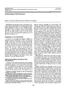
Fro. 1 Median neuropathy associated with Colles’ fracture may involve a prominent volar callus, which in this patient compressed the median nerve proximal to the. carpal tunnel. Surgical release was extended into the distal end of the forearm tO ensure adequate decompression.
Oneof the three patients had had an undisplaced fracture. Our treatment of these patients was difficult and prolonged. Wevariably used nerve and muscle decompression, lysis of tendons and nerves, release or lengthening of
This is more appropriately called upper-limb dystrophy or pain-dysfunction, and was a significant problem in twenty patients, sixteen of whomhad been referred. Four patients had acute symptomswith predominant sympathetic componentsof change in skin temperature, color, and texture; pain and loss of motion in the shoulder; and stiffness of the hand or specific local trigger areas of exquisite pain and tenderness (or both). In one patient it was the result of radial-nerve irritation from pin fixation; in two patients, from excessive wrist flexion which produced acute median neuropathy; and in one, from an unreduced, severely displaced fracture with associated disuse of the limb. Twoof the four patients had one other complication and two had two additional complications. The sixteen referral patients had late upper-limb dystrophy. They had fewer sympathetic components than did the patients with the acute condition, but had longestablished clinical complaints of stiffness and disuse of the shoulder, stiffness of the hand, painful motion, carpal
Figs. 2-A throi~gti 2-D: Maluni0nof the distal end of the radius developedin the wrist of a forty-five-year-old farmer whohad been gored by a bull. During life-saving measures the fracture was overlooked. Figs. 2-A and 2-B: Whentreatment of the malunion was begun at six months, there was marked shortening and radial angulation, median neuropathy, and weakgrip. An open-wedgeosteotomy, a bone graft from the distal end of the ulna, and a small plate were employedto regain length and restore alignment of the forearm. The carpal tunnel ~,as released.
muscle-tendonunits, and tendon transfers whenindicated, and the long-term results in three patients were only fair. The fourth patient had persistent pain and finally had to have a below-the-elbow amputation.
tunnel symptoms,and radiocarpal arthrosis. Ten of the sixteen patients had fracture malunion. Fourteen of them were referred with long-established complaints, but improved on conservative treatment extending for from six
COMPLICATIONS OF COLLES’ FRACTURES
weeks to four months. Six of the sixteen had one other complication and four, two or more complications. Stiff Hands
617
reduction (seven patients), improper immobilization in the cast (four patients), poor mobilization of the joint (eight patiients), and inadequateefforts at rehabilitation.
Stiff hands from arthrofibrosis of the fingers were a severe complication in nine patients. It was manifested by pain and swelling limited to the hand, with a loss of finger motion and occasionally a loss of motion of the wrist. Swelling and pain, particularly in the structures lined with synovial tissue, were the most characteristic findings in seven patients. Swelling of the proximal interphalangeal joint was the major source of pain and resulted in a severe loss of motion. The factor most commonlyassociated with the clinical symptomsand signs was improper application of a cast. Stiff hands occurred most often after improper cast application (seven of nine patients in this category). lack of early motion of the hand was evident in five of the nine patients, and pre-existing degenerative arthritis was present in three patients. Three patients had a mild Dupuytren’s contraeture in the affected hand. Six of the nine patients had full recovery, and the other three had improvedfunction after conservative treatment.
Discussion Severe complications from Colles’ fracture continue to occur frequently. Wefound that there were more patients than we anticipated whose complications required exte.nsive treatment. Possibly the percentage of complications in this report is higher than in other reports because more than 46 per cent of the patients (sixty of 128):with complications were referred for treatment. Wehave divide, d the complications into nine groups, of which the largest was the neuropathies. Compression neuropathies occurred both early (within the first two weeks) and later during the period treatment. Whenthe median nerve was involved, early recognition was common. However, in some "patients, whenthe radial or ulnar nerve was compressed, the diagnosis was delayed because the physicians failed to appreciate or suspect that the nerve was compressed, stretched, or irritated. This failure was especially evident whenfixation pins were utilized. Delay in diagnosis usuMultiple Complications ally lied to complicationssuch as a stiff handor carpal tunA study of the patients whohad multiple complicanel syndrome. tions that usually included the shoulder-hand syndromereMedianneuropathy was identified more often in this vealed that the underlying cause of the dystrophy appeared ’~, series of patients than in previously reported series1,6, to be a combinationof predisposing factors in conjunction probably because there is increased recognition of this with difficulties in treatment, such as repeated attempts at condition and because more patients are referred for surgi-
FIG. 2-C
F~G. 2-D
618
W. P.
COONEY, tlI,
J.
H. DOBYNS, AND R. L. LINSCHEID
1°,16 cal decompression. Weagree with previous authors produce a proximal carpal thrust that results in a dorsal that a significant contributor to the neuropathyis the force compressive force leading to collapse and displacement. of fracture reduction and the position of immobilization; Present methodsof fracture reduction and cast supthe higher frequencyof this complicationafter local block, port do not always prevent these potentially deforming with or without systemically administered analgesics, forces, particularly in comminutedfractures. In unstable tends to support this belief. fractures, weprefer to use external pin fixation in order to Post-fracture arthrosis was the second most common maintain a distracting force, prevent collapse, and allow complication in our patients, yet often it went unrecog- the volar fragmentsof the cortex to unite in goodposition. nized for sometime. Subtle forms of this arthrosis are re- Wehave used this methodfor patients in whomreduction sponsible for a large portion of the weaknessof grip and of the fracture was lost after cast immobilizationand also limited motion that are commonlyseen after this fracture. for potentially unstable intra-articular fractures (Frykman Whenthe condition is recognized, the patient often can be Types V through VIII), and have achieved satisfactory reimprovedby conservative measures, such as splinting, the sults 3. ~ local injection of steroids, and the use of salicylates. Open reduction of Coltes’ fracture is rarely advoOperative treatment for radiocarpal arthrosis Wasneces- cated, despite the need for accurate reduction of the fracsary in only nine patients in our series. The radio-ulnar ar- ture s. Becausethe functional results so closely parallel the throsis that was seen in twenty-seven patients mostly anatomical results, it is our practice that whenclosed restemmedfrom the inability to obtain an adequate anatomi- duction, including the use of external pin fixation, is not cal reduction, manifested in two ways. One was successful, open reduction is indicated. Definite criteria malalignmentof the sigmoid notch of the distal end of the for open reduction of Colles’ fractures have not been radius with the ulnar head, owing to radial deviation and completely formulated, but for the present the technique dorsiflexion of the distal radial component.The other was should be more strongly considered for use in youngadults inadequate restoration of length to maintain the normal re- in whomcomminuted, unstable intra-articular fractures lationship of the radio-ulnar joint. This problemwas sig- have been treated unsuccessfully by closed reduction nificant enoughto require surgical treatment in nineteen of techniques. the twenty-seven patients. Webelieve that the common The incidence of complications from Colles’ fractures technique of reduction and immobilization in full prona- reported here does not differ significantly from the types tion with ulnar deviation so that the distal end of the ulna and frequency of problems reported by others. Frykman provides stability is mechanicallyunsound, particularly in noted the significant sequelae of radio-ulnar arthrosis displaced, highly Comminuted fractures. The distal (18.6 per cent), shoulder-hand syndrome(2 per cent), radio-ulnar joint often is unstable, and any radio-ulnar sub- peripheral neuropathy (315 per cent) in his series of 430 luxation or dislocation that exists is only increased by im- "’cases. He found that symptomsat the distal radio-ulnar mobilizing the hand in full pronation. The end result may joint weremost frequently related to fractures into the joint be that rotation of the forearm, especially supination, be- (41 per cent) combinedwith dorsal angulation and shorten/comes severely limited. ing of the distal end of the radius. Lippmanand. Lidstr6m Weagree with Sarmiento et al., and others ~,6, that had similar findings (10 per cent and 15 per cent incithe best position for maintaining normal alignment and dences of radio-ulnar arthrosis, respectively) and stressed minimizing deforming forces is supination. Whenthe that radio-ulnar instability was the most commoncause of proper length of the distal end of the radius is difficult to a poor result. Gartland and Werley reported an incidence maintain, strong, protracted traction and external pin fixa- of arthrosis of 22 per cent. In combiningboth radiocarp~l tion maybe the best form of treatment. and radio-ulnar arthroses, we found symptomsthat were Early loss of reduction and late collapse after Colles’ significant enoughto require surgical treatment in thirtyfracture probably are two commoncomplications that are seven (6.5 per cent) of 526 patients. too readily accepted by treating physicians. To us, each of Shoulder-hand syndrome was present in 1.4 per cent these conditions signifies that the fracture being treated is of patients reviewed by Bacorn and Kurizke, in 3.4 per unstable. It usually has one or more of the following cent in Rosen’sseries, and in 10 per cent in Lidstr6m’sse¯ characteristics: extensive comminution,markeddisplace- ries of 515 patients. The latter included finger-joint stiffment of fragments, or interposition of soft tissue -- and hess and Siideck’s atrophy. Unsatisfactory results were reany one of them can lead to an incomplete reduction. ported in 67 per cent. The incidence in our series was four. Webelieve that whenever a fracture is unstable, no (1.1 per cent) of 356 local patients. Whileaffected patients amountof residual dorsal angulation after reduction is are fewer in number,this complicationis the most difficult permissible. Adequatereduction requires that the full dor- to treat, and prevention by the techniques described by sal length of the radius be restored and maintained. This Mobergshould be studied. requires a stable volar buttress plus dorsal tension by tissue Peripheral neuropathy as a serious complication was or an apparatus that prevents dorsal collapse. Otherwise not noted by others to be as frequent as we have reported it the force of active finger flexor and extensor tendons, to be (forty-five patients over-all and twenty-one[3.7 per combinedwith dorsal translation of the lunate, tends to cent] of patients who were primarily under our care).
COMPLICATIONS OF COLLES ’ FRACTURES
Lidstr6mbelieved that nerve injuries are rare after fractures of the distal end of the radius (slightly morethan per cent). Bacornand Kurtzkereported an incident of 0.2 per cent and Schlesinger and Liss noted only one case per 1,000 fractures. Webelieve that these negative reports weredue in part to a lack of recognition andpossibly more concernwith treatmentof the fracture than with potential sequelae. Lynch and Lipscomb, Frykman, Robbins, and others5 have placed proper emphasison the causes of median neuropathyand the need for aggressive treatment in certain acute as well as late cases. Complicationsrelated to morethan one factor were
619
commonwheneverthree complications -- neuropathy, arthrosis, and shoulder-hand syndrome-- were present. Frykman foundthat of eighty patients with radio-ulnar arthrosis, five (6.3 per cent) had medianneuropathyandfive had shoulder-handsyndrome.Of our twenty patients with shoulder-hand syndrome, sixteen had one or more associated complications(arthrosis in ten patients, median neuropathyin nine patients, malunionin ten patients, and sympatheticdystrophyin five patients). Evidently, these complicationsandothers contributedirectly to the 24 to 27 per cent incidenceof poorfunctional results that has been 3"6"~fromthe treatmentof Colles’ fractures. reported
References 1. BACORN, R. W., and KURTZ~:E,J. F.: Colles’ Fracture: A Study of TwoThousand Cases from the NewYork State Workmen’sCompensation Board. J. Bone and Joint Surg., 35-A: 643-658, July 1953. 2. BOSACCO, D. N., and TRABULSL L. R.: The Colles’ Fracture -- Treatment by Closed Reduction, Internal Fixation and Short Arm Cast Application. In Proceedings of The American Academyof Orthopaedic Surgeons. J. Bone and Joint Surg., 57-A: 1030, Oct. 1975. 3. COONEY, W. P., III; LINSCHEID, R. L.; and DOBYNS, J. H.: External Pin Fixation for Unstable Colles’ Fractures. J. Boneand Joint Surg., 61-A: 840-845, Sept. 1979. 4. DARRACH, W~LL~AM: Partial Excision of LowerShaft of Ulna for Deformity following Colles’s Fracture. Ann. Surg., 57: 764-765, 1913. 5. DoBYNS, J. H., and LINSCHEID, R. L.: Complications of Treatment of Fractures and Dislocations of the Wrist. In Complications in Orthopaedic Surgery, edited by C. H. Epps, Jr. Vol. 1, pp. 271-352. Philadelphia, J. B. Lippincott, 1978. 6. FRYKMAN, G.: Fractur§ of the Distal Radius Including Sequelae -- Shoulder-Hand-Finger SyndromeDisturbance of the Distal Radio-Ulnar Joint and Impairment df Nerve Function. A Clinical and Experimental Study. Acta Ortt~op. gcandina~ica, Suppl¢mentum108, 1967. 7. GARTLAND, J. J., JR., and WERLEY, C. W.: Evaluation of Healed Colles’ Fractures. J. Bone and Joint Surg., 33-A: 895-907, Oct. 1951. 8. KRISTIANSEN, AMUND, and G~ERSOE, E~NAR:Colles’ Fracture. Operative Treatment, Indications and Results. Acta Orthop. Scandinavica, 39: 33-46, 1968. 9. LIDSTR6M, ANDERS: Fractures of the Distal End of the Radius. A Clinical and Statistical Study of End Results. Acta Orthop. Scandinavica, Supplementum 41, 1959. 10. LIPPMAN, R. K.: Laxity of the Radio-ulnar Joint following Colles’ Fracture. Arch. Surg., 35: 772-786, 1937. 11. LYNCH, A. C., and LIPSCOMI~,P. R.: The Carpal Tunnel Syndromeand Colles’ Fractures. J. Am. Med. Assn., 185: 363-366, 1963. 12. MARVEL, J. R., JR.: ComminutedFractures of the Distal End of the Radius Treated by Pins and Plaster Technique. In Proceedings of The American Academyof Orthopaedic Surgeons. J. Bone and Joint Surg., 57-A: 1030, Oct. 1975. 13. M~L¢8,HENRY: Cuff Resection of the Ulna for Malunited Colles’ Fracture. J. Boneand Joint Surg., 23:311-313, April 1941. 14. MOSERG, ERIK: Shoulder-Hand-Finger Syndrome, Reflex Dystrophy, Causalgia [Abstract]. Acta Chir. Scandinavica, 125: 523, 1963. 15. POOL, CHRISTOPHER: Colles’ Fracture. A Prospective Study of Treatment. J. Bone and Joint Surg., 55-B: 540-544, Aug. 1973. 16. RO88INS,J. V.: Logical Reduction of Displaced Colles’ Fractures. NewYork State J. Med., 50: 2959-2962, 1950. 17. ROSEN, ERIK:Fractura Extremitatis Distalis Radii. Ugeskr. Laeger., 109: 603-610, 1947. 18. SARMIE~qTO, At~GUSTO; PRATT,G. W.; BERRY,N. C.; and S~NCLAm, W. F.: Cg~lles’ Fractures. Functional Bracing in Supination. J. Boneand Joint Surg., 57-A: 311-317, April 1975. 19. S¢I-IECK, MAx:Long-TermFollow-up of Treatment of ComminutedFractures of the Distal End of the Radius by Transfixation with Kirschner Wires and Cast. J. Bone and Joint Surg., 44-A: 337-351, March 1962. 20. SC8LES~NGER, E. B., and L~ss, H. R.: Fundamentals, Fads and Fallacies in the Carpal Tunnel Syndrome. Am.J. Surg., 97: 466-470, 1959. 21. SMAI.LL,G. B.: Long-TermFollow-up of Colles’ Fracture. J. Bone and Joint Surg., 47-B: 80-85, Feb. 1965.