Christina Boulton, 2004
May 2004
Gillian Lieberman, MD
Imaging of Pelvic Fractures Christina Boulton Harvard Medical School, Year III Gillian Lieberman, MD Images: Borril et al. Orthoteers Orthopedic Education Resource. http://www.orthoteers.co.uk/. Wheeless CR. Wheeless’ Wheeless’ Texbook of Orthopaedics. Orthopaedics. http://www.wheelessonline.com http://www.wheelessonline.com
1
Christina Boulton, 2004 Gillian Lieberman, MD
Outline Pelvic
Anatomy Review Introduction to Pelvic Fractures, Associated Injuries and Complications Our Patient Pelvic Imaging Modalities Pelvic Fracture Classification Summary Image: Wheeless CR. Wheeless’ Wheeless’ Texbook of Orthopaedics. Orthopaedics. http://www.wheelessonline.com http://www.wheelessonline.com
2
Christina Boulton, 2004 Gillian Lieberman, MD
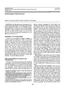
Pelvic Anatomy Review - Bones ILIUM
SI Joint
Sacral Strut
SPR
Posterior Column
Anterior Column
IPR
Neural Foramen Obturator Foramen Image: Gray, Henry. 1918.
3
Christina Boulton, 2004 Gillian Lieberman, MD
Pelvic Anatomy Review - Ligaments Iliolumbar Ligament
Ventral Sacroiliac Ligaments Dorsal Sacroiliac Ligaments Sacrospinous Ligament Sacrotuberous Ligament Image: Gray, Henry. 1918.
4
Christina Boulton, 2004 Gillian Lieberman, MD
Pelvic Anatomy Review – III
The bony pelvis lies in close proximity to numerous vascular, neural and soft tissue structures that are vulnerable during pelvic ring disruption. Images: Gray, Henry. 1918.
5
Christina Boulton, 2004 Gillian Lieberman, MD
Introduction to Pelvic Fractures
3-4% of all Fx, usually associated with significant trauma Types of Pelvic Fracture – Lower Energy: Avulsion & Acetabular Fx – Higher Energy: Ring Disruptions Stable vs. Unstable (33%), Open vs. Closed Rule of Rings – usually >1 disruption, breaks occur at weakest points Mechanism of Injury – Requires significant force, e.g. MVA (50-60%), Motorcycle Crash (1020%), Pedestrian Struck (10-2-%), Fall (8-10%), Crush (3-6%) Morbidity & Mortality – Adult mortality 10-15% (secondary to retroperitoneal hemorrhage, infection) – Mortality approaches 50% if hypotensive on presentation, ~30% if open fracture – Significant decrease in morbidity and mortality with prompt stabilization of unstable pelvic fractures – Pelvic Fx more common in patients 55yrs 6
Christina Boulton, 2004 Gillian Lieberman, MD
Associated Injuries & Complications
Vascular Injuries + Hemorrhage (40%) Nerve (10%) – Sciatic, Femoral, Obturator, Lumbosacral Plexus esp. roots S1-5 Organ Injuries (rare) – Liver, Spleen, GI GU system: Bladder/Urethral Rupture (10-15%) Chronic Pain Arthritis Immobility – increase risk for PE, all pelvic fracture patients should be anticoagulated once hemorrhage stabilized. 7
Christina Boulton, 2004 Gillian Lieberman, MD
Our Patient: MS, 77yo F struck by car Documented
20min LOC Intubated at scene and transported to BIDMC ED via Life Flight Hypotensive during transport and given IVFs, stable VS on arrival at ED In ED: multiple injuries noted to head, Cspine, upper and lower extremities, pelvis noted as stable to palpation 8
Christina Boulton, 2004 Gillian Lieberman, MD
Patient MS: Trauma Series
Findings: – Minimally displaced fractures of R inferior and superior pubic rami – Sacrum not well visualized – Sacral Fx seen on CT scout Image: PACS, BIDMC
9
Christina Boulton, 2004 Gillian Lieberman, MD
Pelvic Imaging Techniques
Plain Films - Trauma Series - Inlet & Outlet Views - Judet (Oblique Views) - acetabular fractures Pelvic CT Ortho Protocol – highlights bone detail MRI – usually modality of choice for detailed imaging of joints and ligaments in other circumstances FAST Exam – utility only for intra-abdominal or retroperitoneal blood collections Other Studies - Retrograde urethrogram or CT cystogram if exam suggests injury to GU system - Arteriography for minor persistent hemorrhage or hematoma
10
Christina Boulton, 2004 Gillian Lieberman, MD
Pelvic Imaging – Plain Films
Trauma Series – Used for initial evaluation – Detects pelvic fx 90% of time – Good for: rami fx, pubic symphysis, large hematomas – Bad for: ligamentous injuries, subtle displacements esp. axial, sacral fractures Supplemental Case 1: Symphyseal Diastasis, Sacral Fx
Image: Wheeless CR. Wheeless’ Wheeless’ Texbook of Orthopaedics. Orthopaedics. http://www.wheelessonline.com http://www.wheelessonline.com
11
Christina Boulton, 2004 Gillian Lieberman, MD
Pelvic Imaging – Plain Films II
Inlet View –
–
Outlet View – –
–
best demonstrates ring configuration of pelvis, narrowing/widening of diameter of ring evaluates for posterior displacement of pelvic ring or opening of pubic symphysis
evaluates for vertical shift of pelvis proximal/distal displacements of anterior or posterior portion of ring sacrum appears in its longest dimension, w/ neural foramina evident.
Judet (Oblique) Views – External View: - shows ilioischial line posterior column & anterior wall. – Internal View: - shows iliopectineal line anterior column & posterior wall.
Images: Borril et al. Orthoteers Orthopedic Education Resource. http://www.orthoteers.co.uk/.
12
Christina Boulton, 2004 Gillian Lieberman, MD
Retrograde Urethrogram Retrograde injection of contrast material into bladder via urethra
Supplemental Case 2: Bladder rupture with contrast extravasation Image: Wheeless CR. Wheeless’ Wheeless’ Texbook of Orthopaedics. Orthopaedics. http://www.wheelessonline.com http://www.wheelessonline.com
13
Christina Boulton, 2004 Gillian Lieberman, MD
Pelvic Imaging - CT
Uses - Good visualization of subtle fractures, AP displacement - Useful if pt. not able to be positioned adequately for special views - CT faster now than in past so not contra-indicated in early trauma work up - Many multi-trauma pts will already be bound for CT - Coronal and saggital reconstructions useful for understanding spacial relationships of fragments - CT Cystogram alternative to retrograde urethrogram to r/o bladder injury - Operative planning
Image: PACS, BIDMC
14
Christina Boulton, 2004 Gillian Lieberman, MD
Our Patient (MS): Pelvic CT
Comminuted Fx R Sacral Ala with mild distraction of fragments, small surrounding hematoma, R SI joint slightly ectatic posteriorly Image: PACS, BIDMC
15
Christina Boulton, 2004 Gillian Lieberman, MD
Our Patient (MS): Pelvic CT
R Sacral Fx with posterior displacement of sacral wing, hematoma Image: PACS, BIDMC
16
Christina Boulton, 2004 Gillian Lieberman, MD
Our Patient (MS): Pelvic CT
Comminuted Fx of R inferior pubic ramus, mild distraction of fragments Image: PACS, BIDMC
17
Christina Boulton, 2004 Gillian Lieberman, MD
Our Patient (MS): Pelvic CT
L pubic bone fx @ pubic symphysis + associated small hematoma Image: PACS, BIDMC
18
Christina Boulton, 2004 Gillian Lieberman, MD
Our Patient (MS): Pelvic CT Coronal Reconstruction
R Sacral Ala Fx, superior displacement, Fx R inferior pubic ramus Image: PACS, BIDMC
19
Christina Boulton, 2004 Gillian Lieberman, MD
Our Patient (MS): Arteriogram
Bilateral iliac artery arteriogram Crisp, uniform vessel outline No contrast extravasation
Arteriogram of small branch of R internal iliac Contrast extravasation vessel embolized Images: PACS, BIDMC
20
Christina Boulton, 2004 Gillian Lieberman, MD
Pelvic Imaging – Follow UP Purpose: To determine if pelvic re-alignment procedures have brought bones back to anatomic position and evaluate callous formation & healing
Post ORIF Plain Films – Often done in OR
CT or Plain Films post external fixation – To check correct anatomic relationships
Artifact Reduction CT – Subtracts hardware artifact
Judet (Oblique) Views
Intra-operative Portable Supine Pelvic Xray of Unstable Fx s/p ORIF
– To follow acetabular fractures Image: Wheeless CR. Wheeless’ Wheeless’ Texbook of Orthopaedics. Orthopaedics. http://www.wheelessonline.com http://www.wheelessonline.com
21
Christina Boulton, 2004 Gillian Lieberman, MD
Pelvic Fx Classification System Multiple
classification systems in use Young System: 4 general categories, based on mechanism of injury – Lateral Compression (LC I-III) – Anterior/Posterior Compression (APC I-III) – Vertical Shear (VS) – Combined Mechanical (CM) Hemorrhage
predominantly with APC & VS injuries, rare with LC injuries 22
Christina Boulton, 2004 Gillian Lieberman, MD
Lateral Compression Fractures KEY: Transverse Pubic Rami Fx/s + Ipsi- or Contralateral Posterior Injury
LC I
Mech: lateral compression to sacrum Post. Injury = ipsilateral sacral strut compression fractures STABLE
LC II
Mech: lateral compression to ilium Post. Injury = ipsilateral posterior iliac fracture USUALLY STABLE
LC III
Mech: rollover Post. Injury = LC I/II + contralateral AP compression UNSTABLE
Images: Borril et al. Orthoteers Orthopedic Education Resource. http://www.orthoteers.co.uk/.
23
Christina Boulton, 2004 Gillian Lieberman, MD
A/P Compression Fractures KEY: Syphysis diastasis OR Longitudinal Rami Fx +/- Post. Ligament Tear
APC I
Mech: low/mod AP force (e.g. sports) Injury: PS2cm/ PR Fx + anterior SI ligament tear + torn ST, SS ligaments UNSTABLE, “open book”
APC III
Mech: very high energy AP force Post. Injury: anterior AND posterior SI, SS & ST ligaments torn VERY UNSTABLE
Images: Borril et al. Orthoteers Orthopedic Education Resource. http://www.orthoteers.co.uk/.
24
Christina Boulton, 2004 Gillian Lieberman, MD
Vertical Shear Fractures
S/I Displacement often evident on PEx Mechanism: fall from significant height Injury: can be ligamentous only or involve fractures also UNSTABLE
Images: Borril et al. Orthoteers Orthopedic Education Resource. http://www.orthoteers.co.uk/.
25
Christina Boulton, 2004 Gillian Lieberman, MD
Combined Mechanical
Mechanism: multiple forces from multiple directions, usually VS + LC Injury: combination High association with severe trauma, hemorrhage and multiple other injuries UNSTABLE Images: Borril et al. Orthoteers Orthopedic Education Resource. http://www.orthoteers.co.uk/.
26
Christina Boulton, 2004 Gillian Lieberman, MD
Our Patient (MS) Summary
MS’s Injuries – – – –
R superior & inferior pubic rami fx L pubic symphysis fx R sacral fx + R sacroiliac joint widening Pelvic hematoma
Classification – CM pelvic ring disruption Complications – bleeding/hematoma, CT Cysto showed no bladder injury (not shown) Interventions – arterial embolization, external pelvic fixation 27
Christina Boulton, 2004 Gillian Lieberman, MD
Pelvic Fractures: Summary
Associated Morbidity & Mortality = High Associated Injuries & Complications = Common + Dangerous – Injury to vessels, nerves, bladder and other organs common – Longterm Fx complications e.g chronic pain, immobility, arthritis Mechanism of injury can predict fracture pattern + associated injuries if you understand the anatomy Classification System: – Low Energy vs. High Energy (Ring Disruption) – Stable vs. Unstable, Open Vs. Closed – Lateral Compression, A/P Compression, Vertical Shear 28
Christina Boulton, 2004 Gillian Lieberman, MD
Pelvic Imaging: Summary
Trauma Series KUB - Initial detection of Fx Inlet & Outlet Plain Films
Judet Plain Films
Pelvic CT Ortho Protocol
CT Cystogram or Retrograde Urethrogram
– Primarily used in characterization of fracture architecture and for follow up – Oblique views used for evaluation and follow up of acetabular fractures
– Further characterization of Fx, esp. degree of displacement and detection of subtle sacral fractures – Coronal & saggital reconstructions for specific positional relationships – Artifact reduction protocol for evaluations s/p ORIF – Sensitive for detection of hematoma, organ injuries Arterogram – Detection and embolization of active bleeds – Rule out bladder & urethral injury
29
Christina Boulton, 2004 Gillian Lieberman, MD
References
Borril J, Funk L, Deakin S. Orthoteers Orthopedic Education Resource (Online Textbook). Weblink: http://www.orthoteers.co.uk/. Demetrios D, Karaiskakis M, Toutouzas K. Pelvic Fractures: Epidemiology and Predictors of Associated Abdominal Injuries and Outcomes. J Am Coll Surg, 2002; 195:1-10. Gonzales RP, Fried PQ, Bukhalo M. Utility of Clinical Examination in Screening for Pelvic Fractures in Blunt Trauma. J Am Coll Surg, 2002; 194: 121-125. Gray, Henry. 1918. Anatomy of the Human Body. Bartelby.com Online Publishing. Weblink: http://www.bartleby.com/107/ Henry SM, Pollack AN, Jones AL, et al. Pelvic Fracture in Geriatric Patients: A Distinct Clinical Entity. J Trauma, 2002; 53:15-20. McCabe C. Trauma: an annotated bibliography of the recent literature, 2002. Am J Emerg Med 2003, 21(7): 585-603. Mirvis SE & Young JWR. Imaging in Trauma & Critical Care; Chapter 7: Pelvic Fractures. Williams & Wilkins © 1992. Baltimore MD, USA. Novelline, R. 1997. Squire’s Fundamentals of Radiology. 5th Edition. Harvard University Press. Routt ML, Nork SE, Mills WJ. Treatment of Complex Fractures. Orthopedic Clinics of North America. Jan 2002; 33(1). Wheeless CR. © 1996. Wheeless’ Texbook of Orthopaedics. Duke University Medical Center & Datatrace Internet Publishing. Weblink: http://www.wheelessonline.com
30
Christina Boulton, 2004 Gillian Lieberman, MD
Acknowledgements Larry
Barbaras Fabio Komlos, MD Pamela Lepkowski Gillian Lieberman, MD
31