Jean-Marc Gauguet, HMS IV Gillian Lieberman, MD
Radiology of Pediatric Fractures Jean-Marc Gauguet, HMS Year IV Gillian Lieberman, MD 9/14/06
Jean-Marc Gauguet, HMS IV Gillian Lieberman, MD
Overview • Osteology • Salter-Harris Fractures • Greenstick Fractures • Torus Fractures
Jean-Marc Gauguet, HMS IV Gillian Lieberman, MD
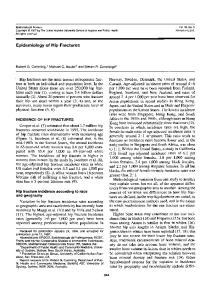
Longitudinal Bone Growth Provisional Calcification Hypertrophing Proliferating Resting Pediatric Fractures and Dislocations
• • • •
Modified from: http://www.unu.edu/Unupress/food2/UID06E/uid06e0u.htm
The physis is divided into metaphyseal and epiphyseal sections. Mitosis of chondrocytes in the epiphyseal section and growth in the metaphyseal section leads to longitudinal growth. The resting and proliferating zones contain high amounts of chondoitin sulfate, which imparts strength. The hypertrophing zone lacks chondroitin sulfate or calcium and is the weakest layer - this is the site of physeal fractures .
Jean-Marc Gauguet, HMS IV Gillian Lieberman, MD
Kids bones are special • Epiphyseal growth plates in children have not completely ossified. • The periostium in children is thicker, more metabolically active, less easily torn, and more easily stripped from the bone.
Jean-Marc Gauguet, HMS IV Gillian Lieberman, MD
Endplate Closure
Pediatric Fractures and Dislocations
• Prior to endplate closure there is a balance between chondrocyte proliferation and bone mineralization. • Once proliferation slows and ceases mineralization can be completed. • Endplate closure occurs when mineralization crossed into the epiphysis.
Jean-Marc Gauguet, HMS IV Gillian Lieberman, MD
Salter-Harris (SH) Overview • Developed 5 level classification system in 1963. • Fracture involving injury to the physis of long bones before complete closure occurs. • 35% of all skeletal injuries in children. • 75% of cases occur in kids between 10-15 years. • Most common sites: wrist 50%, ankle 30%. • The growth plate is 2-5 times weaker than joint capsule or ligaments.
Jean-Marc Gauguet, HMS IV Gillian Lieberman, MD
Type I • • • •
• • www.emedicine.com
Line of cleavage confined to the physis. Accounts for 6% of cases. Most commonly occurs in the phalanges. Growth disturbance uncommon because germinal layer and vessels not disturbed. Excellent prognosis, treat with closed reduction and immobilization. Patients often treated if no overt signs of fracture, but there is soft tissue swelling or patient is symptomatic.
Jean-Marc Gauguet, HMS IV Gillian Lieberman, MD
Type I continued • Best identified by comparing to normal joint and looking for widening of the physis.
SH I
Normal
Swischuk, L.E., et al.
•Swischuk, L.E., et al., Frequently missed fractures in children (value of comparative views). Emergency Radiology. (2004) 11: 22-28.
Jean-Marc Gauguet, HMS IV Gillian Lieberman, MD
Type II
SH Type II
www.emedicine.com
• • • • •
Boston Children’s Hospital
Fracture line through the physis and extends through margin of metaphysis - “corner sign” Most common type (75% of cases) Distal radius most common site Minimal shortening, good outcomes. Treat with closed reduction and immobilization.
Jean-Marc Gauguet, HMS IV Gillian Lieberman, MD
Type III
SH Type III
www.emedicine.com
• • • • •
Boston Children’s Hospital
Fracture runs through epiphysis then horizontally through physis. Accounts for 8% of cases. Typically seen after partial epiphyseal plate closure in tibia and distal femur. Some deformity, but most problems arise from fracture entry into joint space. Treat with closed reduction and immobilization, occasional ORIF.
Jean-Marc Gauguet, HMS IV Gillian Lieberman, MD
Type IV
SH Type IV
www.emedicine.com
• • • •
www.emedicine.com
Vertically oriented fracture through epiphysis, physis, and metaphysis. 10% of injuries. Most commonly in distal femur and distial tibia. Requires surgical repair, and often causes disability and joint deformity from damage to germinal layer and epiphyseal blood supply.
Jean-Marc Gauguet, HMS IV Gillian Lieberman, MD
Type V
SH Type V
www.emedicine.com
• • • •
Crush injury of physis typically from axial loading, no injury to epiphysis or metaphysis. Accounts for 2mm and fragment could not reduced closed, decision made to surgically repair
Jean-Marc Gauguet, HMS IV Gillian Lieberman, MD
Case I: Treatment Plain Film
Boston Children’s Hospital
A screw was placed through displaced fragment and the patient was casted.
Plain Film
Boston Children’s Hospital
3 months later, cast removed, good fracture healing. Patient did well.
Jean-Marc Gauguet, HMS IV Gillian Lieberman, MD
Complications of SH Fractures: Case Ia Plain Film 12 year old boy with a triplane fracture: - SH-II of medial tibial - SH-III of lateral epiphysis Patient still has occasional pain 7 months after injury.
Boston Children’s Hospital
Orthopedic surgeons requested a CT to evaluate patient for epiphyseal plate damage.
Jean-Marc Gauguet, HMS IV Gillian Lieberman, MD
Case Ia: Bone Bridge Formation
Coronal CT
Bone bridge spanning from metaphysis to epiphysis.
Boston Children’s Hospital
•
When area of growth arrest less than 50%, then outcomes are good with surgical intervention1.
1
Williamson, R.V., Partial physeal growth arrest: treatment by bridge resection and fat interposition. J Pediatric Orthopaedics (1990) 10: 769-776.
Patient currently has no signs of growth arrest, he will be closely followed. No surgery planned at this time.
Jean-Marc Gauguet, HMS IV Gillian Lieberman, MD
Use of MRI in Salter Harris Evaluation • Not widely used, however, powerful when plain films equivocal as in one study, radiographs detected 9/14 cases while MRI detected 14/14 cases, this improved sensitivity led to a change in management in 5/14 patients.1 • Also used to evaluate damage to physis.
Normal knee?
No! SH IV Fracture
Spin Echo T1 Carey, et al. 1
Carey, J. et al. MRI of pediatric growth plate injury: correlation with plain film radiographs and clinical outcome. Skeletal Radiology. (1998) 27: 250-255.
Jean-Marc Gauguet, HMS IV Gillian Lieberman, MD
Case II • A 13 year old male presents to the ED with wrist pain after falling off bicycle onto outstretched hand. • PE reveals deformity of forearm proximal to right wrist, normal distal pulses. • Radiographs were taken to evaluate patient.
Jean-Marc Gauguet, HMS IV Gillian Lieberman, MD
Case II: Plain Flims
AP
Boston Children’s Hospital
Lat
Boston Children’s Hospital
Incomplete Greenstick fracture of distal radius with palmar angulation. Patient was casted and recovered well.
Jean-Marc Gauguet, HMS IV Gillian Lieberman, MD
Greenstick Fracture • Derived from “green” tree branches that bend, but do not completely break. • Commonly results from a twisting motion on an outstretched hand. • The genuine greenstick fracture involves disruption of cortex and periostium on covex side of fracture. • Due to the strength of the periostium and incomplete mineralization of bone. Pediatric Fractures and Dislocations
Jean-Marc Gauguet, HMS IV Gillian Lieberman, MD
Treatment/Prognosis • Often very easy to reduce and immobilize in a cast. • Some fractures require completion of the fracture prior to reduction and immobilization. • The prognosis is also very good for these types of fractures. • May be complicated by recurrent deformity while casted.
Jean-Marc Gauguet, HMS IV Gillian Lieberman, MD
Case III • 14 year old male presents to the ED after falling onto his left forearm while snowboarding. • Pain is localized to the forearm, denies any loss of sensation. • Plain films were ordered.
Jean-Marc Gauguet, HMS IV Gillian Lieberman, MD
Case III: Plain Films
Non-displaced torus fracture through distal metaphysis of L radius Boston Children’s Hospital
Patient was casted and recovered well.
Jean-Marc Gauguet, HMS IV Gillian Lieberman, MD
Torus Fracture • Torus derived from Latin (tori), which means a swelling or protuberance. • A buckling of the cortex on the compression side, typically 2-3cm from physis. • Stable, non-displaced fracture, treat with casting. • Important radiologic distinction between torus and greenstick since risk of recurrent deformation in cast higher in patients with greenstick fractures.
Jean-Marc Gauguet, HMS IV Gillian Lieberman, MD
Summary • Pediatric bones are immature with incomplete growth plate closure and a strong periosteum. • Salter-Harris fractures are common fractures in children and classification of the type of fracture has important implications on treatment and outcome. • Plain films are the standard of care for SH fractures, while CT and MRI may be necessary in more complicated cases. • Torus and Greenstick fractures are “incomplete” fractures unique to children.
Jean-Marc Gauguet, HMS IV Gillian Lieberman, MD
Acknowledgements • • • • •
Dr. Josh Nagler - Boston Children’s Hospital Dr. Stefanie Koch - Boston Children’s Hospital Dr. Jim Wu - BIDMC Pamela Lepkwoski - BIDMC Dr. Gillian Lieberman - BIDMC
Jean-Marc Gauguet, HMS IV Gillian Lieberman, MD
References • • • • • • • • • • • • •
Brown, J.H., Growth plate injuries: Salter-Harris classification. Am Fam Physicians. (1992) 46: 1180-1184 Brown, SD, et al., Analysis of 51 tibial triplane fractures using CT with multiplanar reconstruction. AJR (2004) 183: 1489-95 Carey, J. et al. MRI of pediatric growth plate injury: correlation with plain film radiographs and clinical outcome. Skeletal Radiology. (1998) 27: 250-255. Close, B.J., Strouse, P.J. MR of physeal fractures of the adolescent knee. Pediatric Radiology. (2000) 30: 756-762. eMedicine, Salter-Harris Fractures Hubner, U., et al., Ultrasound in the diagnosis of fractures in children. J Bone Joint Surg Br. (2000) 82: 1170-1173. Rodriguez-Merchán, E.C., et al., Pediatric skeletal trauma: A review and historical perspective. Clin Orthop Relat Res. (2006) 432: 8-13. Rogers, L.F., Poznanski, A.K., Imaging of Epiphyseal Injuries. Radiology. (1994) 191: 297308. Sailhan, F., et al., Three-dimensional MR imaging in the assessment of physeal growth arrest. Eur Radiol. (2004) 14: 1600-1608. Swischuk, L.E., et al., Frequently missed fractures in children (value of comparative views). Emergency Radiology. (2004) 11: 22-28. von Laer, L., Pediatric Fractures and Dislocations, New York, NY, 2004 www.wheelessonline.com (Wheeless’ Textbook of Orthopedics Online) http://www.unu.edu/Unupress/food2/UID06E/uid06e0u.htm