Wo m e n ’s I m a g i n g • P i c t o r i a l E s s ay Hedegard et al. Breast Reconstruction Flap
Downloaded from www.ajronline.org by 37.44.207.101 on 01/25/17 from IP address 37.44.207.101. Copyright ARRS. For personal use only; all rights reserved
Women’s Imaging Pictorial Essay
Breast Reconstruction With a Deep Inferior Epigastric Perforator Flap: Imaging Appearances of the Normal Flap and Common Complications Wade Hedegard1 Bethany Niell1 Michelle Specht 1 Jonathan Winograd2 Elizabeth Rafferty 1 Hedegard W, Niell B, Specht M, Winograd J, Rafferty E
Keywords: breast reconstruction, deep inferior epigastric perforator flap, DIEP flap DOI:10.2214/AJR.12.9270 Received May 16, 2012; accepted after revision July 3, 2012. 1
Avon Comprehensive Breast Evaluation Center, Massachusetts General Hospital, Wang Bldg Ste 240, Boston, MA 02114. Address correspondence to B. Niell (
[email protected]).
2
Department of Plastic Surgery, Massachusetts General Hospital, Wang Ambulatory Care Center, Boston, MA. WEB This is a Web exclusive article. AJR 2013; 200:W75–W84 0361–803X/13/2001–W75 © American Roentgen Ray Society
OBJECTIVE. The purpose of this essay is to illustrate the normal imaging appearance of deep inferior epigastric perforator (DIEP) flap breast reconstruction and common postoperative complications. CONCLUSION. Familiarity with the anatomy and normal imaging appearance of a DIEP flap reconstruction will help the breast imager recognize normal postsurgical findings and common postoperative complications.
B
reast reconstruction after mastectomy can be divided into two categories: implant and autogenous tissue flap reconstruction. Approximately 70% of breast reconstructions are performed with an implant inserted in either a primary or two-stage procedure [1]. The imaging characteristics and complications of implant reconstruction have been previously reported [2, 3]. The potential benefits of flap reconstruction compared with implant reconstruction include a more natural cosmetic outcome, avoidance of placement of a foreign body, and the opportunity for simultaneous abdominal contouring at the donor site. The earliest and most commonly used flap reconstruction is the pedicled transverse rectus abdominis myocutaneous (TRAM) flap [1]. In that procedure, the abdominal skin, subcutaneous fat, and rectus abdominis muscle with its associated native vasculature are used to create a neobreast. The rectus abdominis muscle is elevated, pedicled on the deep superior epigastric vessels, and tunneled subcutaneously into the mastectomy defect. One of the morbid conditions associated with pedicled TRAM reconstruction is fat necrosis of the neobreast, which can lead to palpable lesions and falsepositive breast imaging findings. Morbidity also can occur at the donor site, patients becoming prone to the development of abdominal wall weakness and hernia [4]. In a free TRAM flap, the infraumbilical portion of the rectus abdominis muscle and the deep inferior epigastric vasculature are divided and reanastomosed to the recipient vessels of the chest. The muscle is not tun-
neled under the skin, so the plastic surgeon can sacrifice less of the rectus muscle. The normal anatomy and imaging characteristics of TRAM flap reconstruction have been previously described [4]. With the goal of equal or superior cosmesis in combination with decreased morbidity, newer muscle-sparing techniques for autogenous tissue reconstruction after mastectomy have been developed. In a perforator flap, the skin and subcutaneous fat overlying the muscle are harvested, and the underlying muscle is left intact except for an incision through the muscle to remove the vessels of interest. The deep inferior epigastric perforator (DIEP) flap is the most common perforator flap breast reconstruction. The frequency of DIEP flap procedures increased 16% between 2009 and 2010 [5]. DIEP flap reconstructions may be imaged for a number of reasons, including suspected recurrence, annual screening of the contralateral breast, and further evaluation of an area of clinical concern. The objective of this essay is to illustrate the normal imaging appearance of a DIEP flap and to review the common postoperative complications. Deep Inferior Epigastric Perforator Flap Reconstruction In a DIEP reconstruction, an elliptic flap of abdominal skin and subcutaneous fat is elevated from the rectus fascia, leaving the rectus abdominis muscle in situ and largely intact with only an incision through the infraumbilical portion (Fig. 1). One or more lateral or medial perforator vessels are identified and dissected through the rectus sheath and muscle
AJR:200, January 2013 W75
Downloaded from www.ajronline.org by 37.44.207.101 on 01/25/17 from IP address 37.44.207.101. Copyright ARRS. For personal use only; all rights reserved
Hedegard et al. [6]. The vascular dissection is performed until the deep inferior epigastric artery and vein are exposed. These vessels are ligated, and the flap is ready for transfer. No rectus muscle is included in the DIEP flap (Fig. 1). At the mastectomy defect in the chest, the recipient site is prepared by resection of 2–3 cm of costal cartilage from the third or fourth rib and opening of the posterior perichondrium to expose the internal mammary vessels (Fig. 1). The harvested tissue flap is weighed, and effort is taken to make the flap similar in weight and shape to the original breast. The flap is rotated 90° with the vascular pedicle pointing toward the recipient site. Flap vessels are anastomosed to the internal mammary or thoracodorsal vessels. A surgical drain is placed, and the skin island is sutured into place. Nipple reconstruction and any cosmetic revisions are performed at a second-stage operation, usually 6–12 weeks after the initial reconstruction. DIEP reconstructions were developed to provide excellent cosmesis while decreasing postoperative pain, recovery time, and the risk of abdominal wall weakness and hernias [7]. Despite these potential advantages, DIEP flaps remain less common than TRAM flaps because of longer operative time (6 hours or more), increased cost, higher rate of blood transfusion (as high as 20%), and the high level of technical expertise in microsurgery required to complete the vascular anastomoses [6, 7]. Preoperative vascular mapping with Doppler ultrasound or CT or MR angiography is used to assesses the quality, course, and branching of perforator vessels arising from the deep inferior epigastric artery for surgical planning [8]. At our institution, all patients considering a DIEP flap undergo preoperative CT vascular mapping (Fig. 2). As many as 10% of patients may be excluded from a DIEP flap reconstruction because of inadequate perforator vessels [8]. Adequate assessment of the size and quality of venous communications on preoperative maps is also critical to avoiding venous congestion and subsequent flap failure. Use of preoperative CT or MR angiography has decreased the duration of the surgical procedure and postoperative morbidity [8]. Deep Inferior Epigastric Perforator Flap: Normal Appearance In a DIEP flap, the normal glandular breast parenchyma is completely replaced by abdominal adipose tissue. The absence of atrophied rectus abdominis muscle and its vascular ped-
W76
icle in the reconstructed breast differentiates a DIEP from a TRAM flap (Fig. 3). A contact line separates the residual native breast skin and thin rim of subcutaneous fat from the abdominal fat of the tissue flap (Fig. 4). DIEP flap vascular anastomoses with either the internal mammary or the thoracodorsal artery in the chest may be demarcated at breast MRI by susceptibility artifacts on gradient-echo images. The artifact is likely generated by the plastic and metal coupling device deployed at the venous anastomosis. In contrast, the pedicled TRAM flap is supplied by the native superior epigastric artery, so susceptibility artifact is not present adjacent to the internal mammary vessels. The presence of atrophied rectus muscle may also be used to differentiate a pedicled or free TRAM flap from a DIEP flap (Fig. 5). Complications of Deep Inferior Epigastric Perforator Flap Reconstruction Fat Necrosis Fat necrosis occurs in 6–18% of DIEP reconstructions as a result of suboptimal blood supply from the inferior epigastric perforator [9]. In contrast, the published rate of fat necrosis in a pedicled TRAM flap ranges from 10% to 24% [1]. Fat necrosis becomes evident as a lucent centered oil cyst, dystrophic calcifications, or an area of focal asymmetry on a mammogram. Fat necrosis presenting as a palpable finding may prompt sonographic evaluation. The ultrasound appearance of fat necrosis is variable, ranging from an anechoic cyst to a mixed-echogenicity mass or extensive acoustic shadowing [10]. Given the nonspecific sonographic and mammographic appearances, MRI may be helpful for further evaluation. The characteristic appearance on MR images includes an irregular or lobular mass that is hyperintense on non–fat-saturated T1-weighted images and hypointense on fat-saturated images, confirming the presence of fat (Fig. 6). Enhancement patterns vary, minimal rim enhancement around the fat-containing mass being common [11]. Fat necrosis occasionally presents as architectural distortion or an irregular mass that is difficult to differentiate from tumor recurrence with mammography and MRI [10, 11]. Percutaneous biopsy may be required to confirm the diagnosis. Hematoma or Seroma Seromas or hematomas can occur in the immediately postoperative period, occasionally becoming large enough to be palpable
(Fig. 7). Seromas and hematomas are reported to occur less frequently in DIEP flaps than in TRAM flaps because of the lesser extent of dissection in DIEP flap reconstruction [12]. These fluid collections present as circumscribed low-density to isodense masses on mammograms. Sonographically, they contain a variable amount of echogenic debris. At MRI, seromas and hematomas are typically nonenhancing, T2 hyperintense, and of variable signal intensity on T1-weighted images because of the amount of proteinaceous debris they contain (Fig. 7). Percutaneous or surgical intervention for a postoperative fluid collection is needed in rare instances. Abdominal Donor Site Abdominal wall hematomas or seromas can occur and occasionally require drainage. Despite the muscle-sparing technique, abdominal wall asymmetry or hernias requiring mesh placement may still occur in a DIEP flap, albeit at a lower rate than with TRAM flaps [1] (Fig. 8). These complications may result from the fascial incision and intramuscular dissection required to free the vascular pedicle in a DIEP reconstruction. Vascular Compromise Immediate flap failure can occur as the result of venous congestion or arterial occlusion; the most common cause is venous occlusion at the vascular anastomosis. The reported incidence of DIEP flap loss is 1–4% [9]. The results of a Doppler ultrasound examination of the anastomoses may confirm the diagnosis. Newer anastomotic venous coupling devices include an internal Doppler probe for continuous monitoring. Tumor Recurrence After flap reconstruction, 2.3% of patients have locoregional tumor recurrence. The recurrence rate depends mainly on the grade and stage of the primary tumor and the presence of clear margins [7]. The most common site of tumor recurrence is the contact line (Fig. 9). The superficial location of the contact line allows detection of many local recurrences by palpation. Controversy exists regarding the utility of posttreatment imaging of a breast that has been reconstructed with an autogenous flap, but limited data suggest that routine imaging may depict a small number of nonpalpable recurrent tumors [13]. At our institution, autogenous tissue flaps are routinely imaged with annual screening mammography. The mammographic and sonographic findings
AJR:200, January 2013
Downloaded from www.ajronline.org by 37.44.207.101 on 01/25/17 from IP address 37.44.207.101. Copyright ARRS. For personal use only; all rights reserved
Breast Reconstruction Flap of tumor recurrence are often nonspecific. Contrast-enhanced breast MRI may help to differentiate early recurrence from postoperative scar. Although both recurrent tumor and scar can produce architectural distortion and irregular masses, postoperative fibrosis typically exhibits minimal enhancement, whereas tumor recurrence usually exhibits pronounced, rapid enhancement [14, 15].
1. Serletti JM, Fosnot J, Nelson JA, Disa JJ, Bucky LP. Breast reconstruction after breast cancer.
Plast Reconstr Surg 2011; 127:124e–135e 2. Yang N, Muradali D. The augmented breast: a pictorial review of the abnormal and unusual. AJR 2011; 196:987; [web]W451–W460 3. Venkataraman S, Hines N, Slanetz PJ. Challenges in mammography. Part 2. Multimodality review of breast augmentation—imaging findings and complications. AJR 2011; 197:1323; [web]W1031– W1045 4. Devon RK, Rosen M, Mies C, Orel SG. Breast reconstruction with a transverse rectus abdominis myocutaneous flap: spectrum of normal and abnormal MR imaging findings. RadioGraphics 2004; 24:1287–1299 5. American Society of Plastic Surgery website. 2010 reconstructive plastic surgery trends. www. plasticsurgery.org/Documents/news-resources/ statistics/2010-statisticss/Member-SurgeonData/2010-reconstructive-trends-statistics2.pdf. Accessed April 11, 2012 6. Craigie JE, Allen RJ, DellaCroce FJ, Sullivan SK. Autogenous breast reconstruction with the deep inferior epigastric perforator flap. Clin Plast Surg 2003; 30:359–369 7. Gill PS, Hunt JP, Guerra AB, et al. A 10-year retrospective review of 758 DIEP flaps for breast reconstruction. Plast Reconstr Surg 2004; 113:1153–1160 8. Karunanithy N, Rose V, Lim AK, Mitchell A. CT
angiography of inferior epigastric and gluteal perforating arteries before free flap breast reconstruction. RadioGraphics 2011; 31:1307–1319 9. Lipa JE. Breast reconstruction with free flaps from the abdominal donor site: TRAM, DIEAP, and SIEA flaps. Clin Plast Surg 2007; 34:105–121 10. Taboada JL, Stephens TW, Krishnamurthy S, Brandt KR, Whitman G. The many faces of fat necrosis in the breast. AJR 2009; 192:815–825 11. Daly CP, Jaeger B, Sill DS. Variable appearances of fat necrosis on breast MRI. AJR 2008; 191:1374–1380 12. Chen CM, Halvorson EG, Disa JJ, et al. Immediate postoperative complications in DIEP versus free/muscle-sparing TRAM flaps. Plast Reconstr Surg 2007; 120:1477–1482 13. Lee JM, Georgian-Smith D, Gazelle GS, et al. Detecting nonpalpable recurrent breast cancer: the role of routine mammographic screening of TRAM flap reconstructions. Radiology 2008; 248:398–405 14. Peng C, Chang CB, Tso HH, Flowers CL, Hylton NM, Joe BN. MRI appearance of tumor recurrence in myocutaneous flap reconstruction after mastectomy. AJR 2011; 196:989; [web]W471–W475 15. Chansakul T, Lai KC, Slanetz PJ. The postconservation breast. Part 2. Imaging findings of tumor recurrence and other long-term sequelae. AJR 2012; 198:331–343
A
B
C
Conclusion Autogenous breast reconstruction with a DIEP flap has become increasingly common after mastectomy. Familiarity with the normal imaging appearance of a DIEP flap reconstruction is critical because it allows the breast imager to differentiate normal postsurgical findings from tumor recurrence and postoperative complications. Acknowledgments We thank Susanne Loomis and the Massachusetts General Hospital Imaging REMS Media Services for assistance in the preparation of Figure 1. References
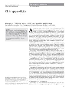
Fig. 1— Diagrams show anatomy of deep inferior epigastric perforator (DIEP) flap reconstruction after mastectomy. A, Inferior epigastric artery originates from external iliac artery and courses superiorly within rectus abdominis muscle. DIEP vessels provide vascular supply to abdominal tissue flap. Tissue flap is anastomosed to either internal mammary artery or thoracodorsal artery in chest. B, Harvested tissue flap containing skin and subcutaneous fat is removed from abdomen, rotated, and placed at mastectomy defect. C, Postoperative appearance after DIEP flap reconstruction with incisions at donor and recipient sites.
AJR:200, January 2013 W77
Downloaded from www.ajronline.org by 37.44.207.101 on 01/25/17 from IP address 37.44.207.101. Copyright ARRS. For personal use only; all rights reserved
Hedegard et al.
A
B
Fig. 2— 42-year-old BRCA2 mutation carrier undergoing CT angiography for preoperative planning before bilateral prophylactic mastectomies with deep inferior epigastric perforator (DIEP) flap reconstruction. A, Reconstructed sagittal oblique maximum-intensity-projection (MIP) image shows inferior epigastric arteries (arrowheads) originating from external iliac artery. B, Axial MIP image through pelvis shows one right lateral and one right medial DIEP (arrows).
W78
AJR:200, January 2013
Downloaded from www.ajronline.org by 37.44.207.101 on 01/25/17 from IP address 37.44.207.101. Copyright ARRS. For personal use only; all rights reserved
Breast Reconstruction Flap Fig. 3— Normal postoperative mammographic appearance of deep inferior epigastric perforator flap and transverse rectus abdominis myocutaneous (TRAM). A and B, 53-year-old woman with history of extensive left ductal carcinoma in situ. Mediolateral oblique (A) and craniocaudal (B) mammograms of reconstructed left breast and DIEP flap reconstruction show complete fatty replacement of normal breast parenchyma. C and D, 49-year-old woman who has undergone mastectomy and TRAM flap reconstruction for multicentric invasive ductal carcinoma. Mediolateral oblique (C) and craniocaudal (C) mammograms of reconstructed left breast show atrophy of rectus abdominis muscle (circle) in flap.
A
B
C
D
AJR:200, January 2013 W79
Downloaded from www.ajronline.org by 37.44.207.101 on 01/25/17 from IP address 37.44.207.101. Copyright ARRS. For personal use only; all rights reserved
Hedegard et al.
A
B
Fig. 4— Normal postoperative MRI appearance of deep inferior epigastric perforator (DIEP) flap. A, 51-year-old woman who underwent right mastectomy with DIEP flap for grade 3 invasive ductal carcinoma. Axial T1-weighted spoiled gradient-recalled echo (SPGR) MR image shows fatty replacement of normal breast parenchyma and contact line (arrow) at junction of residual subcutaneous breast adipose tissue and implanted abdominal adipose tissue. B, 48-year-old woman who underwent left mastectomy with DIEP flap for multicentric ductal carcinoma in situ. Axial T1-weighted SPGR MR image shows susceptibility artifact in medial aspect at internal mammary anastomosis site (circle).
A
B
Fig. 5— Comparison of deep inferior epigastric perforator (DIEP) flap versus transverse rectus abdominis myocutaneous (TRAM) flap at MRI. A, 52-year-old woman with left DIEP flap reconstruction after mastectomy for invasive lobular carcinoma. Axial T1-weighted spoiled gradient-recalled echo (SPGR) MR image shows susceptibility artifact in medial aspect at internal mammary anastomosis (circle). Absence of atrophied rectus abdominis muscle in flap is evident. B, Same patient as in A. Sagittal T1-weighted unenhanced fat-saturated MR image shows complete fatty replacement of parenchyma and absence of atrophied muscle in neobreast. (Fig. 5 continues on next page)
W80
AJR:200, January 2013
Downloaded from www.ajronline.org by 37.44.207.101 on 01/25/17 from IP address 37.44.207.101. Copyright ARRS. For personal use only; all rights reserved
Breast Reconstruction Flap
C
D
Fig. 5 (continued)— Comparison of deep inferior epigastric perforator (DIEP) flap versus transverse rectus abdominis myocutaneous (TRAM) flap at MRI. C and D, 49-year-old woman with left TRAM reconstruction after mastectomy for invasive ductal carcinoma. Axial T1-weighted SPGR (C) and sagittal T1-weighted gadolinium-enhanced fat-saturated (D) MR images show atrophied rectus muscle (circles) in posterior aspect of neobreast.
A
B
Fig. 6— Fat necrosis in two patients with deep inferior epigastric perforator (DIEP) flap reconstructions. A, 39-year-old woman who underwent left mastectomy with DIEP reconstruction for multifocal invasive ductal carcinoma. Axial T1-weighted spoiled gradient-recalled echo MR image shows hyperintense mass (circle) in lateral aspect of reconstructed breast. B, Same patient as in A. Axial T1-weighted fat-saturated gadolinium-enhanced image shows decreased signal intensity in mass, confirming presence of central fat in area of fat necrosis (circle). (Fig. 6 continues on next page)
AJR:200, January 2013 W81
Hedegard et al.
Downloaded from www.ajronline.org by 37.44.207.101 on 01/25/17 from IP address 37.44.207.101. Copyright ARRS. For personal use only; all rights reserved
Fig. 6 (continued)—Fat necrosis in two patients with deep inferior epigastric perforator (DIEP) flap reconstructions. C, Same patient as in A and B. Left mediolateral oblique mammogram shows extensive coarse dystrophic calcification in area of fat necrosis. D, 46-year-old woman who underwent left mastectomy with implant reconstruction and subsequent revision with DIEP flap reconstruction. Left mediolateral mammogram shows irregular mass (circle) in inferior aspect of left breast. Percutaneous sampling revealed fat necrosis. MR images show atrophied rectus muscle (circles) in posterior aspect of neobreast.
C
D
A
B
Fig. 7— Postoperative seroma and hematoma after deep inferior epigastric perforator (DIEP) flap reconstruction. A, 34-year-old woman 1 month after left mastectomy and DIEP flap reconstruction for extensive ductal carcinoma in situ. Axial T1-weighted gadolinium-enhanced fat-saturated MR image shows mass with thin rim of peripheral enhancement (circle) in posterior aspect of left reconstructed breast. B, Same patient as in A. Axial STIR image shows that mass is T2 hyperintense, most consistent with postoperative seroma. (Fig. 7 continues on next page)
W82
AJR:200, January 2013
Downloaded from www.ajronline.org by 37.44.207.101 on 01/25/17 from IP address 37.44.207.101. Copyright ARRS. For personal use only; all rights reserved
Breast Reconstruction Flap
D
C Fig. 7 (continued)—Postoperative seroma and hematoma after deep inferior epigastric perforator (DIEP) flap reconstruction. C, 51-year-old woman 2 months after right mastectomy and DIEP flap reconstruction for multifocal invasive ductal carcinoma. Right craniocaudal mammogram shows elongated mass (circle) in medial aspect of breast. D, Same patient as in C. Sonographic image shows complicated fluid collection. Subsequent percutaneous sampling showed organized hematoma.
A
B
Fig. 8— 43-year-old BRCA mutation carrier with abdominal discomfort 2 days after bilateral prophylactic mastectomies with deep inferior epigastric perforator (DIEP) flap reconstruction. A and B, Axial (A) and sagittal (B) CT images obtained with IV and oral contrast administration show large left rectus hematoma (circle). C, Sonographic image shows complex fluid collection along incision at abdominal donor site. Finding is consistent with rectus abdominis hematoma. Hematoma required surgical drainage.
C
AJR:200, January 2013 W83
Downloaded from www.ajronline.org by 37.44.207.101 on 01/25/17 from IP address 37.44.207.101. Copyright ARRS. For personal use only; all rights reserved
Hedegard et al.
A
C
B
D
Fig. 9—Two patients with tumor recurrence after autogenous tissue flap reconstruction. A and B, 55-year-old woman with history of left mastectomy with deep inferior epigastric perforator flap reconstruction for invasive ductal carcinoma (IDC) and ductal carcinoma in situ (DCIS) and normal result of sentinel node biopsy. Axial (A) and sagittal (B) T1-weighted gadolinium enhanced fat-saturated screening breast MR images 3 years after surgery show newly enlarged axillary lymph nodes in left axilla (circle). C, Same patient as in A and B. Sonographic image of axilla shows enlarged lymph nodes with complete loss of normal fatty hilum. Axillary lymph node core biopsy revealed IDC. D, 48-year-old woman 4 years after right mastectomy and transverse rectus abdominis myocutaneous flap reconstruction for DCIS and IDC. Gadolinium-enhanced subtracted maximum-intensity-projection screening MR image shows two superficial enhancing masses (circle) along contact line in lateral aspect of reconstructed breast. Subsequent ultrasound core biopsy revealed recurrent IDC.
W84
AJR:200, January 2013