Pictorial essay
Med Ultrason 2013, Vol. 15, no. 3, 230-236 DOI: 10.11152/mu.2013.2066.153.ts1ln2
Point-of-care ultrasound in the diagnosis of upper extremity fracture-dislocation. A pictorial essay. Turandot Saul1, Lorraine Ng2, Resa E Lewiss1 Department of Emergency Medicine, Division of Emergency Ultrasound, St. Luke’s/Roosevelt Hospital Center, 2Division of Pediatric Emergency Medicine, Division of Emergency Ultrasound, Columbia University College of Physicians and Surgeons, New York-Presbyterian Morgan Stanley Children’s Hospital, New York, USA 1
Abstract Patients commonly present with orthopedic injuries to the emergency department (ED). Although radiographs are the standard of care for evaulating these injuries, point-of-care ultrasound is being increasingly used to make the diagnosis. This modality can be useful in patients who are too clinically unstable to leave the acute care ED and in nonverbal pediatric or geriatric patients who are unable to isolate their injuries. Published case series and prospective studies highlight the emergency physician’s (EP) ability to detect fractures with point-of-care ultrasound with good accuracy. The American College of Emergency Physicians ultrasound guidelines advocate fracture identification as within the EP’s scope of practice. This pictorial essay reviews how to use point-of-care ultrasound to diagnose fractures and dislocations of the upper extremity. Keywords: ultrasonography, point-of-care ultrasound, orthopedic trauma, upper extremity, fracture, dislocation
Introduction Patients commonly present with orthopedic injuries to the emergency department (ED). Although radiographs are the standard of care for evaluating these injuries, point-of-care ultrasound is being increasingly used to make the diagnosis. The literature to date highlights certain advantages of musculoskeletal point-of-care ultrasound, particularly in radiation sensitive pediatric populations, in the pre-hospital environment, in austere environments, in the pregnant patient, and to reduce serial radiograph utilization in fracture reduction. Musculoskeletal sonographic evaluation can be useful in identifying fractures in patients who are too clinically unstable to leave the acute care ED and in nonverbal pediatric or Received 30.05.2013 Accepted 13.06.2013 Med Ultrason 2013, Vol. 15, No 3, 230-236 Corresponding author: Turandot Saul, MD, RDMS St. Luke’s / Roosevelt Hospital Center Department of Emergency Medicine Division of Emergency Ultrasound 1000 Tenth Avenue, Room GE-01 New York, New York 10019 Tel.: 212-523-6745 Email:
[email protected]
geriatric patients who are unable to isolate their injuries. Published case series and prospective studies highlight the emergency physician’s (EP) ability to detect fractures with point-of-care ultrasound with good accuracy [1,2]. The American College of Emergency Physicians (ACEP) ultrasound guidelines advocate fracture identification as within the EP’s scope of practice [3]. Normal sonographic anatomy When visualized with ultrasound, bone cortex appears as a bright white echogenic line with posterior shadowing as the bone reflects the sound waves back to the transducer and blocks further transmission. This property, however, makes the superficial cortex of the bone easily imaged. Disruption of the typically smooth, linear bony cortex can be demonstrated when a fracture is present. The location to perform sonographic evaluation as well as clinical correlation of findings can be determined by the patient’s point of maximal tenderness. Associated findings such as soft tissue swelling, adjacent and sub-periosteal hematomas, foreign bodies or bone fragments and proximity to surrounding vascular or neural structures are similarly identified. Care should be taken when scanning potentially displaced or shortened fractures as the normal cortex may
Med Ultrason 2013; 15(3): 230-236
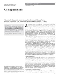
Fig 1. Ultrasound of the normal open physis (arrow) of a skeletally immature patient.
end abruptly at the site of displacement and this could be mistaken for a joint space. In skeletally immature children with open physes, ultrasound has been found to be less sensitive in the identification of fractures adjacent to joints (fig 1) [4]. Comparing findings to those of the contralateral unaffected side can help avoid this pitfall. Examination technique and technical requirements The point-of-care ultrasound examination, in most patients, can be performed utilizing a high frequency (7-10 MHz) linear transducer or probe. The probe is placed at the area of maximal or point tenderness and evaluation is performed in two orthogonal planes. By applying a thick layer of conducting gel, the sonologist can minimize any pressure placed on the injured area, thereby minimizing pain and potentially improve the resolution of superficial structures by bringing them into the focal zone. Using copious amounts of conducting gel can also help maintain probe contact over bony prominences. Alternatively, a water bath can be used for patient comfort and improved visualization of the superficial structures. The need for conducting gel or pressure on the skin is obviated. Long bones are easiest to image using ultrasound as they have a regular linear cortex. However, the smaller bones in the hands and the feet can be imaged as well. Visualized structures should be viewed in two orthogonal planes to assist in understanding the 3-dimensional relationships of structures utilizing 2-dimensional imaging. The unaffected contralateral side should be imaged to determine normal tissue architecture. In the pediatric population, the unaffected side may be imaged first to familiarize the child with the gel, transducer and mild pressure applied before approaching the painful extremity.
Fig 2. (a) Placement of the ultrasound transducer along the long axis of the right clavicle. (b) Ultrasound of a left clavicle fracture (arrow) in the longitudinal plane.
Pathologic conditions of the upper extremity Clavicle fracture Clavicle fractures usually occur after a direct blow to the lateral shoulder. They account for 2.6-5% of all adult fractures and 35-44% of all shoulder girdle injuries [5]. Clavicle fractures are also the most common fracture in the pediatric population. Point-of-care ultrasound is performed with a high frequency linear array transducer in both the longitudinal (fig 2) and transverse planes along the length of the clavicle. The middle of the shaft of the clavicle is the most commonly fractured portion of the clavicle as it is the thinnest, least supported site. Care must be taken to evaluate for injuries to surrounding vascular and pleural structures, particularly for proximal and medial fractures. Point-of-care ultrasound evaluation can be of great use as a targeted examination to identify isolated clavicle fractures that do not present with skin tenting, vascular or pleural compromise. Acromio-clavicular joint separation Injuries to the acromio-clavicular joint usually occur as a result of force directed down on the acromion such as a fall onto the dome of the shoulder or a fall on the elbow forcing the humeral head into the joint. Pain originating in this area may be difficult to localize given the joint’s dual innervation by the lateral pectoral and suprascapular nerves and in many cases, gross deformity may not be present [6]. Radiographic imaging of the shoulder can be challenging in the diagnosis of this injury due to the tissue penetration ability of X-rays. In addition, stress views have fallen out of favor as their usefulness does not justify the added cost and patient discomfort [7]. Point-ofcare ultrasound can be of great use as a targeted examination to identify acromio-clavicular joint space widening [7]. It is generally accepted that the width of a normal acromio-clavicular joint is less than 3 mm [8]. The severity of injury is classified by the degree of displacement of
231
232
Turandot Saul et al
Point-of-care ultrasound in the diagnosis of upper extremity fracture-dislocation
the acromion and distal clavicle, as well as the direction in which the clavicle dislocates. The point-of-care ultrasound examination is performed by placing a high frequency linear transducer over the acromio-clavicular joint following the long axis of the clavicle (fig 3a). Once the distal clavicle and acromion are visualized, the distance between the two articular surfaces is measured to evaluate for joint space widening. Sonographically, a normal acromio-clavicular joint will demonstrate alignment of the distal clavicle and acromion with the articular surfaces in close approximation (fig 3b). In acromio-clavicular joint dislocation, there is disruption and widening of the joint space > 3 mm and there may be displacement of the distal clavicle (fig 3c). Gleno-humeral joint dislocation The gleno-humeral joint is flexible enough to allow for the wide range of movements of the arm. For this reason it is inherently unstable, and is a commonly dislocated joint. Point-of-care ultrasound can be used in the evaluation of joint dislocation, and, in addition, post-reduction evaluation has been reported in the literature [9]. Since many gleno-humeral joint reductions require conscious sedation, this technique can avoid resedation of the patient and reduce the need for additional hemodynamic monitoring and personnel as well as the need for the sedated patient to leave the acute care area. Point-of-care ultrasound evaluation of this dislocation is most often performed with the high frequency linear transducer. However, utilizing a lower frequency curvilinear transducer with a larger footprint allows visualization of the deeper anatomic relationships of the joint. Multiple approaches have been described, including placing the transducer at the anterior and lateral aspect of the shoulder [10], as well as posteriorly on the patient’s back inferior to the lateral portion of the scapular spine [9,11]. Since the joint can be visualized by any of these methods, the technique used should be one that is comfortable for the patient and should depend on patient positioning which varies based on reduction technique. The transducer is placed on the anterior shoulder with the probe marker to the patient’s head following the long axis of the humerus (fig 4a). Normal sonographic anatomy shows the acromion visualized on the left of the screen with the humeral head on the right (fig 4b). Sonographic features of a gleno-humeral dislocation include disappearance of the humeral head with an empty glenoid fossa (fig 4c). The split-screen function available on most ultrasound machines can be used to
Fig 3. (a) Transverse placement of the ultrasound transducer over the acromio-clavicular joint. (b) Ultrasound of the acromion and clavicle in normal approximation (arrow) and (c) widening of the joint space in acromio-clavicular joint dislocation (arrow).
Fig 4. (a) Anterior placement of the ultrasound transducer over the gleno-humeral joint. Ultrasound demonstrating the unaffected (b) and affected sides (c) in a patient with gleno-humeral joint dislocation.
Fig 5. Ultrasound demonstrating the unaffected (a) and affected sides (b) in a patient after gleno-humeral joint relocation
compare the affected side to the unaffected side. Visualizing the humeral head below the coracoid process has also been described [10]. Once reduction is performed, the area can be re-evaluated for successful relocation (fig 5). Humerus fracture Proximal humeral fractures account for up to 5% of all fractures that present to the ED and are the third most common fracture in the elderly [12]. Mid-shaft
Med Ultrason 2013; 15(3): 230-236
Fig 6. (a) Placement of the ultrasound transducer longitudinally on the humerus. (b) Ultrasound of a humeral fracture (arrow) in the longitudinal plane.
Fig 7. (a) Placement of the ultrasound transducer longitudinally on the distal humerus. Ultrasound demonstrating the unaffected (b) and affected sides (c) in a patient with an elbow dislocation.
Fig 8. (a) Placement of the ultrasound transducer over the olecranon fossa in the transverse view. Ultrasound demonstrating the unaffected fat pad (b, arrow) and affected fat pad, the “overflowing cup” (c, arrow) in a patient with a supracondylar fracture.
fractures may be easier to diagnose with point-of-care ultrasound than those at the ends of the bone. Marshburn et al, studied physicians’ ability to sonographically evaluate patients with upper arm injuries after one hour ultrasound training in long bone fracture evaluation [2]. Of the thirteen upper extremity fractures, one humerus fracture was missed which involved the distal humerus. All other upper extremity ultrasound findings were consistent with radiography results. Sonographic evaluation is performed using a high frequency linear transducer by
visualizing the bone in two orthogonal planes, longitudinal (fig 6) and transverse. Elbow dislocation The elbow is the second most commonly dislocated major joint in adults, and the most commonly dislocated major joint in the pediatric population [13]. Such injuries are usually the result of a fall on an outstretched hand. When the elbow joint dislocates, the proximal ulna displaces posterior to the distal humerus and this abnormal relationship can be visualized sonographically [14]. To perform the examination, the patient’s elbow is flexed to 90 degrees. The probe is placed on the posterior aspect of the distal humerus with the probe marker to the patient’s head (fig 7a) and is then slid distally towards the elbow until the olecranon is visualized. The normal anatomic relationship is the distal humerus on the left of the screen aligned with the olecranon on the right of the screen (fig 7b). In cases of elbow dislocation, this anatomic relationship is disrupted and the proximal ulna is seen posterior to the distal humerus (fig 7c). Visualization of a hypoechoic hematoma or hypoechoic interstitial fluid as soft tissue swelling occurs may also be noted. Pediatric elbow injuries Supracondylar fractures are the most common fractures involving the elbow in pediatric patients [15]. In younger children, radial head subluxation, or Nursemaid’s elbow, is another common elbow injury. Point-of-care ultrasound evaluation has emerged as a new modality in screening for elbow fractures and differentiating them from radial head subluxations [16]. The elbow ultrasound is performed in the longitudinal plane, as described in the above section, as well as in transverse. The probe is slid distally towards the elbow until the olecranon fossa is visualized (fig 8a). The normal, physiologic posterior fat pad is recessed into the deep olecranon fossa (fig 8b) and should not extend above the borders of the distal humeral line in the longitudinal view or above a line connecting the lips of the olecranon fossa, in the transverse view. In the transverse view, when the posterior fat pad is found to ‘overflow,’ this is suggestive of an occult elbow fracture and further imaging and consultation is warranted (fig 8c). Greenstick or buckle fracture The pediatric periosteum is thick and strong, which maintains fracture stability in comparison to adult fractures, and makes greenstick fractures and torus (buckle) fractures a unique pediatric pathological entity. Depending on where the probe is placed, a greenstick fracture can appear as a cortical disruption or have a normal cortex if the probe is placed on the side of incomplete
233
234
Turandot Saul et al
Point-of-care ultrasound in the diagnosis of upper extremity fracture-dislocation
fracture. A buckle fracture will look like a mild elevation or angling in the periosteum without an obvious cortical disruption (fig 9). Minor fractures may be more difficult to detect, and visualization of a hematoma or surrounding soft tissue swelling may help localize a fracture, particularly in greenstick fractures. Distal radius fracture/Ulna fracture The distal radius is the most common site of fracture in the upper extremity [17]. In one study, point-of-care ultrasound detected distal radius and distal ulna fractures with a sensitivity of 71% and 50% and a specificity of 81% and 95%, respectively [18]. Ultrasound is performed with a high frequency linear transducer in the longitudinal (fig 10a) and transverse planes along the length of the bone of interest, evaluating for a cortical disruption (fig 10b). Scaphoid fracture The scaphoid is the most commonly fractured carpal bone, usually sustained by a fall on an outstretched hand. The blood supply to the scaphoid is tenuous, and avascular necrosis and non-union can lead to a poor functional outcome. Diagnosis of these fractures can be difficult as plain radiographs of the wrist even with a dedicated scaphoid view may not reveal the diagnosis. Computerized tomography, magnetic resonance imaging or bone scan may have limited availability in the emergency department setting. There are a few studies evaluating ultrasound as an initial imaging modality. In one study by Munk et al, ultrasound examinations performed in an orthopedic clinic had a sensitivity of 50% and specificity of 91% when compared to radiograph performed on the day of the clinic visit and at 10-14 day follow up [19]. In an ED population, one study found pointof-care ultrasound to have a sensitivity of 78% and a specificity of 89% when compared to radiograph performed on presentation then every month thereafter as long as the patient continued to be symptomatic [20]. Ultrasound is performed by placing a high frequency linear transducer on the dorsum of the wrist following the long axis of the distal radius with the probe marker aimed cephalad (fig 11a). The distal radius is seen on the left of the screen and the scaphoid on the right of the screen (fig 11b). The bony cortex of the scaphoid should be evaluated in two orthogonal planes and a fracture will appear as a cortical disruption (fig 11c). Metacarpal (Boxer’s) fracture Boxer’s fractures are fractures of the neck of the metacarpal bones, typically of the 4th or 5th digit as a result
Fig 9. Ultrasound demonstrating angling of the periosteum (arrow) in a patient with a buckle fracture
Fig 10. (a) Placement of the ultrasound transducer longitudinally on the distal radius. Ultrasound demonstrating a distal radius fracture (b, arrow).
Fig 11. (a) Placement of the ultrasound transducer over the scaphoid. Ultrasound demonstrating the unaffected (b) and affected side (c) in a patient with a scaphoid fracture (arrow).
of axial loading such as striking an object with a closed fist. Ultrasound imaging has been shown to have excellent sensitivity and specificity in the diagnosis of hand fractures [21]. The bone is visualized in two orthogonal planes, longitudinal (fig 12) and transverse. A standoff pad or a water bath is particularly useful with hand fractures to bring superficial structures into the focal zone of the transducer. Evaluating the degree of palmar angulation with ultrasound has been described, which can be an important factor in the ultimate functional outcome of the patient [22].
Med Ultrason 2013; 15(3): 230-236
Acknowledgment: The authors would like to thank Sarah E. Frasure, MD for her assistance with photo acquisition.
References Fig 12. (a) Placement of the ultrasound transducer longitudinally over the metacarpal. Ultrasound demonstrating the unaffected (b) and affected side (c) in a patient with a metacarpal head fracture (arrow).
Fig 13. (a) Placement of the ultrasound transducer longitudinally over the phalanx. Ultrasound demonstrating the unaffected (b) and affected side (c) in a patient with a phalanx fracture (arrow).
Phalanx fracture Fractures of the middle and proximal phalanges are commonly caused by a direct blow, rotary force or hyperextension of the phalanx. Distal phalanx fractures account for approximately half of all hand fractures and are commonly caused by trauma or crush injuries. Ultrasound has shown to have a high sensitivity and specificity in the detection of phalangeal fractures in adults and pediatrics, with a sensitivity of 90% and 50% and a specificity of 98% and 97%, respectively [18,21]. The bone of interest is evaluated in two orthogonal planes, longitudinal (fig 13) and transverse. A standoff pad or a water bath may be used. Care should be taken to differentiate the joint spaces from a cortical disruption or step-off. Conclusion Point-of-care ultrasound can be a valuable tool in the evaluation of the patient with an upper extremity injury. Published case series and prospective studies highlight the emergency physician’s ability to detect fractures with point-of-care ultrasound with good accuracy.
1. Chen L, Kim Y, Moore CL. Diagnosis and guided reduction of forearm fractures in children using bedside ultrasound. Pediatr Emerg Care 2007; 23: 523-531. 2. Marshburn TH, Legome E, Sargsyan A, et al. Goal-directed ultrasound in the detection of long-bone fractures. J Trauma 2004; 57:329-332. 3. Emergency Ultrasound Guidelines. ACEP Policy Statement 2008. www.acep.org/content.aspx?id=32878 4. Hubner U, Schlicht W, Outzen S, Barthel M, Halsband H. Ultrasound in the diagnosis of fractures in children. J Bone Joint Surg Br 2000; 82: 1170-1173. 5. Kopp DA, Char D. Clavicle Fractures. In: Essential Emergency Imaging, 1st ed. Lippincott Williams & Wilkins, Philadelphia 2011: 581-584. 6. DeLee: DeLee and Drez’s Orthopaedic Sports Medicine, 2nd ed. Saunders Elsevier, 2003. 7. Kijowski, R and De Smet, A. The role of ultrasound in the evaluation of sports medicine injuries of the upper extremity. Clin Sports Med 2006; 25: 569-590. 8. Zanca, P. Shoulder pain: Involvement of the acromioclavicular joint. (Analysis of 1,000 cases). Am J Roentgenol Radium Ther Nucl Med 1971; 112: 493-506. 9. Halberg MJ, Sweeney TW, Owens WB. Bedside ultrasound for verification of shoulder reduction. Am J Emerg Med 2009; 27: 134.e5-134.e6. 10. Yuen CK, Mok KL, Kan PG, Wong YT. Ultrasound diagnosis of anterior shoulder dislocation. Hong Kong J Emerg Med 2009; 16: 29-34. 11. Blakeley CJ, Spencer O, Newman-Saunders T, Hashemi K. A novel use of portable ultrasound in the management of shoulder dislocations. Emerg Med J 2009; 26: 662-663. 12. Court-Brown CM, Garg A, McQueen MM. The epidemiology of proximal humeral fractures. Acta Orthop Scand 2001; 72: 365-371. 13. Kuhn MA, Ross G. Acute elbow dislocations. Orthop Clin North Am 2008; 39: 155-161. 14. De Maeseneer M, Marcelis S, Cattrysse E, Shahabpour M, De Smet K, De Mey J. Ultrasound of the elbow: a systematic approach using bony landmarks. Eur J Radiol 2012; 81: 919-922. 15. English J, McManemy J. Supracondylar Fractures. In: Essential Emergency Imaging, 1st ed. Lippincott Williams & Wilkins, Philadelphia 2011: 558-561. 16. Rabiner J, Khine H, Avner JR, Friedman LM, Tsung JW. Accuracy of point-of-care ultrasound for diagnosis of elbow fractures in children. Am Emerg Med 2013; 61: 9-17. 17. Chung KC, Spilson SV. The frequency and epidemiology of hand and forearm fractures in the United States. J Hand Surg Am. 2001;26(5):908.
235
236
Turandot Saul et al
Point-of-care ultrasound in the diagnosis of upper extremity fracture-dislocation
18. Weinberg ER, Tunik MG, Tsung JW. Accuracy of clinicianperformed point-of-care ultrasound for the diagnosis of fractures in children and young adults. Injury 2010; 41: 862-868. 19. Munk B, Bolvig L, Kroner K, Christiansen T, Borris L, Boe S. Ultrasound for the diagnosis of scaphoid fractures. J Hand Surg Br 2000; 25: 369-371. 20. Senall JA, Failla JM, Bouffard A, van Holsbeeck M. Ultrasound for the early diagnosis of clinically suspected
scaphoid fracture. J Hand Surg Am 2004; 29: 400-405. 21. Tayal VS, Antoniazzi J, Pariyadath M, Norton HJ. Prospective use of ultrasound imaging to detect bony hand injuries in adults. J Ultrasound Med 2007; 26: 1143-1148. 22. Hennecke F, Kluge S, Kreutziger J, Jenzer A, Vogelin E. Ultrasonic and radiographic quantification of palmar angulation in metacarpal IV and V neck fractures. Handchir Mikrochir Plast Chir 2011; 43: 39-45.