Downloaded from www.ajronline.org by 37.44.207.34 on 01/23/17 from IP address 37.44.207.34. Copyright ARRS. For personal use only; all rights reserved
Vascular Imaging Gotway et al. Imaging of Takayasu’s Arteritis
Pictorial Essay Michael B. Gotway1,2 Philip A. Araoz3 Thanila A. Macedo3 Anthony W. Stanson3 Charles B. Higgins1 Ernest J. Ring1,2 Samuel K. Dawn1,2 W. Richard Webb1 Jessica W. T. Leung1 Gautham P. Reddy1 Gotway MB, Araoz PA, Macedo TA, et al.
Imaging Findings in Takayasu’s Arteritis OBJECTIVE. The objective of our study was to evaluate the clinical usefulness of crosssectional imaging for establishing the diagnosis of Takayasu’s arteritis (TA), an inflammatory vascular disorder that produces arterial stenoses and aneurysms primarily involving the thoracoabdominal aorta and its branches and the pulmonary arteries. CONCLUSION. CT and MRI findings of TA include vascular wall thickening and enhancement early in the disease, and arterial stenoses, occlusions, and aneurysms later in the disease. Cross-sectional imaging is useful for establishing the diagnosis of TA and for showing response to nonsurgical therapy or for planning a surgical intervention. akayasu’s arteritis (TA) is an idiopathic inflammatory vascular disorder that may involve the thoracoabdominal aorta and its branches and the pulmonary arteries. TA has a much higher incidence in women than in men and is most frequently found in Asian patients, although the condition may occur in North American, European, African, and Middle Eastern patients [1].
T
Received June 28, 2004; accepted after revision September 27, 2004. 1Department
of Radiology, University of California, San Francisco, 505 Parnassus Ave., San Francisco, CA 94143.
2Department of Radiology, San Francisco General Hospital,
1001 Potrero Ave., Rm. 1X 55A, Box 1325, San Francisco, CA 94110. Address correspondence to M. B. Gotway.
3Department
of Radiology, Mayo Clinic, 200 First St. SW, Rochester, MN 55905.
AJR 2005;184:1945–1950 0361–803X/05/1846–1945 © American Roentgen Ray Society
AJR:184, June 2005
Pathophysiology TA causes arterial media destruction, leading to aneurysm formation and, uncommonly, rupture of involved arteries. Histopathologically, early TA changes consist of an adventitial mononuclear infiltrate with perivascular cuffing of the vasa vasorum, followed by medullary mononuclear inflammation, occasionally accompanied by granulomatous changes [2]. Because histopathologic specimens are seldom available as a result of the large vessels commonly affected and the fact that the histopathologic appearance of TA can mimic other arteritides, the diagnosis of TA is largely based on the combination of clinical information, laboratory evaluation, and diagnostic imaging. Therefore, knowledge of the radiologic features of TA is essential for accurate diagnosis
and early treatment. Although angiography has been widely used for the diagnosis of TA, increasingly CT and MRI are used as the diagnostic techniques of choice. Clinical Presentation TA has traditionally been divided into an early, “prepulseless” systemic phase, and a late, occlusive phase. In the early systemic phase, diagnosis is difficult and symptoms are usually nonspecific and constitutional, including fever, myalgias, weight loss, and arthralgias. In the occlusive phase, ischemic symptoms dominate, including angina, claudication, syncope, and visual impairment [3]. Late-phase TA may be further subclassified as classic pulseless disease (type 1), a mixed type (type 2), an atypical coarctation type (type 3), and a dilated type (type 4) [1]. Most patients present with a form of late-phase disease. Treatment High-dose corticosteroids are the mainstay of TA therapy. Treatment of symptomatic fibrotic lesions (stenoses or occlusions) requires either interventional or surgical therapy. This can be achieved by angioplasty with or without stenting or, in severe cases, by vascular resection and surgical placement of composite grafts [4, 5] (Fig. 1).
1945
Downloaded from www.ajronline.org by 37.44.207.34 on 01/23/17 from IP address 37.44.207.34. Copyright ARRS. For personal use only; all rights reserved
Gotway et al.
A
B
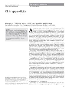
Fig. 1.—45-year-old-woman with Takayasu’s arteritis and graft placement for abdominal aortic occlusion. A and B, Coronal maximum-intensity-projection (MIP) (A) and volume-rendered (B) images show supraceliac aorta-to-right iliac artery bypass graft (single large arrow) and supraceliac aorta-to-right renal artery bypass graft (double small arrows) with occlusion (single small arrow) of midportion abdominal aorta below superior mesenteric artery origin, best shown on MIP image (A). Left renal artery is supplied via bypass graft (single arrowhead). Superior mesenteric artery is supplied from left iliac artery bypass graft (double arrowheads).
a
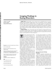
Fig. 2.—50-year-old woman with Takayasu’s arteritis (TA). Catheter angiography shows mild infrarenal abdominal aortic stenosis (arrow). Although atherosclerosis commonly affects infrarenal abdominal aorta, atherosclerosis usually produces abrupt caliber changes just beyond stenosis sites; the smooth tapered nature of this stenosis favors TA.
1946
Fig. 3.—45-year-old woman with Takayasu’s arteritis. Catheter angiography shows severe infrarenal abdominal aortic stenosis (arrows).
Fig. 4.—45-year-old woman with Takayasu’s arteritis. Oblique projection catheter angiogram shows segmental stenosis of origin of right upper lobe pulmonary artery (single arrow). Other stenoses are also visible (double arrows).
AJR:184, June 2005
Downloaded from www.ajronline.org by 37.44.207.34 on 01/23/17 from IP address 37.44.207.34. Copyright ARRS. For personal use only; all rights reserved
Imaging of Takayasu’s Arteritis CT Because the basic pathologic feature of early-phase TA is great vessel wall thickening, CT is useful for early diagnosis because it allows evaluation of wall thickness rather than merely the luminal diameter [6], which is especially important because early diagnosis and treatment are associated with improved prognosis. The spectrum of findings on CT angiography includes stenoses; occlusions; aneurysms (Figs. 7–9); and concentric arterial wall thickening affecting the aorta and its branches, the pulmonary arteries, and occasionally the coronary arteries [1–3, 5–8] (Fig. 9). In the later stage of disease, extensive vascular calcification may occur (Fig. 10). Limitations of CT include the need for iodinated contrast material and ionizing radiation; the latter may limit the utility of CT for following up patients undergoing treatment.
Fig. 5.—52-year-old woman with Takayasu’s arteritis. Catheter angiogram shows severe infrarenal abdominal aortic stenosis, ending in aortic occlusion (arrow), with extensive collateral vessel formation.
Fig. 6.—38-year-old woman with Takayasu’s arteritis. Catheter angiogram shows severe infrarenal abdominal aortic stenosis (arrowhead) with extensive collateral vessel formation by enlarged pancreaticoduodenal artery between superior mesenteric and celiac arteries (arrows).
Imaging Findings Angiography Angiography, particularly digital subtraction angiography, has traditionally been the procedure of choice for the diagnostic evaluation of TA [6, 7]. Angiography often shows long, smooth, tapered stenoses ranging from mild (Fig. 2) to severe (Figs. 3 and 4) or frank occlusions (Fig. 5); collateral vessels (Fig. 6), or the subclavian steal phenomenon [4], are also well shown. Angiography is useful in guiding interventional procedures such as angioplasty or stent placement. However, angiography is invasive, carries a substantial radiation dose, may require a large amount of iodinated contrast material, and can be difficult to perform in patients with long-segment stenoses or heavy arterial calcification. Furthermore, angiography does not depict wall architecture changes as effectively as cross-sectional techniques and cannot differentiate vascular narrowing due to acute mural inflammation from stenoses due to chronic
transmural fibrosis. Finally, the frequency of ischemic complications resulting from angiography in patients with TA has been shown to be high, and cross-sectional techniques are therefore attractive for diagnosis and disease monitoring [8].
AJR:184, June 2005
Sonography Sonography reveals homogeneous circumferential thickening of affected vessels (indistinguishable from atherosclerotic plaque), vascular occlusions and dilation, and flow velocity elevations beyond stenotic lesions in patients with TA. Limitations of sonography include lack of a consistent acoustic window to allow visualization of the root of the great vessels, obscuration of abdominal vasculature by overlying bowel gas, and technically limited studies in obese patients. Invasive imaging with transesophageal echocardiography and intravascular sonography provides extremely high spatial resolution and allows recognition of subtle wall changes in aortic segments that appear normal with other imaging techniques.
MRI MRI advantages include the lack of need for ionizing radiation and iodinated contrast material; therefore, MRI is ideal for serial evaluation of patients with TA who are undergoing treatment. Furthermore, MRI provides the ability to view vessels in any desired plane, and techniques like cine MRI can detect cardiovascular functional and hemodynamic changes, such as aortic regurgitation, in patients with TA [2]. As with CT, MRI is useful for early diagnosis because of its ability to evaluate wall thickness rather than just the luminal narrowing [1, 2, 8]. Findings of TA on MRI include mural thrombi, signal alterations within and surrounding inflamed vessels (Figs. 11 and 12), fusiform vascular dilation, thickened aortic valvular cusps, multifocal stenoses (Figs. 13 and 14), and concentric thickening of the aortic wall [1, 2, 8] (Fig. 12). MRI may also reveal pericardial effusions and signal alterations within the pericardial sac, representing fluid and granulation tissue. Disadvantages of MRI include difficulty in visualizing small branch vessels and poor visualization of vascular calcification. MRI is also expensive and is often less available in regions where TA is most prevalent. MR angiography provides detailed vascular information, including the location, degree, extent of stenoses and dilation, and patency of collateral vessels and surgical bypass grafts (Fig. 15). Limitations of MR angiography include the possibility that vascular branch points may be improperly interpreted as occlusions (breath-hold techniques have lessened this problem) and that maximum-intensity-projection images may
1947
Downloaded from www.ajronline.org by 37.44.207.34 on 01/23/17 from IP address 37.44.207.34. Copyright ARRS. For personal use only; all rights reserved
Gotway et al.
B
A
Fig. 7.—40-year-old woman with aneurysmal form of Takayasu’s arteritis. A, Thoracic CT angiogram shows aneurysm of left subclavian artery (arrow) at origin of left vertebral artery. B, Thoracic CT angiogram obtained caudal to A shows large aneurysm (arrow) of brachiocephalic artery and proximal right subclavian artery. C, Volume-rendered image shows right subclavian artery aneurysm (arrowheads) to advantage.
C
A Fig. 8.—45-year-old woman with aneurysmal form of Takayasu’s arteritis. Axial CT image shows aneurysm of proximal descending thoracic aorta (arrow). Differential diagnosis should include atherosclerosis, mycotic aneurysm, and Behçet's syndrome.
1948
B
Fig. 9.—40-year-old woman with aneurysmal form of Takayasu’s arteritis. A, Thoracic CT angiogram obtained through heart shows aneurysm (arrow) of left anterior descending coronary artery. B, Thoracic CT angiogram obtained caudal to A shows aneurysm (arrow) of left circumflex coronary artery.
AJR:184, June 2005
Imaging of Takayasu’s Arteritis
Downloaded from www.ajronline.org by 37.44.207.34 on 01/23/17 from IP address 37.44.207.34. Copyright ARRS. For personal use only; all rights reserved
Fig. 10.—50-year-old woman with Takayasu’s arteritis in late stage of disease. Unenhanced CT scan shows extensive calcification (arrow) composing abdominal aortic lumen.
A
B
Fig. 11.—19-year-old man with early Takayasu’s arteritis. A, Axial T1-weighted MR image (TR/TE, 500/20) obtained through superior mediastinum shows thickening of right brachiocephalic artery wall (arrowheads). B, Axial T1-weighted gadolinium-enhanced MR image (500/20) shows extensive enhancement of abnormally thickened right brachiocephalic artery wall (arrowheads).
A
B
Fig. 12.—32-year-old woman with Takayasu’s arteritis. A, Axial T1-weighted MR image (TR/TE, 500/20) shows concentric thickening of infrarenal abdominal aorta (arrow). B, Axial T1-weighted MR image (500/20) with fat saturation after gadolinium administration shows extensive enhancement of thickened abdominal aorta (arrow).
AJR:184, June 2005
1949
Downloaded from www.ajronline.org by 37.44.207.34 on 01/23/17 from IP address 37.44.207.34. Copyright ARRS. For personal use only; all rights reserved
Gotway et al.
Fig. 13.—50-year-old woman with Takayasu’s arteritis. Coronal MR angiogram shows short-segment occlusion of infrarenal abdominal aorta (long arrow). Occlusion of right common femoral artery (short arrows) is also evident.
Fig. 14.—59-year-old woman with multifocal great vessel stenoses due to Takayasu’s arteritis (TA). Maximum-intensity-projection coronal MR angiogram shows occlusion of right brachiocephalic artery (arrow), severe stenosis of right subclavian artery (single arrowhead), and occlusion of proximal left subclavian artery (double arrowheads). Subclavian arteries are reconstituted by collateral vessel formation bilaterally. Note relatively proximal great vessel involvement; whereas giant cell arteritis may have imaging appearance similar to that of TA, lesions are usually located more distally in latter disorder.
falsely accentuate the degree of vascular stenoses. Regarding the latter, we believe it is advisable to assess the degree of vascular stenoses from the MR angiography source images.
References
Conclusion Angiographic and cross-sectional imaging findings of TA include vascular stenoses, occlusions, and aneurysm formation. Familiarity with the demographic and imaging features of TA will facilitate accurate diagnosis and allow early treatment, improving patient outcome.
1950
1. Matsunaga N, Hayashi K, Sakamoto I, Ogawa Y, Matsumoto T. Takayasu arteritis: protean radiologic manifestations and diagnosis. RadioGraphics 1997;17:579–594 2. Matsunaga N, Hayashi K, Sakamoto I, et al. Takayasu arteritis: MR manifestations and diagnosis of acute and chronic phase. J Magn Reson Imaging 1998;8:406–414 3. Kerr GS, Hallahan CW, Giordano J, et al. Takayasu arteritis. Ann Intern Med 1994;120:919–929 4. Park JH, Han MC, Kim SH, Oh BH, Park YB, Seo JD. Takayasu arteritis: angiographic findings and results of angioplasty. AJR 1989;153:1069–1074
Fig. 15.—55-year-old woman with Takayasu’s arteritis, infrarenal abdominal aortic occlusion, and surgical arterial bypass graft. Coronal maximum-intensity-projection MR angiogram shows occluded infrarenal abdominal native aorta (arrow) and surgical bypass graft (arrowheads) with reimplanted abdominal vasculature.
5. Miyata T, Sato O, Koyama H, Shigematsu H, Tada Y. Long-term survival after surgical treatment of patients with Takayasu’s arteritis. Circulation 2003;108:1474–1480 6. Park JH, Chung JW, Lee KW, Park YB, Han MC. CT angiography of Takayasu arteritis: comparison with conventional angiography. J Vasc Interv Radiol 1997;8:393–400 7. Paul JF, Fiessinger JN, Sapoval M, et al. Followup electron beam CT for the management of early phase Takayasu arteritis. J Comput Assist Tomogr 2001;25:924–931 8. Yamada I, Numano F, Suzuki S. Takayasu arteritis: evaluation with MR imaging. Radiology 1993; 188:89–94
AJR:184, June 2005