Cover
Restoring Speech to Tracheostomy Patients Linda L. Morris, PhD, APN, CCNS Ana M. Bedon, MSN, APN, AGCNS-BC, CWON Erik McIntosh, RN, MSN, ACNP-BC Andrea Whitmer, RN, MSN, ACNP-BC
Tracheostomies may be established as part of an acute or chronic illness, and intensive care nurses can take an active role in helping restore speech in patients with tracheostomies, with focused nursing assessments and interventions. Several different methods are used to restore speech, whether a patient is spontaneously breathing, ventilator dependent, or using intermittent mechanical ventilation. Restoring vocal communication allows patients to fully express themselves and their needs, enhancing patient satisfaction and quality of life. (Critical Care Nurse. 2015;35[6]:13-28)
T
racheostomy is one of the most common procedures performed in critically ill patients and is becoming more commonplace in the intensive care unit (ICU).1 Indications for tracheostomy include prolonged intubation with unsuccessful weaning, management of bronchial hygiene, obstruction of the upper airway, and airway protection.1,2 Patients with head and neck trauma and/or surgery or those who have airways that cannot be managed via endotracheal intubation may also require a tracheostomy. When the tracheostomy tube is initially placed, the cuff at the distal end of the tube is inflated to protect the airway and provide effective ventilation.2 Because inflation of the cuff does not permit the passage of air up through the larynx, the patient cannot phonate (ie, produce speech sounds).3 The inability to communicate via speech places a great amount of stress on an already critically ill patient. Patients with tracheostomies report feelings of frustration, fear, anxiety, and powerlessness related to the loss of voice.3-5 Donnelly and Wiechula6 have described how patients experience the loss of voice as a form of torture, more so than the physical discomfort the patients feel from the tracheostomy or other procedures performed in the ICU.
CE Continuing Education This article has been designated for CE credit. A closed-book, multiple-choice examination follows this article, which tests your knowledge of the following objectives: 1. Identify the potential effects of the inability to communicate for a patient with a tracheostomy 2. Examine methods to restore phonation for patients with a tracheostomy 3. Discuss the role of critical care nurses in restoring phonation ©2015 American Association of Critical-Care Nurses doi: http://dx.doi.org/10.4037/ccn2015401
www.ccnonline.org
Morris12_15pgs.indd 13
CriticalCareNurse
Vol 35, No. 6, DECEMBER 2015
13 11/9/15 4:46 PM
For patients in the ICU, the inability to express themselves and to actively participate in their plan of care can lead to depression, disengagement in their recovery, and nonadherence with their therapeutic plan.4,7-9 A tracheostomy tube, however, does not prevent phonation. Phonation can enhance the ability of patients with a tracheostomy to express their needs and wishes fully and effectively, allowing the patients to participate in their plan of care and converse with their loved ones.7,9 Critical care nurses are in an ideal position to coach and guide tracheostomy patients to phonate, but nurses may not be aware of all the options available. In this article, we provide inforInability to speak can lead to depression, mation that disengagement, and nonadherence to will enable the therapeutic plan. nurses to take an active role in restoring phonation in these patients. We review the different approaches to restore phonation in patients with a tracheostomy, including patients who are spontaneously breathing, are being treated with intermittent mechanical ventilation, or are ventilator dependent. An essential component of successful communication is to determine what option or options are most appropriate for a particular patient. Forms of communication such as lipreading, writing, hand signals, and picture boards can be useful for enabling patients to express basic needs but do not fully encompass the reciprocal nature of human communication.4,7 Visual acuity, language barriers, literacy, physical immobility or weakness, and cognitive deficit can impair the effectiveness of these other forms of communication.3,5 Ideally, critical care nurses can facilitate nonverbal forms of communication. Lipreading is a specialized skill and may be difficult for many nurses to master.9 Coded eye blinking, head and hand gestures, and nodding answers to yes-no questions require collaboration with patients and must be effectively communicated to other caregivers
in order to be consistent9; however, these activities can be time-consuming and can cause efficiency problems in caring for critically ill patients. A patient-specific communication plan should be made available to everyone interacting with a patient; preferably the plan should be at the patient’s bedside or in another centralized location.9 Even with the best intentions, lapses in care may occur, causing patients distress and frustration with caregivers.
Methods to Restore Phonation for Patients With a Tracheostomy Sound is produced as air passes through the vocal cords, causing the cords to vibrate.10 Medical complications of the pharyngeal, laryngeal, and tracheal structures, including glottic or subglottic edema, ulceration of the vocal fold, vocal cord paralysis, tracheal stenosis, and tracheomalacia, can affect the ability to create sound. The tracheostomy tube itself can markedly obstruct the trachea, causing poor airflow, increased airway resistance, and increased work of breathing and can lead to an inability to produce speech. Therefore, the ability to create sound with a tracheostomy tube depends on having an adequate supply of air reach the vocal cords with a minimum of resistance. The diameter, length, and type of tracheostomy tube play important roles in avoiding complications and leading to greater success in phonation. Changing one or all of these components of the tracheostomy tube can lead to less airway resistance and prevent respiratory distress and unsuccessful phonation trials. Methods to restore phonation for a patient with a tracheostomy will also vary, depending on whether or not the patient is ventilator dependent, and, if so, whether the patient is fully or partially dependent on ventilator support. Methods of restoring phonation for patients who are spontaneously breathing, are being treated with intermittent mechanical ventilation, or are fully ventilator dependent are summarized in Table 1.
Authors Linda L. Morris is a tracheostomy specialist/consultant and an associate professor of clinical anesthesiology, Feinberg School of Medicine, Northwestern University, Chicago, Illinois. She is also a member of the board of directors for the Global Tracheostomy Collaborative, an international group of specialists dedicated to research and quality outcomes of patients with tracheostomies. Ana M. Bedon is a certified wound and ostomy care nurse with a background in critical care. She is currently working as the advanced practice nurse for the Digestive Health Institute at Advocate Illinois Masonic Medical Center, Chicago, Illinois. Erik McIntosh is an acute care nurse practitioner on an inpatient internal medicine unit, Rush University Medical Center, Chicago, Illinois. Andrea Whitmer is the acute care nurse practitioner for the intensivist program in the critical care unit at Elkhart General Hospital, Elkhart, Indiana. Corresponding author: Linda L. Morris, PhD, APN, CCNS, FCCM (e-mail:
[email protected]). To purchase electronic or print reprints, contact the American Association of Critical-Care Nurses, 101 Columbia, Aliso Viejo, CA 92656. Phone, (800) 899-1712 or (949) 362-2050 (ext 532); fax, (949) 362-2049; e-mail,
[email protected].
14 Morris12_15pgs.indd 14
CriticalCareNurse
Vol 35, No. 6, DECEMBER 2015
www.ccnonline.org
11/9/15 4:46 PM
Table 1 Technique
Pros
Methods of phonation
Cons
Special considerations
Spontaneously breathing patient Cuff deflation with digital occlusion
Used for assessment of the patient’s ability to tolerate capping or use of a speaking valve May be an option for patients who may not be completely alert and who may not tolerate capping
Bulkiness of the deflated cuff may cause marked airway obstruction and resistance
Before the cuff is deflated, subglottic suctioning should be performed to prevent aspiration of secretions from above the cuff Cuff must be completely deflated before digital occlusion Thorough suctioning before and after cuff deflation can help prevent aspiration, coughing, respiratory distress, which may lead to an unsuccessful digital occlusion trial Ideally, heated aerosol via tracheostomy collar should be used if supplemental oxygen is needed
Capping trials
Capping the tracheostomy tube allows air to be inhaled and exhaled through the natural airway
Possible airway obstruction, with mucus buildup around or within the tube; therefore, frequent monitoring is essential
Capping should be attempted only with a cuffless tube or tight-to-shaft (TTS) tracheostomy tube of appropriate size; external tube diameter should be minimized as appropriate Nasal cannula or face mask should be used if supplemental oxygen is needed while cap is in place Thorough suctioning before and after capping trials can help prevent aspiration, coughing, respiratory distress, which may lead to an unsuccessful capping trial Anxiety may be a factor in unsuccessful capping trials: the unfamiliar feeling of air moving through the upper part of the airway may lead to tachypnea
Speaking valve
Can be used with fully deflated cuff or cuffless tube
See Table 2 for full list of contraindications
Ideally, heated aerosol via tracheostomy collar should be used if supplemental oxygen is needed Cuff must be completely deflated when speaking valve is used Thorough suctioning before cuff deflation can help prevent aspiration, coughing, and respiratory distress, which may lead to an unsuccessful trial of the speaking valve
Tracheostomy button
Fits within the stoma and does not require tracheostomy ties The tracheostomy button is a stent to keep the stoma open for a prescribed period of time
If patients need positive pressure ventilation and/or need suctioning, button should be replaced with a standard tracheostomy tube
Not usually used in critical care but may be an option for patients after discharge
Cuffless fenestrated tracheostomy tube
A fenestrated tracheostomy tube allows air to travel through the fenestration, which decreases airway resistance, improves airflow in the trachea, and facilitates speech
If tube does not fit properly, granulation tissue may grow within the fenestration, making removal a surgical problem
Requires an evaluation by a specialist to fit the tube to ensure that the fenestration lies centrally within the trachea; otherwise, granulation tissue may grow into the fenestration Secretions can also collect in the fenestration, so the patient should have optimal humidification with heated aerosol Continued
www.ccnonline.org
Morris12_15pgs.indd 15
CriticalCareNurse
Vol 35, No. 6, DECEMBER 2015
15 11/9/15 4:46 PM
Table 1 Technique
Pros
Continued
Cons
Special considerations
Spontaneously breathing patient Speak EZ tracheal cannula
Low profile, does not require tracheostomy ties Fits only within the stoma, thereby providing no tracheal irritation
If patients need positive pressure ventilation and/or need suctioning, cannula should be replaced with a standard tracheostomy tube
Most often used for patients who initially received a tracheostomy for vocal cord paralysis or sleep apnea Not commonly used in critical care, but may be an option for patients after discharge
These TTS tubes are singlecannula tubes and may become clogged with secretions
Cuff must be completely deflated when providing intermittent phonation Only sterile water should be used to inflate the Bivona TTS cuff; saline should NOT be used because it damages the cuff; air should not be used because it diffuses through the cuff, causing cuff deflation over time Thorough suctioning before and after cuff deflation can help prevent aspiration, coughing, and respiratory distress, which may lead to an unsuccessful trial Supplemental oxygen should be provided if needed, via nasal cannula when the tube is capped When cuff is inflated, minimal leak technique should be used to prevent complications associated with the high-pressure TTS cuffs
Intermittently ventilator dependent Intermittent phonation
Because the cuff essentially disappears on deflation, the TTS tube can be safely capped When capped, the natural function of the glottis is restored, and this return to natural function often allows patients to remain free from the ventilator Bivona TTS cuff can be inflated to deliver positive pressure ventilation and then deflated for capping
Ventilator dependent Leak speech
Allows speech even though patient cannot be liberated from mechanical ventilation
Patient must be coached to speak on inspiration and may require practice in timing vocalization
Leak speech can be used in a patient who can tolerate cuff deflation Ventilator settings can be adjusted to compensate for tidal volume loss and to improve speech quality Thorough suctioning before and after cuff deflation can help prevent aspiration, coughing, respiratory distress, which may lead to an unsuccessful leak speech trial
Talking tracheostomy tubes
Used for patients who require continuous cuff inflation The air used for speech is completely separate from the air used for breathing Does not require adjustment of ventilator settings; tidal volume is constant
Speech depends on having patient or caregiver occlude the port Accumulation of secretions above the cuff can clog the air supply line, resulting in no airflow for speech Discomfort and drying of mucous membranes can occur with high airflows
Tube has a port attached to an air source located above the cuff Secretions may pool above cuff; suctioning of secretions from air port should be done as needed to maintain patent airway and facilitate good speech quality Voice is adjusted by increasing airflow, often 5-15 L/min, to achieve optimal vocalization; humidified airflow should be provided to mitigate against discomfort with higher airflows Continued
16 Morris12_15pgs.indd 16
CriticalCareNurse
Vol 35, No. 6, DECEMBER 2015
www.ccnonline.org
11/9/15 4:46 PM
Table 1 Technique
Continued
Pros
Cons
Special considerations
Cuffed fenestrated tracheostomy tube
A fenestrated tracheostomy tube allows air to travel through the fenestration above the cuff, decreasing airway resistance, improving airflow in the trachea, and facilitating speech
Risk for aspirating secretions is high; tube may not be tolerated because of small diameter of fenestration, which may markedly increase airway resistance and result in difficulty exhaling, air trapping (auto-PEEP), and decompensation Tube is not tolerated in patients with increased respiratory rate and minute ventilation because of anxiety and respiratory distress due to uncomfortable breathing during attempted speech
Volumes may need to be adjusted on the ventilator for exhaled volumes lost through the upper part of the airway Fit requires an evaluation by a specialist to ensure that the fenestration lies centrally within the trachea; otherwise, granulation tissue may grow into the fenestration Secretions can also collect in the fenestration, so patient should receive optimal humidification with heated aerosol
Blom tracheostomy tube system
The Blom tube has a fenestration just above the cuff to minimize risk of granulation tissue
System requires greater knowledge and assessment skills by staff in order to maximize success and minimize patient’s respiratory discomfort, anxiety, and distress
System uses a special speech cannula with 2 valves; upon inhalation, the flap valve opens, allowing air through the tube; upon exhalation, the flap valve closes and air moves through the fenestration and through a bubble valve A special exhaled volume reservoir can be used to minimize alarms indicating low exhaled volume
Ventilator dependent
Abbreviation: PEEP, positive end-expiratory pressure.
Phonation in Patients Who Are Breathing Spontaneously For patients with tracheostomies who are breathing spontaneously and do not require mechanical ventilation, 3 primary methods of phonation can be used: cuff deflation with digital occlusion of the tracheostomy tube; capping; and use of a speaking valve. Before any method of phonation is started, the patient’s physical and mental condition should be assessed to determine which method would be the most appropriate. The patient must be attempting to communicate verbally and must have intact cognitive function.8 The ability to follow instructions and communicate any difficulty with breathing or phonation is important to success.11 With any of the following methods, nurses should closely monitor patients for signs and symptoms of respiratory distress, including breathing discomfort, increased respiratory rate, use of accessory muscles, inadequate chest inflation or deflation, and difficulty with air exchange.9 Assessing the work of breathing is a better method to determine tolerance of cuff deflation, capping, or use of a speaking valve than is measuring oxygen saturation.
www.ccnonline.org
Morris12_15pgs.indd 17
Cuff Deflation. Generally, a patient must be able
to tolerate cuff deflation or have a cuffless tube in order to phonate via any of the 3 primary methods.8,11 Deflation of the cuff causes airflow to be redirected around the tracheostomy tube and up through the upper part of the airway and may require a period of adjustment for the patient. Pooled secretions above the cuff and movement of the tube during cuff deflation can cause airway irritation, coughing, obstruction of secretions, increased work of breathing, and shortness of breath, which may lead to cardiorespiratory deterioration. Therefore, verifying that emergency equipment is available, including suction equipment and a manual resuscitation bag, is important. Cuff deflation can be an anxiety-filled experience for a patient if it causes respiratory discomfort and distress. Therefore, it is essential to provide adequate assessments as well as proper coaching and preparation of the patient before, during, and after cuff deflation. The following steps can help facilitate a successful cuff deflation trial: First, explain to the patient the steps that go into cuff deflation and the feelings that might occur. Second, ensure the correct position of the patient and the tracheostomy
CriticalCareNurse
Vol 35, No. 6, DECEMBER 2015
17 11/9/15 4:46 PM
tube, with the head of the bed elevated to the best level for breathing comfort and neutral position of the tracheostomy tube to prevent airway irritation, coughing, and obstruction.9 Third, explain to the patient the need to suction the back of the throat to clear secretions that may have pooled above the cuff. Of note, deep subglottic suctioning will not always ensure complete removal of secretions once the cuff is deflated, and suctioning of the mouth and the posterior part of the pharynx may still be required. If the patient is able, have him or her perform this step. Fourth, before the cuff is deflated, have the patient take a deep breath in to maximize air in the lungs to promote a forceful cough if needed to clear any secretions. Just after the patient takes a deep breath in, and as he or she begins to exhale, completely deflate the cuff. After the cuff is deflated, immediately suction as needed through the tracheostomy tube and/or mouth to clear the secretions that might have remained above the cuff. While the cuff is deflated, closely observe the patient for any signs or symptoms of respiratory distress. Increase the fraction of inspired oxygen as needed; cuff deflation causes admixture of room air in patients receiving oxygen therapy. Secretions and coughing also may lower Proper clearance of secretions will oxygenation. prevent triggers of airway irritation Continuand cough, which can initiate ally reassure bronchospasm. the patient through this process to decrease anxiety. Proper clearance of secretions is crucial to successful cuff deflation and will prevent triggers of airway irritation and cough, which can initiate bronchospasm. A manual resuscitation bag and suction devices should be at hand to stabilize the patient’s condition if needed. If cuff deflation is not initially tolerated, despite meticulous attention to proper procedure, another trial with slow deflation of the cuff, perhaps up to several minutes, is recommended.12,13 If respiratory distress, dyspnea, or shortness of breath occurs with cuff deflation, the cuff should be reinflated, and cuff deflation trials can be repeated at another time. Working side by side with speech language pathologists and respiratory therapists can ensure patients’ comfort, tolerability of cuff deflation trials, and quality outcomes.11 After successful cuff deflation, digital occlusion can be attempted for vocalization.
18 Morris12_15pgs.indd 18
CriticalCareNurse
Vol 35, No. 6, DECEMBER 2015
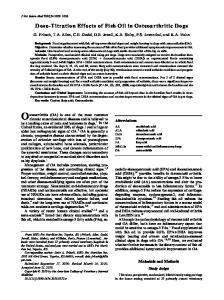
Figure 1 Cuffed tube with deflated cuff versus cuffless tube. Note bulk of deflated cuff on left, compared with cuffless tube on right.
Reproduced from Morris,14 with the permission of Springer Publishing Company, LLC, New York, New York.
Digital Occlusion. Digital occlusion of the tracheostomy tube is used for patients who have a cuffless tube or a cuffed tube with a fully deflated cuff (Figure 1). When the cuff is inflated, the only exit for air from the lungs is out the tracheostomy tube. If the cuff is inflated and the tube is occluded, air cannot move in or out of the lungs. Therefore, digital occlusion should be performed only with the cuff completely deflated. A potential complication with a cuffed tube is the bulkiness of the deflated cuff, which may cause an obstruction while the patient is attempting to breathe around the tube.8 In this case, a smaller diameter tracheostomy tube or a cuffless tube, if appropriate, can be used to facilitate speech.7,8 After cuff deflation, a gloved finger of the caregiver or patient is placed over the opening of the tube. This procedure will redirect air to the upper part of the airway and allow the air to pass through the vocal cords. Many patients lack the dexterity that this method requires,12 but digital occlusion may be an option for patients who may not be completely alert and who may not tolerate capping. Before digital occlusion, a patient’s ability to inhale and exhale around the deflated cuff must be assessed. If the patient has any difficulty, teaching him or her intermittent digital occlusion during the exhalation phase can facilitate speech.9 If a patient is unable to speak or exhale or complains of shortness of breath or trouble breathing, digital occlusion should be stopped.9 Capping. Occluding the opening of the tracheostomy tube with a cap, plug, or cork is another means of producing speech.8 The goal of capping is to prevent
www.ccnonline.org
11/9/15 4:46 PM
Vocal cords
Inspiration
Vocal cords
Expiration
Figure 2 Airflow with cuffless tube, capped. Both inspiration and expiration are around the outside of the tube.
Reproduced from Morris,8 with the permission of Springer Publishing Company, LLC, New York, New York.
Figure 3 Bivona tight-to-shaft (TTS) tracheostomy tube. Note that deflated cuff lies tight to the shaft of the tube, essentially disappearing.
Photo courtesy of Smiths Medical, Norwell, Massachusetts.
air from entering and exiting through the tracheostomy itself; all the airflow is redirected around the tube and up to the vocal cords. When a tracheostomy tube is capped, the patient is not breathing through the tube at all, but completely around the tube (Figure 2). This method requires the ability to tolerate cuff deflation and necessitates maximizing airflow around the tube. With 2 exceptions, cuffed tracheostomy tubes with deflated cuffs should never be capped.8,13 The bulk of the deflated cuff on most tracheostomy tubes creates a great deal of resistance around the tube, potentially interfering with optimal ventilation. The only cuffed tracheostomy tubes that can be safely capped when deflated are a properly fit fenestrated tube or a tight-to-shaft (TTS) tracheostomy tube (Figure 3). With the TTS tube, the deflated cuff flattens completely against the shaft of the tube and mimics a cuffless tracheostomy tube.8,13 The safety implications of both types of tubes are discussed later in this article. Ensuring that the tube is an appropriate size, one that easily allows airflow around it and through the upper part of the airway is important,8 although ease of airflow may not be obvious initially. Tubes that have a large outer diameter should be exchanged for ones with a smaller diameter to allow easy airflow around the tube. The increased resistance caused by a large tracheostomy tube in the airway or one with a bulky deflated cuff can cause anxiety and respiratory distress, which can lead to respiratory compromise.9 One serious complication of capping is obstruction, with mucus buildup around
www.ccnonline.org
Morris12_15pgs.indd 19
or within the tube; therefore, frequent monitoring is essential, especially during the initial capping trials.8 The strength of a patient’s cough and ability to clear the airway of secretions should be assessed before capping is used. A patient with a strong cough might not be able to fully clear the airway of secretions, especially if the secretions are thick and tenacious, because they may be “hanging up” around the tube. Assessment of respiratory rate, oxygen saturation, color, and breathing pattern during a trial are necessary. If any signs of distress or desaturation are noted, the cap should be immediately removed and the patient returned to the humidified tracheostomy collar or mask. Because the patient is breathing around the tube when it is capped, oxygen should be provided as needed by nasal cannula or face mask.8,13 These potential complications reinforce the need to adequately assess a patient’s cognitive status in order for the nurse to detect respiratory distress quickly. Speaking Valve. Use of a speaking valve (Figure 4) is different than capping because the device is a 1-way valve that allows air to enter into the tracheostomy tube but prevents air from being exhaled through it.7,8 A speaking valve can only be used by patients who are able to tolerate total cuff deflation, or ideally have a cuffless tube. While using the valve, the patient inspires through the tube but exhales around it (Figure 5).8 Most speaking valves are flap valves that are placed over the opening to the tracheostomy tube. The flap opens during inhalation
CriticalCareNurse
Vol 35, No. 6, DECEMBER 2015
19 11/9/15 4:46 PM
Table 2
Contraindications to use of a speaking valve
Unstable medical status8,9 Requires significant ventilator support9 Unstable hemodynamic status8 Unconsciousness18 Inflated cuff8,9,18 Total laryngectomy8,9 Severe laryngeal or tracheal obstruction Foam-cuffed (default-inflated) tracheostomy tube8,9,18 Severe risk of aspiration8,18 Nonpatency of upper part of airway (above tracheostomy)
Figure 4 Shiley Phonate speaking valve with additional oxygen port.
Copyright ©2013 Covidien. All rights reserved. Used with permission of Covidien, Boulder, Colorado.
Upper airway stenosis with no voice Craniofacial anomalies that prevent oral or pharyngeal airflow Absence of swallow reflex Reactive airway disease, such as bronchospasm, requiring frequent treatments Paralysis of lips, tongue, and other muscles involved in speech9 Unmanageable secretions that are copious, excessive, or thick9
Figure 5 Airflow with cuffless tube and speaking valve. Inspiration is through the tube, but expiration is around the tube.
Reproduced from Morris,8 with the permission of Springer Publishing Company, LLC, New York, New York.
and closes during exhalation. Closure of the flap on exhalation allows the exhaled air to be directed through the vocal cords, the upper part of the airway, and out the mouth and nose. Because of this path, any supplemental oxygen that is required should be delivered via a humidified tracheostomy collar when a speaking valve is used.8 If the tube or cuff diameter creates a marked obstruction in the trachea, the patient will be unable to exhale freely, and the inability to exhale completely can create adverse effects such as air trapping, lung hyperinflation,
20 Morris12_15pgs.indd 20
CriticalCareNurse
Vol 35, No. 6, DECEMBER 2015
and respiratory distress. Speaking valves should never be placed on a tube with an inflated cuff because inflation of the cuff prevents exhalation, potentially causing barotrauma and other possibly fatal complications.1,9 A wide range of speaking valves is available, each with its own level of resistance and potential to increase work of breathing.15 Table 2 lists several contraindications8,9,16-18 to use of a speaking valve. Patients with poor pulmonary reserve and severe lung disease may not be able to completely inhale and exhale, and hypercarbia can develop; use of a valve may not be appropriate for these patients. Also, patients who have an unstable hemodynamic status, received a total laryngectomy, have an inflated cuff, or have a foamcuffed tube are not candidates for a speaking valve.18 Patients with copious thick secretions or with obstruction of the upper part of the airway should not use a speaking valve.11 Speech language pathologists or respiratory therapists can be resources for determining when a patient may be ready for trials with a speaking valve. Maintaining effective communication between various care providers is important to optimize speaking valve trials.11 Monitoring Patients Who Have a Cap or Speaking Valve. During the initiation of use of a cap
www.ccnonline.org
11/9/15 4:46 PM
or speaking valve, patients should be under close observation to detect signs or symptoms of respiratory distress. If respiratory distress or desaturation occurs, a nurse should immediately remove the cap or valve, suction secretions as needed, and return the patient to use of a high-humidity tracheostomy collar. The patient may need to begin with short sessions of capping or using a speaking valve (as short as 5 or 10 minutes), then gradually increase the duration, and progress toward continuous use. However, overnight use of the Passy-Muir valve is not recommended.8,18 Members of the health care team should be aware of factors that can lead to a patient’s inability to tolerate capping trials, such as inattention to optimum positioning, accumulation of secretions, failure of the valve to open freely on inspiration, and/or patient anxiety related to capping trials. During use of a valve or a cap, promotion of effective coughing and mobility of secretions is important. Some patients do not have an effective cough because of neuromuscular disease or paralysis. With these patients, optimal positioning such as elevating the head of bed are important to maximize breathing comfort and respiratory muscle function. Nurses must be cognizant of any clinical worsening in the patient’s overall status, including during any capping periods. Any new indication of respiratory distress should lead to immediate discontinuation of a capping trial. Proper humidification with a heated aerosol may be necessary to keep secretions thinner and easier to cough out. Patients can be evaluated for use of a smaller tube or decannulation when they can tolerate continuous capping for at least 24 to 48 hours and achieve an acceptable cough strength so that they are able to cough out all their secretions.8,13 Alternative Methods of Phonation. Two other methods of phonation in spontaneously breathing patients may be used by long-term tracheostomy patients in subacute care, home, or rehabilitation settings: the tracheostomy button and the Speak EZ tracheal cannula. Both of these devices eliminate the bulk of a tube within the airway and maintain the patency of the stoma. Neither of these devices requires ties to secure it; therefore, they both require custom fitting to determine the exact length of the stoma. A tracheostomy button (Figure 6) is a device that maintains the patency of the stoma in patients who may require repeated tracheostomies or may need rehabilitation to improve overall strength and meet criteria for
www.ccnonline.org
Morris12_15pgs.indd 21
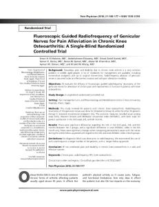
Figure 6 Tracheostomy button in place. The button provides a stent for the stoma with no obstruction within the airway. Image courtesy of Natus Medical Incorporated, Middleton, Wisconsin.
decannulation. The tracheostomy button is a stent that maintains patency of the stoma for a prescribed time, after which the button can be removed, allowing closure of the stoma. If frequent access to the airway is required (eg, for suctioning), the button should be replaced with a tracheostomy tube. The tracheostomy button consists of 3 parts: the tracheal cannula, the closure plug, and spacer rings (Figure 7) that are added to fit the length and width of the stoma exactly. When the closure plug is placed into the cannula, the petals at the distal end splay out to maintain secure position of the cannula within the stoma. Removing the tracheostomy button requires removing the closure plug first, releasing the tension at the distal end of the petals. Then the tracheal cannula can be easily removed. The Speak EZ tracheal cannula (Figure 8) is another stoma maintenance device. This tracheal cannula has the added feature of a built-in speaking valve on the proximal end. The cannula is made of soft silicone and can be used for patients who have vocal cord paralysis or sleep apnea or to maintain the stoma after removal of a tracheostomy tube or T-tube (eg, the Montgomery T-tube). Both the tracheostomy button and the Speak EZ tracheal cannula may be options for spontaneously breathing patients who wish to speak but cannot tolerate capping because of obstruction or resistance to airflow. However,
CriticalCareNurse
Vol 35, No. 6, DECEMBER 2015
21
11/17/15 10:07 AM
Figure 7 Olympic tracheostomy button, with closure plug (top), cannula (middle), and spacers (bottom).
Image courtesy of Natus Medical Incorporated, Middleton, Wisconsin.
Figure 8 Speak EZ tracheal cannula with built-in speaking valve.
Photo courtesy of Technical Products Inc of Georgia, Lawrenceville, Georgia (www.tpi-ga.com).
if a patient requires positive pressure ventilation or frequent suctioning, both the tracheostomy button and the tracheal cannula should be removed and replaced with a standard tracheostomy tube. Phonation in Patients Who Require Intermittent Mechanical Ventilation When a patient requires intermittent mechanical ventilation, an optimal time to begin phonation attempts is when the patient is free from mechanical ventilation. One way to accomplish this freedom is to completely deflate the cuff and use finger occlusion or a speaking valve for phonation. Most often, however, a tube change to a TTS
22
Morris12_15pgs.indd 22
CriticalCareNurse
Vol 35, No. 6, DECEMBER 2015
tube is recommended to allow better airflow around the tube. As discussed in a previous article,13 the cuff of the TTS tube inflates to seal the airway and allow the patient to successfully return to mechanical ventilation, but when deflated, the cuff essentially disappears.8 With the cuff deflated, the TTS tracheostomy tube can also be safely capped, allowing air to pass through the vocal cords, producing speech. An added benefit is that when the tube is capped, the natural function of the glottis is restored, and this return to natural function may allow patients to remain free from mechanical ventilation.8 When a patient is returned to mechanical ventilation, the cuff of the TTS tracheostomy tube should be inflated with sterile water, not physiological saline or air.1,19 Sterile water will distribute pressure evenly and prevent loss of cuff volume that occurs when the cuff is inflated with air, because air diffuses out of the cuff. Also, use of minimal leak technique is important when the cuff of the TTS tube is inflated because the high-pressure cuff will create elevated direct measurements of cuff pressure.8,13,19 Of note, the TTS tracheostomy tube is a single-cannula tube. If a patient has large amounts of thick secretions, he or she may be at risk for obstruction. Therefore, frequent pulmonary hygiene, with suctioning, methods to mobilize secretions, proper humidification, and coughing exercises, are extremely important. These methods include use of a heated aerosol, positioning the patient sitting up or with the head of the bed elevated, optimal fluids to keep secretions thin, and suctioning as needed. If difficulty inserting a suction catheter or mucus plugging occur with an TTS tube, changing to a tube with an inner cannula may be advisable. Phonation in Patients Who Are Ventilator Dependent Restoring speech in patients who are ventilator dependent can be challenging. Approaches vary according to whether or not a patient can tolerate cuff deflation. Ventilator-dependent patients who can tolerate cuff deflation can use leak speech for phonation; those who cannot protect their airway will require approaches that maintain cuff inflation.12 The available devices include talking tracheostomy tubes, cuffed fenestrated tracheostomy tubes, and the Blom tracheostomy tube system. With all of these methods, manipulation of ventilator parameters can facilitate speech in a patient who is ventilator dependent.20
www.ccnonline.org
11/17/15 10:07 AM
Leak Speech. Leak speech can be an effective aid
in communication for patients who are ventilator dependent; however, it cannot be used in patients who cannot tolerate cuff deflation.20 Leak speech is appropriate only for patients who can tolerate cuff deflation or have a cuffless tube. To provide leak speech, the cuff is deflated and the ventilator settings are adjusted to accommodate the air leak that results. Tidal volume delivery can be increased to compensate for the loss of volume during inspiration through the upper part of the airway.20 Humans naturally speak on exhalation, but leak speech is the opposite of normal speech: it occurs on inhalation. The leak during the inspiratory phase allows for phonation, so the patient must be coached to speak on inspiration, as the breath is delivered.19 Leak speech generally results in short phrases followed by long pauses, so increasing the inspiratory time on the ventilator can allow for more syllables per minute.20,21 Some patients have reported anxiety or discomfort with the use of leak speech because of an unfamiliar feeling of airflow through the upper part of the airway. These patients can be taught to push down, hold their breath, or tighten their throat to increase or decrease the volume delivered to the lungs.22 A respiratory therapist can adjust the positive endexpiratory pressure (PEEP) to improve the quality of leak speech and allow phonation during the expiratory phase.20,23 The exhaled air exits through the ventilator circuit instead of the upper part of the airway if the PEEP setting is zero.12 The addition of PEEP can direct exhaled air to pass through the upper part of the airway so that the patient can use 60% to 80% of the breathing cycle for phonation.12 PEEP can also be added to improve voice quality and comfort.20,23 At the end of a leak speech trial, and before cuff reinflation, additional PEEP should be turned off to prevent lung hyperinflation and related adverse effects. Obtaining optimal voice quality is usually a matter of trial and error, so adjustments based on appropriate evaluations by a health care provider can limit or obviate interventions that can cause a patient anxiety or respiratory distress and affect future trials. Patients frequently become frustrated with this method of speech, so practice and patience are essential.20 When the cuff is deflated, supplementing leak speech with the addition of a speaking valve can be beneficial in allowing exhaled air to pass through the upper part of the airway instead of through the ventilator circuit.12 Figure 9
www.ccnonline.org
Morris12_15pgs.indd 23
Passy-Muir speaking valve
Figure 9 Photo of Officer James Mullen who uses leak
speech with a ventilator. (He sustained a gunshot wound to the spinal cord in 1996, resulting in complete paralysis at the level of C1-C2.) Passy-Muir speaking valve is attached within the ventilator circuit and the cuff is deflated. Speech occurs on inspiration as the breath is delivered.
shows a patient using a Passy-Muir speaking valve within the ventilator circuit. Egbers et al23 have reported successful phonation with use of bilevel positive airway pressure and a speaking valve. Some newer ventilator models have speaking modes that can adjust for the change in airflow.8 A speaking valve can improve speech flow and volume in addition to improving voice quality and intelligibility of speech.20,23 Hoit et al21 reported that the most helpful adjustment was increasing the inspiratory time. Patients who use a speaking valve during mechanical ventilation, like their spontaneously breathing counterparts, should be closely monitored. If exhalation is difficult with this method, the patient will not be able to phonate and may not be a good candidate for a speaking valve.9 The valve should be removed immediately if the patient experiences any breathing discomfort. The patient should be assessed for evidence of air trapping, an increased respiratory rate, use of accessory muscles, and other indications of increased work of breathing.9 The speaking valve should be removed for suctioning so that secretions do not occlude the airway during exhalation.9 Talking Tracheostomy Tubes. The most challenging patients for restoration of speech are those who are ventilator dependent and who cannot tolerate cuff deflation. In these patients, one method to consider for speech restoration is the talking tracheostomy tube. A talking tracheostomy tube has an extra port that
CriticalCareNurse
Vol 35, No. 6, DECEMBER 2015
23 11/9/15 4:47 PM
distributes airflow above the inflated cuff (Figure 10). When this port is attached to an airflow source, the air flows through the tubing, and with occlusion of the port, air is directed toward the vocal cords, thereby producing phonation. Voice quality is adjusted by increasing airflow, usually from 5 to 15 L/min for optimal voice quality; variability depends on the individual patient’s needs. Significant increases in voice quality are detectable as airflow increases from 5 L/min to 15 L/min; but even with this system, voice quality can be a whisper, at best.12,24 The advantages of this method are that the air used for speech is completely separate from the air used for breathing and that it does not require manipulation of the ventilator settings.9 Disadvantages of this method include the need for the patient or a caregiver to occlude the port for speech; accumulation of secretions above the cuff, which can clog the air supply line; poor voice quality; and discomfort and drying of mucous membranes with high airflows.8 Also, the port might not be properly fitted in the trachea and may lead to ineffectiveness of the talking tracheostomy tube. Patients also need time and practice to perfect the use of this type of speech.
Air control port
Larynx
Air line to compressed air source
Inflated cuff
Pilot balloon
to Lungs
Figure 10 Illustration of airflow with talking tracheostomy tube. Note that when the air port is occluded, air is directed through the lumen above the cuff, and up to the larynx. Used with permission of Pat Thomas Medical Illustration, East Troy, Wisconsin.
Cuffed Fenestrated Tracheostomy Tubes.
Another method to restore speech in patients who require an inflated cuff is use of a fenestrated tracheostomy tube. A fenestration is an opening on the dorsal side of the shaft of the tube; it is usually placed one-third of the distance down the shaft. This opening allows air to move through the tube and up through the vocal cords.9 When the cuff on a fenestrated tube is inflated, inspiration and expiration occur primarily through Fenestration must fit perfectly in the the tube via center of the trachea so that it does not the venticome in contact with the tracheal wall. lator, but a small amount of air moves through the upper part of the airway and past the vocal cords via the fenestration (Figure 11). As with leak speech, ventilator alarms and settings must be adjusted to accommodate for the exhaled volume lost through the upper part of the airway. An important consideration with the use of a fenestrated tracheostomy tube is that the fenestration must fit perfectly in the center of the trachea so that it does not come in contact with the tracheal wall. Blockage of the fenestration affects breathing, and granulation tissue may form at the fenestration site, causing an
24 Morris12_15pgs.indd 24
CriticalCareNurse
Vol 35, No. 6, DECEMBER 2015
Figure 11 Cuffed fenestrated tube with cuff inflated. Inspiration and expiration is through (but not around) the tube. A small amount of air is directed to the vocal cords on expiration. Reproduced from Morris,8 with the permission of Springer Publishing Company, LLC, New York, New York.
occlusion of the aperture and trauma on removal of the tube. Because an off-the-shelf fenestrated tube might not fit properly, fenestrated tubes may require a custom order. The anterior and posterior tracheal depths must be measured and compared with the position of the fenestration. If these measurements do not match, a custom tube should be ordered.8,25 A simple assessment of proper fit can be done by removing the inner cannula
www.ccnonline.org
11/9/15 4:47 PM
and shining a light into the tube to observe for an open versus a blocked fenestration.8,9 Proper placement of the tube can also be ensured by using bronchoscopy.9 A blockage of the fenestration affects breathing, and granulation tissue may form at the fenestration site, causing occlusion of the aperture and trauma on removal of the tube. Nurses should also be mindful that secretions can occlude the fenestration and cause complications as well as impede optimal phonation. For the fenestrated tube to work properly, the fenestrated inner cannula should be in place when the patient is attempting to phonate. This fenestrated inner cannula is usually identified in some way. For example, the 15-mm connector of the fenestrated inner cannula of the Shiley tracheostomy tube is colored green. However, if a patient with a fenestrated tube requires suctioning, the nonfenestrated inner cannula must be used so that the suction catheter does not get lodged within the fenestration.8 Blom Tracheostomy Tube System. The Blom tracheostomy tube system (Figure 12) was designed for ventilator-dependent patients with no cognitive impairment who require continuous cuff inflation and who desire to speak. The system is a relatively new device that has a fenestrated outer cannula lying just above the cuff; the position is intended to prevent contact with the tracheal mucosa. A total of 4 different types of inner cannulas can be used with the Blom tube: a standard inner cannula, an inner cannula with a subglottic suctioning port, a speech cannula, and a low-profile inner cannula that can be used for patients who do not require ventilator support or who can tolerate cuff deflation. These cannulas have a unique locking mechanism that can be used only with the Blom tube system.26 When phonation is desired with a Blom tube for a patient receiving mechanical ventilation, the uniquely designed speech cannula should be used. This cannula has 2 valves on its soft and flexible shaft. On inhalation, air is delivered to the lungs through a flap valve that opens at the tip of the tube (Figure 13). Upon exhalation, the flap valve closes, and air passes through the fenestration and through a bubble valve along the shaft of the speech cannula. Before the speech cannula is inserted, the patient should be assessed to ensure that he or she is breathing comfortably and that the airway is clear of secretions. After the speech cannula is placed, airflow through the upper part of the airway should be assessed. If the patient has any indications of respiratory distress, the speech cannula
www.ccnonline.org
Morris12_15pgs.indd 25
Figure 12 Blom tracheostomy tube system. From left to right, outer cannula with fenestration above the cuff, speech cannula, and inner cannula with subglottic suctioning port. Reprinted with permission of Pulmodyne, Indianapolis, Indiana.
should be immediately removed and replaced with the standard cannula.26 Patients with thick or copious secretions should not use this type of tube.26 Another unique feature of the Blom system is the exhaled volume reservoir. This small bellows system expands and traps air during the inspiratory phase and then returns that air to the ventilator during the expiratory phase so that the air can be measured as exhaled volume.26,27 This reservoir should be used only while the speech cannula is in place; it should be removed when the speech cannula is not in use.26,27
Conclusion Many different methods can be used to restore phonation in patients who have a tracheostomy, and critical care nurses are the ideal members of the health care team to facilitate a planned and systematic approach to achieving phonation. Coordination of the interdisciplinary team, which includes critical care nurses, respiratory therapists, speech pathologists, advanced practice nurses, and physicians, is essential to the goal of voice restoration. Early involvement of this team can improve clinical outcomes and patient satisfaction by reducing the time needed for phonation.28 Nurses who provide care for patients with tracheostomies need not only focus on the tasks associated with care but also acknowledge that patients with tracheostomies can struggle with loss of the voice. The team must be sensitive to this loss and explore the anger and frustration that can overwhelm patients.5 Nurses can
CriticalCareNurse
Vol 35, No. 6, DECEMBER 2015
25 11/9/15 4:47 PM
Inhalation No air escapes past the cuff, allowing all of the air to fill the lungs
Air to larynx
Air Bubble valve collapsed
Fenestration
Inflated cuff Bubble valve expanded
Flap valve closed Air Exhalation Exhaled air flow is available for phonation Open flap valve
Figure 13 Airflow with the Blom tracheostomy tube system. On inspiration, air flows through the speech cannula, expanding the bubble valve to cover the fenestration and opening the flap valve at the tip. On expiration, the flap valve closes and air flows around it, collapsing the bubble valve, allowing air to flow through the fenestration to the vocal cords. Reprinted with permission of Pulmodyne, Indianapolis, Indiana.
facilitate a method of communication that is ideal for the patient and can be consistent in its implementation until the patient’s voice is restored.5 Patients need a way to communicate their feelings as well as their physical and emotional needs, and in turn, they are grateful to nurses who take the time to be patient, who are creative with methods of communication, and who provide reliable quality care.4 CCN Financial Disclosures
Dr Morris is a coeditor/author of the 2010 edition of Tracheostomies: The Complete Guide. She has been a consultant for Covidien and was the recipient of research funding for a study of outcome evaluation of a structured program of deep breathing and arm exercises for patients with new tracheostomies, funded by an Eleanor Wood-Prince Grant: A Project of the Woman’s Board of Northwestern Memorial Hospital.
Now that you’ve read the article, create or contribute to an online discussion about this topic using eLetters. Just visit www.ccnonline.org and select the article you want to comment on. In the full-text or PDF view of the article, click “Responses” in the middle column and then “Submit a response.”
d tmore
To learn more about patients with a tracheostomy, read “Comparison of Respiratory Infections Before and After Percutaneous Tracheostomy” by Sole et al in the American Journal of Critical Care, November 2014;23:e80-e87. Available at www.ajcconline.org.
26
Morris12_15pgs.indd 26
CriticalCareNurse
Vol 35, No. 6, DECEMBER 2015
References
1. Mitchell RB, Hussey HM, Setzen G, et al. Clinical consensus statement: tracheostomy care. Otolaryngol Head Neck Surg. 2013;148(1):6-20. 2. Bove MJ, Afifi MS. Tracheotomy procedure. In: Morris LL, Afifi MS, eds. Tracheostomies: The Complete Guide. New York, NY: Springer Publishing Co; 2010:7-40. 3. Freeman S. Care of adult patients with a temporary tracheostomy. Nurs Stand. 2011;26(2):49-56. 4. Carroll SM. Silent, slow lifeworld: the communication experience of nonvocal ventilated patients. Qual Health Res. 2007;17(9):1165-1177. 5. Foster A. More than nothing: the lived experience of tracheostomy while acutely ill. Intensive Crit Care Nurs. 2010;26(1):33-43. 6. Donnelly F, Wiechula R. The lived experience of a tracheostomy tube change: a phenomenological study. J Clin Nurs. 2006;15(9):1115-1122. 7. Batty S. Communication, swallowing and feeding in the intensive care unit patient. Nurs Crit Care. 2009;14(4):175-179. 8. Morris LL. Phonation with a tracheostomy. In: Morris LL, Afifi MS, eds. Tracheostomies: The Complete Guide. New York, NY: Springer Publishing Co, 2010:181-209. 9. Grossbach I, Stranberg S, Chlan L. Promoting effective communication for patients receiving mechanical ventilation. Crit Care Nurse. 2011;31(3):46-60. 10. Ahmad M, Dargaud J, Morin A, Cotton F. Dynamic MRI of larynx and vocal fold vibrations in normal phonation. J Voice. 2009;23(2):235-239. 11. Baumgartner CA, Bewyer E, Bruner D. Management of communication and swallowing in intensive care: the role of the speech pathologist. AACN Adv Crit Care. 2008;19(4):433-443. 12. Hess DR. Facilitating speech in the patient with a tracheostomy. Respir Care. 2005;50(4):519-525. 13. Morris LL, McIntosh E, Whitmer A. The importance of tracheostomy progression in the intensive care unit. Crit Care Nurse. 2014;34(1):40-48. 14. Morris L. Downsizing and decannulation. In: Morris LL, Afifi MS, eds. Tracheostomies: The Complete Guide. New York, NY: Springer Publishing Co; 2010:303-322. 15. Prigent H, Orlikowski D, Blumen MB, et al. Characteristics of tracheostomy phonation valves. Eur Respir J. 2006;27(5):992-996. 16. Kazandjian MS, Dikeman KJ. Communication options for tracheostomy and ventilator-dependent patients. In: Myers EN, Johnson JT, eds. Tracheostomy: Airway Management, Communication, and Swallowing. San Diego, CA: Plural Publishing; 2008:187-214.
www.ccnonline.org
11/17/15 10:08 AM
17. Woodnorth G. Assessing and managing medically fragile children: tracheostomy and ventilator support. Lang Speech Hear Serv Schools. 2004,35(4):363-372. 18. Passy-Muir Inc. Passy-Muir Tracheostomy and Ventilator Speaking Valve Resource Guide. Irvine, CA: Passy-Muir Inc; 2003. 19. Bivona TTS Adult Tracheostomy Tube [package insert]. Gary Indiana: Smiths Medical; 2010. 20. MacBean N, Ward E, Murdoch B, et al. Optimizing speech production in the ventilator-assisted individual following cervical spinal cord injury: a preliminary investigation. Int J Lang Commun Disord. 2009;44(3):382-393. 21. Hoit JD, Banzett RB, Lohmeier HL, HIxon TJ, Brown R. Clinical ventilator adjustments that improve speech. Chest. 2003;124:1512-1521. 22. Tippett DC, Vogelman L. Communication, tracheostomy and ventilatordependency. In: Tippett DC, ed. Tracheostomy and Ventilator Dependency. New York, NY: Thieme; 2000:93-142. 23. Egbers PH, Bultsma R, Middelkamp H, Boerma EC. Enabling speech in ICU patients during mechanical ventilation. Intensive Care Med. 2014; 40(7):1057-1058. 24. Leder SB. Verbal communication for the ventilator-dependent patient: voice intensity with the Portex Talk tracheostomy tube. Laryngoscope. 1990;100(10, pt 1):1116-1121. 25. Morris LL. Fitting and changing a tracheostomy tube. In: Morris LL, Afifi MS, eds. Tracheostomies: The Complete Guide. New York, NY: Springer Publishing Co; 2010:115-158. 26. Kunduk M, Appel K, Tunc M, et al. Preliminary report of laryngeal phonation during mechanical ventilation via a new cuffed tracheostomy tube. Respir Care. 2010;55(12):1661-1670. 27. Leder SB, Pauloski BR, Rademaker AW, et al. Verbal communication for the ventilator-dependent patient requiring an inflated tracheotomy tube cuff: a prospective, multicenter study on the Blom tracheotomy tube with speech inner cannula. Head Neck. 2013;35(4):505-510. 28. Freeman-Sanderson A, Togher L, Phipps P, Elkins M. A clinical audit of the management of patients with a tracheostomy in an Australian tertiary hospital intensive care unit: focus on speech-language pathology. Int J Speech Lang Pathol. 2011;13(6):518-525.
www.ccnonline.org
Morris12_15pgs.indd 27
CriticalCareNurse
Vol 35, No. 6, DECEMBER 2015
27 11/10/15 11:58 AM
CE Test Test ID C1562: Restoring Speech to Tracheostomy Patients
Learning objectives: 1. Identify the potential effects of the inability to communicate for a patient with a tracheostomy 2. Examine methods to restore phonation for patients with a tracheostomy 3. Discuss the role of critical care nurses in restoring phonation
1. Which of the following are indications for tracheostomy placement? a. Confirmed ventilator-associated pneumonia b. Prolonged intubation with unsuccessful weaning c. Prolonged need for vasoactive medications d. Two or more self-extubations
7. Leak speech is most appropriate for which of the following patients? a. Those who no longer require mechanical ventilation b. Those who require mechanical ventilation at night c. Those who can tolerate cuff deflation without signs of distress d. Those who have thick secretions that can be expelled easily
2. Which of the following statements best describes the difference between capping a tracheostomy and using a speaking valve? a. For a cuffless tube, a speaking valve should not be used and only a cap is appropriate. b. Capping a tracheostomy should never be done on a fenestrated tube while a speaking valve can be used on any tube. c. A speaking valve will allow air to enter into the tracheostomy tube while capping will not. d. There is no difference between capping a tube and using a speaking valve.
8. Which of the following adjustments to a mechanical ventilator can improve leak speech quality? a. Decreasing tidal volume c. Decreasing inspiratory time b. Increasing positive end-expiratory pressure d. Increasing oxygen
3. Which of the following devices should be used to provide supplemental oxygen for a patient with a speaking valve? a. Nasal cannula b. High-flow nasal cannula c. Venturi mask d. Humidified tracheostomy collar 4. Which of the following statements do the authors suggest as rationale for avoiding the use of a speaking valve on an inflated cuffed tube? a. The cuffed tube will not allow for air to escape during exhalation, which could lead to barotrauma. b. The rate of inhalation of air through the speaking valve may damage the cuff. c. The speaking valve can prevent the expectoration of mucus when the cuff is inflated. d. The cuffed tube may prevent adequate inhalation of supplemental oxygen through the speaking valve.
9. Which of the following statements describes the differences between normal speech and leak speech? a. Normal speech often is comprised of short phrases whereas leak speech generally has long phrases with pauses. b. here is no difference in quality between normal speech and leak speech. c. The use of leak speech often requires supplemental oxygen while the oxygen demands do not increase with normal speech. d. Leak speech occurs during inhalation while normal speech occurs during exhalation. 10. The role of critical care nurses in restoring phonation in patients with a tracheostomy includes which of the following? a. Deferring the decisions regarding devices to a speech therapist b. Monitoring for complications of respiratory distress exclusively c. Reporting patient frustrations of inability to communicate to the health care provider d. Serving as a member of the interdisciplinary health care team to assist in the coordination of care
5. Which of the following do the authors suggest as contraindications for the use of a speaking valve? a. Previous failed attempt at using a speaking valve b. Total laryngectomy c. Supplemental oxygen requirements d. Acute delirium
11. Proper steps involved in cuff deflation include which of the following? a. Prior coaching and prepping of the patient, deep oropharyngeal suctioning, cuff deflation, observe for symptoms of respiratory distress b. Ask patient to take deep breath, cuff deflation, deep subglottic suctioning, increase fraction of inspired oxygen c. Slow cuff deflation over several minutes, increase fraction of inspired oxygen, reassure patient, deep oropharyngeal suctioning d. Deep subglottic suctioning, cuff deflation, ask patient to take a deep breath, observe for respiratory distress
6. Advantages to using the tracheostomy button and the Speak EZ tracheal cannula include which of the following? a. Having the ability to custom fit the tracheostomy ties b. Reducing the need for mechanical ventilation c. Eliminating the bulk of the tube in the airway d. Decreasing the supplemental oxygen requirement
12. Considerations for safe capping of a tracheostomy tube include which of the following? a. Use of a standard cuffed tube with the cuff deflated b. Use of a cuffless, tight-to-shaft, or fenestrated tube of appropriate size c. Use of a Speak EZ tracheal cannula d. Use of humidified tracheostomy collar
Test answers: Mark only one box for your answer to each question. You may photocopy this form.
1. q a qb qc qd
2. q a qb qc qd
3. q a qb qc qd
4. q a qb qc qd
5. q a qb qc qd
6. q a qb qc qd
7. q a qb qc qd
8. q a qb qc qd
9. q a qb qc qd
10. q a qb qc qd
11. q a qb qc qd
12. q a qb qc qd
Test ID: C1562 Form expires: December 1, 2018 Contact hours: 1.0 Pharma hours: 0.0 Fee: AACN members, $0; nonmembers, $10 Passing score: 9 correct (75%) Synergy CERP Category A Test writer: Jodi Berndt, PhD, RN, CCRN, PCCN, CNE
Program evaluation
For faster processing, take this CE test online at www.ccnonline.org or mail this entire page to: AACN, 101 Columbia Aliso Viejo, CA 92656.
Yes No Objective 1 was met ❑ ❑ Objective 2 was met ❑ ❑ Objective 3 was met ❑ ❑ Content was relevant to my nursing practice ❑ ❑ My expectations were met ❑ ❑ This method of CNE is effective for this content ❑ ❑ The level of difficulty of this test was: ❑ easy ❑ medium ❑ difficult To complete this program, it took me hours/minutes.
Name
Member #
Address City Country E-mail RN Lic. 1/St
State
ZIP
Phone
RN Lic. 2/St
Payment by: ❑ Visa ❑ M/C ❑ AMEX ❑ Discover ❑ Check Card #
Expiration Date
Signature
The American Association of Critical-Care Nurses is accredited as a provider of continuing nursing education by the American Nurses Credentialing Center’s Commission on Accreditation. AACN has been approved as a provider of continuing education in nursing by the State Boards of Nursing of California (#01036) and Louisiana (#LSBN12). AACN programming meets the standards for most other states requiring mandatory continuing education credit for relicensure.
Morris12_15pgs.indd 28
11/9/15 4:47 PM