Pain Physician 2018; 21:169-177 • ISSN 1533-3159
Randomized Trial
Fluoroscopic Guided Radiofrequency of Genicular Nerves for Pain Alleviation in Chronic Knee Osteoarthritis: A Single-Blind Randomized Controlled Trial Eman H. El-Hakeim, MD1, Abdelraheem Elawamy, MD2, Emad Zarief Kamel, MD2, Samar H. Goma, MD1, Rania M. Gamal, MD1, Abeer M. Ghandour, MD1, Ayman M. Osman, MD2, and Khalid M. Morsy, MD2 From: 1Department of Rheumatology, Rehabilitation, and Physical Medicine, Assiut University Hospital, Assiut, Egypt; 2Department of Anesthesia, ICU, and Pain.,Faculty of Medicine, Assiut University, Egypt. Address Correspondence: Dr. Emad Z. Kamel, MD Department of Pain, Anesthesia, and Intensive Care Faculty of Medicine Assiut University Hospital Assiut, Egypt. E-mail:
[email protected] Disclaimer: There was no external funding in the preparation of this manuscript. Conflict of interest: Each author certifies that he or she, or a member of his or her immediate family, has no commercial association (i.e., consultancies, stock ownership, equity interest, patent/licensing arrangements, etc.) that might pose a conflict of interest in connection with the submitted manuscript. Manuscript received: 08-02-2017 Revised manuscript received: 10-01-2017, 11-29-2017 Accepted for publication: 12-08-2017 Free full manuscript: www.painphysicianjournal.com
Background: Nowadays, pain and disability due to chronic knee arthritis is a very common problem in middle aged people. A lot of modalities for management are available, including conservative analgesics and up to surgical interventions. Radiofrequency ablation of genicular nerves is assumed to be an effective less invasive and safe pain alleviation modality. Objectives: To evaluate the efficacy of fluoroscopic guided radiofrequency neurotomy of the genicular nerves for alleviation of chronic pain and improvement of function in patients with knee osteoarthritis. Study Design: A single-blind randomized controlled trial. Setting: Pain management unit, and Rheumatology and Rehabilitation clinics of Assiut University hospitals, Assiut, Egypt. Methods: This study involved 60 patients with chronic knee osteoarthritis. Radiofrequency neurotomy of the genicular nerves was done for 30 patients (Group A) while the other 30 patients (Group C) received conventional analgesics only. The outcome measures included visual analog scale (VAS), Western Ontario and McMaster Universities Index (WOMAC), and Likert scale for patient satisfaction in the 2nd week,3rd, and 6th months. Results: There were significant differences regarding the VAS in the 2nd week, 3rd, and 6th months between the 2 groups, and a significant difference in total WOMAC index in the 6th month only. There were significant changes when comparing pretreatment values with the values during the whole follow-up period with regard to the VAS and total WOMAC index in both groups. Limitations: No diagnostic block was done prior to radiofrequency. We recommend the use of such a technique on a larger number of OA patients, with a longer follow-up period. Conclusion: RF can ameliorate pain and disability in chronic knee osteoarthritis in a safe and effective manner. Keywords: Chronic pain, radiofrequency (RF), knee osteoarthritis. Clinical trial number and registry: ClinicalTrials.gov Identifier is NCT03224637. Pain Physician 2018; 21:169-177
O
steoarthritis (OA) is one of the most common chronic forms of arthritis affecting patients usually aged above 45 years. It affects the
patient’s physical activity as it causes pain, fatigue, and functional limitations. Not only does it affect the patient, but also OA has impacts on the economy
www.painphysicianjournal.com
Pain Physician: March/April 2018; 21:169-177
in various ways as it causes increased health care utilization, decreased health-related quality of life and increased economic costs (1, 2). As a result of increased obesity and population aging, the burden of OA is expected to increase (3,4). Knee joint is the commonest joint to be affected by OA, and the prevalence is both symptomatically and radiographically evident. A recent meta-analysis proved that females have a higher prevalence than males, and also suffer a greater risk than males in developing prevalent and incident knee OA. This gender difference increases with age> 55 years (5,6,7). Clinical manifestations of OA include pain, stiffness, decreased range of movement, alteration of proprioception, and weakness of the quadriceps (8). Progressive loss of joint function eventually leads to significant decrease in the strength of the muscle groups involving this joint. All these manifestations make it difficult for the patient to do simple daily activities such as walking, climbing the stairs, and even getting up from a chair (4). Individuals with OA could suffer from poor alignment of the limb, crepitation during movements, limp while walking, stiffness, and instabilities (3,9). The conservative treatment of OA usually includes physical therapy, analgesics such as nonsteroidal antiinflammatory drugs (NSAIDs), intra-articular steroids, and hyaluronan injections (10). Although conservative management is effective in most OA patients, it may not be effective in a small percentage of patients, and some of them have serious adverse effects; hence, prompting the researchers to investigate further treatment methods. Minor and major surgical interventions have also been applied for the management of knee OA. The role of arthroscopy as one of these methods is controversial. It was reported that arthroscopy would not be useful in cases where findings of a meniscal rupture or a recent trauma does not exist (11). Total knee replacement is also another choice. Despite the good results achieved by a total knee replacement, some patients continue to suffer from pain (12). Radiofrequency (RF) has been used for the treatment of chronic pain, that has been unresponsive to conservative therapies (13). It has been used for several painful conditions such as trigeminal neuralgia, cancer pain, and spinal pain. It acts by disrupting the transmission of pain signals by means of thermal lesion production in order to interrupt nociceptive signals (14). There have been few attempts to use RF current in the treatment of painful conditions of joints of the extremi-
170
ties. Case studies of RF treatment for hip joint pain have been reported (15–19). In patients where surgical treatment is contraindicated, radiofrequency (RF) neurotomy might be a successful alternative treatment with few complications. Nerve supply of the knee joint consists of branches from various nerves including the common peroneal, saphenous, tibial, femoral, and the obturator nerves, all together known as genicular nerves (20, 21). Any of these nerves can be approached percutaneously under fluoroscopic guidance, and ablation of the lateral superior, medial superior, and medial inferior genicular nerves can alleviate this category of knee pain (22). The aim of this study is to evaluate the efficacy of RF ablation of genicular nerves for chronic pain alleviation in patients with knee OA in comparison to conventional pharmacologic treatment.
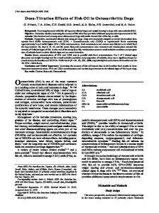
Methods This is a single-blind randomized controlled trial, approved by Faculty of Medicine, Assiut University local ethics committee, and registered in clinical trials under the number of (NCT03224637). It was carried out in accordance with the CONSORT Statement for Reporting Trials as shown in Fig. 1, and involved 60 adult patients with chronic knee OA. Patients were consented to take part in this study, then randomly allocated by computer generated tables into one of two groups; Group A; where RF ablation of genicular nerves was utilized, and Group C; for those who received conventional analgesic therapy. The study was conducted in the Pain management unit and Rheumatology and Rehabilitation clinics of Assiut University hospitals, Assiut, Egypt, between July 2014, and December 2015. The patients included in this study were diagnosed with knee osteoarthritis according to the American College of Rheumatology criteria (23) by a consultant of rheumatology , and confirmed radiologically to be in stage 3 or 4 of the Kellgren-Lawrence classification (24). Exclusion criteria included patients with other causes of pain such as radiculopathy, neurological disorders, or intermittent claudication, those who received intraarticular steroid or hyaluronic acids during the previous three months, previous knee surgery, or the presence of any contraindication for the invasive interventions such as coagulation disorders, systemic or local infection, presence of connective tissue disease affecting the knee, associated radicular pain, and or psychiatric disorders. In Group A, the intervention was performed by
www.painphysicianjournal.com
Radiofrequency for Pain Alleviation in Chronic Knee Osteoarthritis
Fig.1. CONSORT flow chart of the study.
the same physician under complete aseptic conditions, with vital signs monitoring. The patient rested in a supine position under fluoroscopy (Ziem C-arm). The affected knee joint was flexed to 15 degrees by putting a pillow under it. The anteroposterior fluoroscopic view of the tibiofemoral joint was obtained and showed an open tibiofemoral joint space with equal width interspaces on both sides as possible. Skin and soft tissues were infiltrated and anesthetized with 2 mL lidocaine 1%, then a 10 cm 22-gauge RF cannula with 10 mm active tip (NeuroThermTM, Medpoint GmbH, Hamburg, Germany) was inserted. Under fluoroscopic guidance,
www.painphysicianjournal.com
the cannula was advanced percutaneously towards the junction of the shaft and the epicondyle using the end on tunnel technique until bone contact was obtained, then, in the lateral fluoroscopic view we stopped the cannula tip at the junction of the anterior two thirds and posterior third of the bone. This was done upon the three genicular nerves (upper medial, upper lateral and lower medial), but not performed in the lower lateral genicular nerve to avoid injury to the common peroneal nerve located at the fibular head. Sensory stimulation (50 Hz) was done to detect the nerve position, with its threshold < 0.6 V (meaning
171
Pain Physician: March/April 2018; 21:169-177
that the distance between the active tip and genicular nerve was < 0.3 mm). In order to avoid inadvertent motor nerves ablation, the nerve was checked for the absence of fasciculation on its corresponding area of lower limb with a current of 2.0 V at 2 Hz. A volume of 2 lidocaine 2% was injected before the start of the RF (NeuroTherm TM, Morgan automation LTD, Liss, UK); then, the electrode was inserted through the cannula and tip temperature was raised up to 80 °C for 270 seconds (3 cycles of 90 sec.). The patient was discharged and instructed to rest for 24 hours with the use of topical ice. Paracetamol supplementation was allowed if they experienced pain in the treated region. Participants were advised to mobilize the next day, whenever they felt comfortable. The conventional Group C patients were also assessed and prescribed analgesics as following: oral paracetamol (maximally 1gram / 6hours), nonsteroidal anti-inflammatory Diclofenac sodium 75 mg 2 times a day, and physiotherapy program if needed. All participants were assessed by visual analog scale (VAS) for pain, Western Ontario McMaster Universities OA index (WOMAC) for disability (25), and LIKERT scale for patient satisfaction (26) in the preintervention visit, then by the end of the 2nd week, 3rd, and 6th months consequently. WOMAC Index of OA is commonly used for assessment of improvement in the quality of life (QOL). The score includes 3 domains; pain (5 questions, possible subscale score 0−20), stiffness (2 questions, 0−8), and physical functioning (17 questions, 0−68), and accordingly has a minimum score of 0 (the best), and a maximum score of 96 (the worst). This was done by a pain physician who was
blind to grouping. Any complication related to RF ablation such as infection, hemorrhage, thermal injury, loss of motor, and or sensory control in the corresponding area of the involved nerves was recorded within the follow-up period.
Statistical analysis We determined a sample size of 30 patients per group to obtain a study power of 80% to detect a difference at the significance level of 5% in the VAS. The data were tested through the Anderson-Darling test for normality and homogeneity variances as a 1st step. Categorical variables were described as number and ratio, whereas continuous variables were described as mean and standard error (Mean ± SE). Chi-square test was utilized to compare categorical variables, whereas continuous variables were compared by independent Samples T Test, and ANOVA. Nominal and non-normally distributed variables were analyzed using Mann-Whitney U test. A 2-tailed P < 0.05 was considered statistically significant. Statistical analysis was done using the computer program IBM, SPSS (Statistical Package for Social Sciences), Version 23, 2015.
Results Sixty osteoarthritic patients who met the inclusion criteria were included in this study. Demographic data, clinical presentation, and duration of the diseases are presented in (Table 1) with insignificant differences between them. Some of the participants have an old history (>3 month ago) of intra-articular steroid injection (once in 36.7% of participants, and twice in 56.7% of them) . VAS scale values were significantly lower in RF
Table 1. Demographic and clinical data.
Group A (n = 30)
Group C (n=30)
Age (years)
62 ± 7.37
56.87 ± 6.53
0.9
Male/Female
9/21
12/18
0.4
BMI (Kg/m2 )
32.02 ± 6.26
30.21 ± 3.69
0.08
Bilateral
20 (66.7%)
24 (80.0%)
Rt. knee
4 (13.3%)
2 (10.0%)
Lt. knee
6 (20.0%)
2 (10.0%)
Disease Duration (month)
7.6 ± 3.14
5.7 ± 5.1
0.1
17/13
18/12
0.06
Variables
P value
Complaint
X-ray grading : Grade 3/ Grade 4
0.73
Data are presented as mean ± SD, ratio, or percentage. Group A; the radiofrequency treated patients. Group C; the conventional medically treated participants. P < 0.05 is considered statistically significant.
172
www.painphysicianjournal.com
Radiofrequency for Pain Alleviation in Chronic Knee Osteoarthritis
group during the whole follow-up period. In the same time, follow-up VAS scales showed significant decreases when compared to their corresponding basal value in each group (Table 2). The total WOMAC index showed significant difference between the two groups by the 6th month only; however, WOMAC domains (pain, and stiffness) showed significant differences in the 3rd, and 6th months, with lower values in Group A. Difficulties as a domain of WOMAC was significantly lower in the group A in the 6th month. Overall, the WOMAC index and its domains showed significant decreases (improvement) in comparison to their basal value in
each Group (Table 2). There was an improvement in the quality of life for all participants in consequence to improvement of pain and WOMAC index. Patient`s satisfaction (Likert scale) as well, showed significantly higher values in Group A in comparison to Group C in the 3rd and 6th months as shown in figure 2. Follow-up values of the Likert scale showed significant increases (better satisfaction) when compared to their basal value in each group. A high percentage ratio of the patients (63.3%) in the conventional Group C received physiotherapy during the follow-up period. Finally, none of the patients in Group A has devel-
Table 2. Follow-up of study scales in both groups.
Variables
Group A (n=30)
Group C (n=30)
P value
Pre-intervention
7.07±0.2
6.9±0.2
0.622
2nd week
2.47±0.3¥
3.63±0.27 ¥
0.004*
3rd month
2.83±0.5¥
4.93±0.2¥