The Effect of Bracing on Patella Alignment and Patellofemoral Joint Contact Area CHRISTOPHER M. POWERS1,2, SAMUEL R. WARD3, LI-DER CHAN1, YU-JEN CHEN1, and MICHAEL R. TERK1 Department of Biokinesiology and Physical Therapy, University of Southern California, Los Angeles, CA; 2Department of Radiology, Keck School of Medicine University of Southern California, Los Angeles, CA; and 3Department of Orthopaedics, University of California San Diego, San Diego, CA
1
ABSTRACT POWERS, C. M., S. R. WARD, L.-D. CHAN, Y.-J. CHEN, and M. R. TERK. The Effect of Bracing on Patella Alignment and Patellofemoral Joint Contact Area. Med. Sci. Sports Exerc., Vol. 36, No. 7, pp. 1226 –1232, 2004. Purpose: To examine the influence of two patellofemoral braces on pain response, patellar alignment, and patellofemoral joint contact area in persons with patellofemoral pain. Methods: Fifteen women between the ages of 18 and 45 yr with a diagnosis of patellofemoral pain participated. After the assessment of pain response using a visual analog scale, subjects underwent axial plane magnetic resonance imaging of patellofemoral joint at 0°, 20°, 40°, and 60° of knee flexion. Imaging was done with the knee extensors contracted (25% body weight) under three conditions: 1) no brace, 2) On-Track brace, and 3) Patellar Tracking Orthosis (PTO). Measures of mediolateral patellar displacement and tilt and medial and lateral facet contact area were obtained from the magnetic resonance images. Results: On average, the On-Track brace reduced symptoms by 50%, whereas the PTO reduced pain by 44%. When averaged across all knee flexion angles, the PTO and the On-Track brace significantly increased total patellofemoral joint contact area by 52.0 mm2 (21%) and 59.3 mm2 (24%), respectively, when compared with the no-brace condition. Bracing had no influence on lateral patellar tilt; however, small but significant changes in lateral patellar displacement were observed. Conclusion: Large changes in pain and contact area occurred without sizable changes in patellar alignment. The results of this study suggest that changes in patellar alignment by itself may not be responsible for pain alleviation after patellar bracing. Key Words: PATELLOFEMORAL BRACING, MAGNETIC RESONANCE IMAGING, PATELLOFEMORAL PAIN, PATELLAR KINEMATICS
P
clear. It is assumed that decreases in pain are associated with changes in patellar alignment or tracking; however, radiographic studies do not universally support this premise. Despite the fact that some studies have shown that wearing a brace influences patellar alignment (18,20), other studies have shown that wearing a brace does not influence patellar alignment (6,11,16). Comparison of these studies is difficult because of differences in radiologic methods (radiographs, computerized tomography (CT) scans, magnetic resonance imaging (MRI), or kinematic MRI scans), measuring methods (quantitative vs qualitative analysis), and the braces evaluated. Powers et al. (16) proposed that changes in symptoms with patellar bracing may be the result of changes in patellofemoral joint contact area and not necessarily changes in patellar alignment. Increases in contact area may be important clinically as a larger contact area or change in contact area location could serve to distribute forces over a greater surface area and/or shift contact from sensitive to less irritated areas. The purpose of the current investigation was to examine the influence of two patellofemoral braces on pain, patellar alignment, and patellofemoral joint contact area (as measured by MRI) in subjects with patellofemoral pain. It was hypothesized that both braces would reduce pain and increase contact area without a substantial change in patellar alignment.
atellofemoral pain is one of the most common disorders affecting the lower extremity (7,21). Although the etiology of patellofemoral pain continues to be debated, abnormal patellar tracking is thought to be the primary cause (1,8 –10,13,22). As such, numerous braces have been designed with the purpose of centralizing the patella within the trochlear groove (11,12,18,20,23). Results of several investigations have shown that wearing a brace reduces patellofemoral pain. For example, Palumbo (12) reported that 93% of patients with patellofemoral pain reported a decrease in pain, whereas Shellock et al. (19) reported that two-thirds of their patients had alleviation of symptoms. Moreover, Moller and Krebs (8) and Greenwald et al. (2) reported that wearing a patellofemoral brace resulted in an 88% and 73% reduction in symptoms, respectively. Although wearing a brace appears to be effective in reducing patellofemoral pain, the underlying mechanism is not entirely
Address for correspondence: Christopher M. Powers, Ph.D., P.T., Department of Biokinesiology and Physical Therapy, University of Southern California, 1540 E. Alcazar St., CHP-155, Los Angeles, CA 90036; E-mail:
[email protected]. Submitted for publication March 2003. Accepted for publication March 2004. 0195-9131/04/3607-1226 MEDICINE & SCIENCE IN SPORTS & EXERCISE® Copyright © 2004 by the American College of Sports Medicine
MATERIALS AND METHODS Subjects. Fifteen women between the ages of 18 and 43 yr with a diagnosis of patellofemoral pain participated in the
DOI: 10.1249/01.MSS.0000132376.50984.27
1226
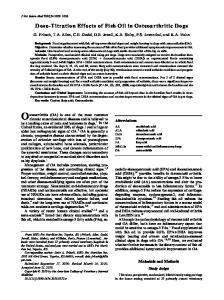
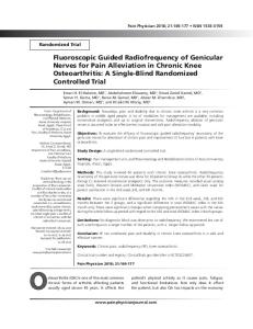
current study. The average age, height, and weight of the subjects was 31.1 ⫾ 7.5 yr, 164.1 ⫾ 4.9 cm, and 57.3 ⫾ 7.0 kg, respectively. Only women were studied because of the higher incidence of patellofemoral pain in this population and the potential biomechanical differences between sexes. Subjects were admitted to this study if their pain originated from the patellofemoral joint articulation (only subjects with overuse or insidious onset were accepted) and was readily reproducible with activities commonly associated with patellofemoral pain (squatting, stair climbing, knee extension) (14,15). The subjects were recruited from the author’s institution and were screened by physical examination to exclude those whose pain was not related to the patellofemoral joint articulation (i.e., ligamentous instability, torn meniscus and/or loose bodies, patellar tendonitis). Subjects were excluded if they reported having previous knee surgery or a history of traumatic patellar dislocation. Instrumentation. Axial plane images of the patellofemoral joint were obtained with a 1.5-T magnet (GE Medical Systems, Milwaukee, WI), using a fat suppressed FSPGR pulse sequence (TR: 8.2 ms, TE: 1.5 ms, NEX: 1, and spectral inversion for fat suppression). The image field of view was 20 cm ⫻ 20 cm with a 512 ⫻ 512 matrix. The slice thickness was 1 mm. Using this pulse sequence, the patellar and femoral cartilage was observed to be bright white, and any separation between the cartilage surfaces appeared as a dark line. In order to capture the entire patella in sequential axial slices, 50 – 60 images typically were obtained for each subject. The number of slices per subject varied based on patella length. Given a slice thickness of 1 mm, this would roughly equate to an axial field of view of approximately 5– 6 cm. Resistance to the extensor mechanism was accomplished using a custom-built, nonferromagnetic loading apparatus that resembled a leg press machine (Captain Plastic, Seattle, WA). This device allowed the subjects to perform unilateral knee extension in the supine position (Fig. 1). Resistance to knee extension was accomplished by pushing against a footplate that was connected (through a pulley system) to a moveable carriage containing epoxy weights. The footplate was only free to move linearly, and subjects were required to hold this position during imaging (thereby requiring muscle contraction). Two patellofemoral orthoses were evaluated in the current study, the On-Track patellar brace (Don Joy Inc, Vista, CA) and the Patellar Tracking Orthosis (PTO; Breg Inc., Vista, CA). Although both braces were similar in that they consisted of an elastic sleeve and were designed to apply a medial force to the patella, the primary functional difference between the two was the method in which the medial force was applied. The On-Track brace was comprised of a 5-mm neoprene knee cuff with a patellar cutout. A self-adhesive Velcro® patch (Velcro U.S., Manchester, NH) placed directly over the patella was used to secure a 5-mm neoprene pull strap, which applied a constant medial pull on the patella (Fig. 2A). The PTO consisted of an elastic sleeve with rigid medial and lateral bars. The medial bar incorporated a cantilever device that translated posteriorly with PATELLOFEMORAL BRACING AND CONTACT AREA
FIGURE 1—Photograph showing the subject set-up on the nonferromagnetic loading device used for imaging. This device allowed the subjects to do unilateral knee extension in the supine position. Resistance to knee extension was accomplished by pushing against a footplate that was connected (through a pulley system) to a moveable carriage containing concrete weights. Reprinted with permission from Salsich, G.B., S.R. Ward, M.R. Terk, and C.M. Powers. In vivo assessment of patellofemoral joint contact area in individuals who are pain free. Clin. Orthop. 417:277–284, 2003.
knee extension. Nonelastic straps connected this cantilever to a rigid lateral patella buttress plate that was integrated within the knee sleeve (Fig. 2B). During knee extension, tension was applied progressively to the lateral buttress plate via the cantilever device, thereby producing a medially directed force on the patella that peaked at full knee extension. Procedures. Before each subject agreed to participate, all procedures were explained, and informed written consent
FIGURE 2—The two patellofemoral braces evaluated in the current study were (A) On-Track Patellar Tracking System (Don Joy Inc. Vista, CA) and (B) Dynamic Patellar Orthosis (PTO; Breg Inc, Vista, CA). Medicine & Science in Sports & Exercise姞
1227
was obtained as approved by the University of Southern California, Institutional Review Board. Subjects were positioned supine on the loading device and Velcro® straps were placed across hip and shoulders to stabilize the trunk and pelvis. Two, 5-inch receive-only coils were placed on each side of the knee (with the patella centered between) and secured with tape. Starting with the knee fully extended, subjects were instructed to place the foot of the symptomatic side (or in the case of bilateral symptoms, the most painful side) on the footplate with a normal amount of toe out (i.e., 3–5°). Once the device was moved into the MRI bore and loaded to 25% body weight, imaging commenced. After imaging at 0°, the subject was removed from the MRI bore and repositioned on the loading device. Magnetic resonance images were obtained at 0°, 20°, 40°, and 60° knee flexion (as measured by standard goniometer). Images were obtained statically under three conditions: 1) no brace, 2) On-Track brace, and (3) PTO. Before and immediately after the application of each brace, subjects were asked to rate their perceived pain (visual analog pain scale) while doing an activity that reproduced their symptoms (unilateral squat or deep knee bend). The order of bracing conditions was randomized for each subject. The total imaging time was 60 s at each knee flexion angle. Data analysis. All images were analyzed by one investigator using Scion Image software (Scion Corp., Frederick, MD). For measures of mediolateral patellar displacement and mediolateral patellar tilt, only images containing the midsection of the patella (the image containing the maximum patellar width) were used for analysis. Although the investigator taking the measurements was not informed as to whether a particular set of images being analyzed was from a brace or no-brace condition, true blinding was virtually impossible as soft tissue compression around the thigh and patella (resulting from elastic sleeve compression) was visible on all braced images. However, it was not possible for this investigator to ascertain which brace was being analyzed in a particular set of images. Mediolateral patellar displacement was assessed using the bisect offset index as described by Powers et al. (16,17). The bisect offset was measured by drawing a line connecting the posterior femoral condyles and then projecting a perpendicular line anteriorly through the deepest point (apex) of the trochlear groove. This line intersected the patellar width line, which connected two widest points of the patella (Fig. 3). To obtain data when the trochlear groove was flattened, the perpendicular line was projected anteriorly from the bisection of the posterior condylar line (17) (Fig. 3). The bisect offset was representative of the extent of the patella lateral or medial to the midline and was expressed as a percentage of the total patellar width. Mediolateral patellar tilt was measured as the angle formed by the lines joining the maximum with of the patella and the line joining the posterior femoral condyles (16,17) (Fig. 4). All tilt measurements were reported in degrees. Contact area was measured from the sequential axial plane images of the patellofemoral joint. Images were displayed for analysis using Scion Medical Imaging Software 1228
Official Journal of the American College of Sports Medicine
FIGURE 3—The method used to measure mediolateral displacement using the bisect offset index. Right. When the trochlear groove was observable, this measurement was obtained by drawing a line connecting the posterior femoral condyles (AB) and then projecting a perpendicular line anteriorly through the deepest portion of the trochlear groove (CD) to a point where it bisected the patellar width line (EF). Left. To obtain data when the trochlear groove was flattened, the perpendicular line was projected anteriorly from the bisection of the posterior condylar line. The bisect offset was representative of the extent of the patella lateral or medial to the midline and was expressed as a percentage of the total patellar width (i.e., bisect offset ⴝ EG/EF ⴛ 100). Reprinted with permission of from ref. 16.
(Scion Corp.). The section of the image containing the patella and surrounding portion of the femur was enlarged to 1.5 times normal view to enhance visualization of the articular cartilage. As in previous studies, (3–5) contact was defined as areas of patella and femur approximation in which no distinct separation could be found between the cartilage borders of the two joint surfaces. Because cartilage is relatively bright on fat suppressed fast spoiled gradient echo images, the definition of contact area was defined operatively as white on white (3–5). The determination of noncontact was made when a line of separation could be observed between the articular surfaces of the patella and trochlear groove. This method has been shown to be reliable and comparable with contact area measurements obtained using pressure-sensitive film in cadaver specimens (5). The line of contact (curvilinear) between the patella and femur was measured and recorded using the electronic calipers feature within the Scion software. To obtain the contact area for each slice, the length of the line of contact was multiplied by the 1-mm slice thickness (3–5). To quantify medial and lateral facet contact area for each slice, the
FIGURE 4 —The method used to assess patellar tilt is shown. This angle was defined by lines joining the maximum width of the patella (AB) and the posterior femoral condyles (BC). All tilt measurements were reported in degrees. Reprinted with permission from ref. 16. http://www.acsm-msse.org
FIGURE 6 —Comparison of the bisect offset index among no-brace, On-track brace, and the Patellar Tracking Orthosis (PTO) conditions from 0° to 60° knee flexion. 夝 On-Track and PTO significantly less than the no brace condition (P < 0.05). Error bars indicate 1 SD. FIGURE 5—The method used to measure patellofemoral contact area is shown. The contact area was measured from the sequential axial plane images of the patellofemoral joint. Contact was defined as areas of patella and femur approximation in which no distinct separation could be found between the cartilage borders of the two joint surfaces (curvilinear lines). The apex of the patella served as the point of separation between the medial and lateral facets (vertical line). Reprinted with permission from Salsich, G.B., S.R. Ward, M.R. Terk, and C.M. Powers. In vivo assessment of patellofemoral joint contact area in individuals who are pain free. Clin. Orthop. 417:277–284, 2003.
length of each respective line of contact was multiplied by the same 1-mm slice thickness. The median ridge of the patella served as the point of separation between the medial and lateral facets (Fig. 5). The areas of contact from each sequential image were summed to obtain the patellofemoral joint contact area for each facet. Total contact area was calculated by summing the medial and lateral facet contact areas at each knee flexion angle. Contact area measurements were made twice by the same investigator and averaged for final analysis. All values were reported in square millimeters. Reliability and measurement error. To determine whether consistent data could be obtained with respect to the bisect offset index and patellar tilt, repeated measurements for each of these variables were made on 50 randomly selected images (selected from the 180 images used for these measurements). For contact area, repeated measurements were made on complete data sets (i.e., all slices with patellofemoral contact) for 10 subjects. In all cases, repeated measurements were made 1 wk apart, and the intraclass correlation coefficient (ICC) was used to assess intrarater reliability. Measurement reliability was excellent for the bisect offset index (0.99), patellar tilt (0.96), and total contact area (0.99). In addition, it was determined that the measurement error (standard error of measurement) was 2.5% of patellar width for the bisect offset index, 1.2° for patellar tilt, and 1.3 mm2 for total contact area. Statistical analysis. Comparison of pain response between conditions with and without a brace was made using a one-way ANOVA with repeated measures. To determine whether patellar alignment or contact area varied between PATELLOFEMORAL BRACING AND CONTACT AREA
brace conditions and knee flexion angles, separate 3 ⫻ 4 (brace condition ⫻ knee flexion angle) ANOVA with repeated measures were performed. This analysis was repeated for each dependent variable (bisect offset index, patella tilt angle, medial facet contact area, lateral facet contact area, and total contact area). An a priori power analysis revealed that given 15 subjects and an alpha level of 0.05, there was an 85% chance (power ⫽ 0.85) to detect a 30% decrease in both patellar alignment variables (bisect offset index and patellar tilt angle) and contact area variables (total contact, medial facet and lateral facet). Significant main effects were reported if there were no significant interactions. If a significant interaction was identified, individual main effects were analyzed separately. All statistical analyses were performed using SPSS statistical software (SPSS Inc, Chicago, IL) with a significance level of a probability less than 0.05.
RESULTS Pain response. Before the application of either brace, the mean subjective pain score for the subjects with patellofemoral pain was 4.6 ⫾ 1.9 out of a possible 10. Immediately after the application of the On-Track brace and the PTO, significant reductions in pain were observed when compared with the no brace condition (2.3 ⫾ 1.8 and 2.6 ⫾ 1.9, respectively; P ⬍ 0.05). There was no difference in the pain scores between the two braces. Patellar alignment. There were small but significant differences in the bisect offset index between the braced conditions (F2,3 ⫽ 3.66, P ⫽ 0.039, p2 ⫽ 0.207) and no interaction. Post hoc analysis revealed that the On-Track brace and the PTO reduced lateral patellar displacement when averaged across all knee flexion angles compared to the no brace condition (Fig. 6). On the average, the amount of decrease was 3.6% and 2.4% of patellar width for the On-Track brace and the PTO, respectively. No differences were observed between the two braces at any knee flexion angle (Fig. 6). The greatest decrease in lateral patellar disMedicine & Science in Sports & Exercise姞
1229
FIGURE 7—Comparison of patellar tilt angle among no-brace, OnTrack brace, and the Patellar Tracking Orthosis (PTO) conditions from 0° to 60° knee flexion. Error bars indicate 1 SD.
placement was 4.8% of patellar width, which occurred at 20° using the On-Track brace (Fig. 6). In contrast, there were no differences in the patellar tilt angle between the conditions with and without a brace (no brace effect, no interaction). When averaged across all knee flexion angles, the patellar tilt angle for the no-brace condition was 10° of lateral tilt compared with 9.3° and 8.6° of lateral tilt for the PTO and the On-Track brace, respectively (Fig. 7). Patellofemoral joint contact area. There were significant differences in total contact area between brace conditions (F2,3 ⫽ 26.98, P ⬍ 0.001, p2 ⫽ 0.658), and there was a significant brace by angle interaction (F6,28 ⫽ 2.41, P ⫽ 0.034, p2 0.147). Post hoc analysis revealed that the On-Track brace and the PTO increased contact area at each knee flexion angle when compared with when no brace was worn. On the average, the amount of increase was 59.3 mm2 and 52.0 mm2 for the On-Track brace and the PTO, respectively (Fig. 8). No differences were observed between the two braces at any knee flexion angle. The greatest increase in total contact area was 90.4 mm2, which occurred at 20° using the On-Track brace (Fig. 8). When evaluating lateral facet contact area, statistically significant differences were observed between brace conditions (F2,3 ⫽ 25.41, P ⬍ 0.001, p2 ⫽ 0.645) with no interaction. Post hoc analysis revealed that the On-Track brace and the PTO increased lateral facet contact area at each knee flexion angle when compared with when no brace was worn. On the average, the amount of lateral facet contact area increased 40.0 mm2 and 35.6 mm2 for the PTO and the On-Track brace, respectively (Fig. 9). No differences were observed between the two braces at any knee flexion angle. The greatest increase in lateral facet contact area was 52.1 mm2, which occurred at 20° using the OnTrack brace (Fig. 9). When evaluating medial facet contact area, there was a significant brace effect (F2,3 ⫽ 14.96, P ⬍ 0.001, p2 ⫽ 1230
Official Journal of the American College of Sports Medicine
FIGURE 8 —Comparison of total contact area among no-brace, OnTrack brace, and the Patellar Tracking Orthosis (PTO) conditions from 0° to 60° knee flexion. 夝 indicates PTO and On-Track brace significantly greater than the no brace condition (P < 0.05). Error bars indicate 1 SD.
0.517) and a significant brace by angle interaction (F6,28 ⫽ 3.20, P ⫽ 0.007, p2 ⫽ 0.186). Post hoc analysis revealed that at 20°, application of the On-Track brace and the PTO resulted in significantly greater medial facet contact area (41.6 mm2 and 26.0 mm2, respectively) than when no brace was worn (3.3 mm2) (Fig. 10). There was no difference between the two braces at 20°. At 40°, medial facet contact area significantly increased for the On-Track brace (100.5 mm2) when compared with the PTO (82.9 mm2) and when no brace was worn (71.4 mm2) (Fig. 10). No differences were seen at 40° between the PTO and when no brace was worn. At 60°, the On-Track brace significantly increased contact area when compared with when no brace was worn (127.7 vs 103.4 mm2) (Fig. 10). No differences were seen between the On-Track brace and the PTO or between the
FIGURE 9 —Comparison of lateral facet contact area among nobrace, On-Track brace, and the Patellar Tracking Orthosis (PTO) conditions from 0° to 60° knee flexion. 夝 indicates PTO and On-Track brace significantly greater than the no brace condition (P < 0.05). Error bars indicate 1 SD. http://www.acsm-msse.org
FIGURE 10 —Comparison of medial facet contact area among nobrace, On-Track brace, and the Patellar Tracking Orthosis (PTO) conditions from 0° to 60° knee flexion. 夝 Indicates PTO and On-Track brace significantly greater than the no brace condition (P < 0.05). † Indicates On-Track brace greater than the PTO and the no-brace condition. ‡ Indicates On-Track brace greater than the no brace condition. Error bars indicate 1 SD.
PTO when no brace was worn. There was no difference in medial facet contact area for any of the brace conditions at 0°.
DISCUSSION The results of the current study found that both braces significantly reduced pain immediately after application. On the average, the On-Track brace reduced symptoms by 50%, whereas the PTO reduced pain by 44%. These large changes in pain occurred without significant differences in lateral patellar tilt and small but significant changes in lateral patellar displacement. Although significant differences in the bisect offset index were observed at each knee flexion angle for both braces, the magnitude of these differences was small, ranging from 4.8% to 1.8% of patellar width. Given a measurement error for the bisect offset index of 2.5% of patella width, and an average maximum patellar width of 38.8 mm (translating into a range of reduction in lateral patellar displacement from 1.9 to 0.7 mm), the clinical relevance of such findings could be challenged. Despite small decreases in lateral patellar displacement, substantial increases in patellofemoral joint contact area were observed. When averaged across all knee flexion angles, the PTO and the On-Track brace significantly increased total patellofemoral joint contact area by 21% and 24%, respectively. The largest increases in total contact area occurred at 0° and 20° knee flexion where the PTO increased total patellofemoral joint contact area by 43% and 46%, respectively, as compared with 34% and 57%, respectively, for the On-Track brace. The observed increase in total contact area for both braces was primarily the result of an increase in lateral facet contact area. When averaged across all knee flexion angles, the PTO and the On-Track brace significantly increased lateral facet contact area by 20% and 18%, respectively. However, both PATELLOFEMORAL BRACING AND CONTACT AREA
braces also were observed to increase contact medially as shown by increased medial facet contact area. Although both braces failed to significantly increase medial facet contact area at 0°, relatively large increases in medial facet contact area occurred at 20°. At 40° and 60° knee flexion, the On-Track brace was more effective in increasing medial facet contact area than the PTO. The ability of the On-Track brace to increase medial facet contact area at greater knee flexion angles may have been the result of its inherent design. For example, the force provided by the On-Track brace was applied directly over the center of the patella whereas the PTO applied its force primarily along the lateral border of the patella. Given as such, it is not surprising that greater medial facet contact was seen with the On-Track brace. The fact that substantial increases in contact area occurred with only small decreases in lateral patellar displacement suggests that other mechanisms may have been involved. For example, it is likely that the patella was seated more firmly within the trochlear groove after bracing. This could have occurred as a result of the passive compressive force inherent in the knee sleeve that was integrated into both braces. It is possible that a compressive force applied to the patellofemoral joint could increase contact area without causing a change in patellar alignment; however, further research would be necessary to test this hypothesis. Although the exact mechanism of pain alleviation cannot be ascertained from this study, it is possible that the braces evaluated reduced symptoms by shifting contact and/or force from sensitive to less irritated areas. Furthermore, because stress is defined as force per unit area, any increase in contact area could theoretically result in a decrease in stress. However, care must be made in attributing the observed diminution in pain to a decrease in stress, as we did not quantify the effect of bracing on the patellofemoral joint reaction force. It is conceivable that the overall stress between the braced and nonbraced conditions was similar, as any increase in contact area with bracing could have been offset by an increase in the compressive force on the joint. Whether or not any increase in the joint reaction force as a result of compression would negate any potential benefits of an increase or shift in the location of contact area has yet to be determined. A limitation of this study was that only partial blinding of the investigator taking the MRI measurements was feasible. Although it was not possible for the investigator to determine which brace was being analyzed in a particular set of images, the application of both braces resulted in observable soft tissue deformation around the knee when compared with the images obtained during the no-brace condition. Therefore, comparisons between the brace and no-brace conditions may be viewed with a certain degree of caution. However, we feel that the between brace comparisons were not influenced by investigator bias as true blinding was achieved. Another limitation of this study was that only lateral displacement and lateral tilt were evaluated. It is possible that the patella flexion angle or anteroposterior position of the patella may have been changed by bracing. Future inMedicine & Science in Sports & Exercise姞
1231
vestigations may want to consider how bracing influences other degrees of freedom with respect to patellar alignment and their effects on patellofemoral joint contact area. The two patellofemoral braces evaluated in the current study significantly decreased pain and increased patellofemoral joint contact area. These results were achieved with only small decreases in lateral patellar displacement. Increases in contact area mostly were the result of increased
lateral facet contact; however, shifts toward increased medial facet contact also were observed. The findings of this study suggest that changes in patellar alignment by itself may not be responsible for pain alleviation after patellar bracing. The current study was funded in part by grants from the Breg Inc. and Don Joy Inc.
REFERENCES 1. FULKERSON, J. P., and K. P. SHEA. Mechanical basis for patellofemoral pain and cartilage breakdown. In: Articular Cartilage and Knee Joint Function, J. W. Ewing (Ed.). New York: Basic Science and Arthroscopy, 1990, pp. 93–101. 2. GREENWALD, A. E., A. M. BAGLEY, E. P. FRANCE, L. E. PAULOS, and R. M. GREENWALD. A biomechanical and clinical evaluation of a patellofemoral knee brace. Clin. Orthop. 324:187–195, 1996. 3. HEINO-BRECHTER, J., and C. M. POWERS. Patellofemoral stress during walking in persons with and without patellofemoral pain. Med. Sci. Sports Exerc. 34:1582–1593, 2002. 4. HEINO-BRECHTER, J., and C. M. POWERS. Patellofemoral stress during stair ascent and descent in persons with and without patellofemoral pain. Gait Posture 16:115–123, 2002. 5. HEINO-BRETCHER, J., C. M. POWERS, S. R. WARD, and M. R. TERK. Quantification of patellofemoral joint contact area using magnetic resonance imaging. Magn. Res. Imaging 21:955–959, 2004. 6. KOSKINEN, S. K., and U. M. KUJALA. Effect of patellar brace on patellofemoral relationships. Scand. J. Med. Sci. Sports 1:119 – 122, 1991. 7. MALEK, D. L., and R. MANGINE. Patellofemoral pain syndromes: a comprehensive and conservative approach. J. Orthop. Sports Phys. Ther. 2:108 –116, 1981. 8. MOLLER, B. N., and B. KREBS. Dynamic knee brace in the treatment of patellofemoral disorders. Arch. Orthop. Trauma Surg. 104:377– 379, 1986. 9. MOLLER, B. N., F. MOLLAR-LARSON, and L. H. FRICH. Chondromalacia induced by patellar subluxation in the rabbit. Acta Orthop. Scand. 60:188 –191, 1989. 10. MORRISH, G. M., and R. C. WOLEDGE. A comparison of the activation of the muscles moving the patella in normal subjects and in patients with chronic patellofemoral problems. Scand. J. Rehabil. Med. 29:43– 48, 1997. 11. MUHLE, C., G. BRINKMANN, A. SKAF, H. HELLER, and D. RESNICK. Effect of a patellar realignment brace on patients with patellar subluxation and dislocation. Am. J. Sports Med. 27:350 –353, 1999. 12. PALUMBO, P. M. Dynamic patellar brace: a new orthosis in the management of patellofemoral disorders. Am. J. Sports Med. 9:45– 49, 1981.
1232
Official Journal of the American College of Sports Medicine
13. POWERS, C. M. Patellar kinematics, part I: the influence of vastus muscle activity in subjects with and without patellofemoral pain. Phys. Ther. 80:956 –964, 2000. 14. POWERS, C. M., R. LANDEL, and J. PERRY. Timing and intensity of vastus muscle activity during functional activities in subjects with and without patellofemoral pain. Phys. Ther. 76:946 –955, 1996. 15. POWERS, C. M., J. PERRY, A. HSU, and H. J. HISLOP. Are patellofemoral pain and quadriceps strength associated with locomotor function? Phys. Ther. 77:1063–1074, 1997. 16. POWERS, C. M., F. G. SHELLOCK, T. V. BEERING, et al. Effect of bracing on patellar kinematics in patients with patellofemoral joint pain. Med. Sci. Sports Exerc. 31:1714 –1720, 1999. 17. POWERS, C. M., F. G. SHELLOCK, and M. PFAFF. Quantification of patellar tracking using MRI. J. Magn. Reson. Imaging 8:724 –732, 1998. 18. SHELLOCK, F. G., J. H. MINK, A. L. DEUTSCH, et al. Effect of a patellar realignment brace on patellofemoral relationships: evaluation with kinematic MR imaging. J. Magn. Reson. Imaging 4:590 –594, 1994. 19. SHELLOCK, F. G., J. H. MINK, A. L. DEUTSCH, and T. MOLNAR. Effect of a newly designed patellar realignment brace on patellofemoral relationships. Med. Sci. Sports Exerc. 27:469 – 472, 1995. 20. SHELLOCK, F. G., M. MULLIN, K. R. STONE, R. COLEMAN, and J. V. CRUES. Kinematic magnetic resonance imaging of the effect of bracing on patellar position: qualitative assessment using an extremity magnetic resonance system. J. Athletic Training 35:44 – 49, 2000. 21. SHELTON, G. L., and L. K. THIGPEN. Rehabilitation of patellofemoral dysfunction: a review of literature. J. Orthop. Sports Phys. Ther. 14:243–349, 1991. 22. THOMEE, R., J. AUGUSTSSON, and J. KARLSSON. Patellofemoral pain syndrome: a review of current issues. Sports Med. 28:245–262, 1999. 23. WORRELL, T., C. D. INGERSOLL, K. BOCKRATH-PUGLIESE, and P. MINIS. Effect of patellar taping and bracing on patellar position as determined by MRI in patients with patellofemoral pain. J. Athletic Training 33:16 –20, 1998.
http://www.acsm-msse.org