SURGICAL
TREATMENT
OF
THE
SYMPTOMATIC
ACCESSORY
NAVICULAR M.
From
The
accessory
Princess
MACNICOL,
Margaret
S.
Rose
measures
fail,
is occasionally
VOUTSINAS
Orthopaedic
Hospital,
Edinburgh
of pain and local tenderness over the instep. If and the results of 62 operations to one or both feet in 47 patients are reported. Twenty-six patients were treated by the Kidner operation, in which the main insertion of the tibialis posterior is re-routed; in the remaining 21 the ossicle was merely excised. Excision was as effective as the Kidner technique, provided that the medial surface of the main navicular bone was contoured to prevent any residual prominence. Both procedures were successful in relieving symptoms in the majority of cases and failures resulted from errors in the selection of patients or in the surgical technique. Correction of any associated fiat foot was secondary to growth and maturation of the foot rather than to the operation; hence the Kidner procedure does not confer any particular advantages over simple excision. conservative
navicular
F.
surgical
the source
treatment
may
be required
Pain and tenderness in relation to the medial arch of the foot may be produced by an accessory navicular bone (synonyms : os tibiale externum ; navicular secundum; prehallux) which was first clearly described by Bauhin in 1605 (Froelich 1909). Whether this minor anomaly alters
out from the tibialis the tendon was also
the suspensory mechanism has been a subject of some
The
often
associated
feet that istic
(1909)
produced
it may
apart
also
from
(Giannestras
gested ment
than
simple
the
arch
present
Sullivan
malleolus
of the ossicle
imbalance even
lead
MD, Keratsini, for reprints
1984 British 0301 -620X/84/2027
218
Research Piraeus, should
then
of the
tarsus.
the peroneal
overactive.
He
of
to subluxation
the
ofthe
foot,
metatarsal
ofBone
Joint
normal
or talonavicularjoints
rationale
and
of this 1973;
1979) although
number
of orthopaedic
excision
was recommended.
have
been
1978;
Sullivan
and
Veitch
the procedure
is still described
textbooks.
for the
of the
cuneiform-
operation
Leonard
supported the use of the operation, recommended the Kidner procedure simple
of pull
of the
also
efficacy
Miller
line
fusion
in a
et a!. (1965)
and Chater (1962) for adults, reserving
child.
Both the Kidner procedure and simple excision have been used in Edinburgh in patients whose symptoms have not indications
responded to conservative for the two operations have
measures. The been comparable,
that and
and
operashelled
Consultant
Ortho-
Fairmilehead,
Edin-
certain
radiographic
features
CLINICAL
will
be discussed.
Surgery
MATERIAL
Between
1962 and
1978,
76 patients
accessory surgically.
navicular Patients
in with
one or a fused
prominent case
navicular
notes
excluded
M. F. Macnicol. and
the a limited
questioned(Giannestras
details, to Mr
of restoring
Occasionally,
talonavicular
Fellow Greece. be sent
intention
tendon.
but the main portion of under the navicular, with
although in general the Kidner procedure has been reserved for the more severe flat foot. In all patients the source of symptoms was considered to be the result of a prominent accessory navicular, though flat foot was an associated feature in a significant proportion of cases. The results of the two operations have been reviewed
He
muscles
hypothesised
pronation
Editorial Society $2.00
sug-
in medial displaceposterior, thus
M. F. Macnicol, BSc, MCh Orth, FRCS Ed(Orth) paedic Surgeon, Part-time Senior Lecturer Princess Margaret Rose Orthopaedic Hospital, burgh EH 10 7ED, Scotland. S. Voutsinas. I 2 Thiras,
it was
1933)
of the foot became proupon the deltoid ligament
produced
might
when
(1929,
as an elevator
occurred
reflexly
navi-
recommended
Kidner therefore advocated a more complex tion in which not only was the accessory navicular
©
in and
accessory
but
Kidner
that adduction if impingement
become
indeed joint.
excision
its action and
or medial
that
the bony anomaly resulted the tendon of tibialis
compromising
Requests
foot it is
the character-
1973;
medial
Subsequently,
that of
considered nounced,
considered
a flattened
symptomatic.
this
normal
prominence
no more
would
planus,
arch of the for although
1979). Froelich
cular
pes
are relatively
medial
Miller
with
of the medial controversy,
the
posterior, re-routed
were
from
the
radiographs
performed
study
as the
a symptomatic
not
included. and
pre-operative
photographs
were
JOURNAL
21 were OF BONE
treated AND
The
29 patients
clinical
considered
remaining 47 patients clinic. In 26 the Kidner
; the remaining THE
were
retrospectively
and
be incomplete. The examined at a special was
tuberosity
reviewed
with
both feet were treated accessory navicular or a
by excision JOINT
to
were all procedure
SURGERY
SURGICAL
TREATMENT
OF
THE
SYMPTOMATIC
ACCESSORY
219
NAVICULAR
Pain Pain
interfering
Pain
atthe
Rest
pain
Pain
since
with
end ofan
sport
day
ordinary
trauma
Prominence
Localised
Shoe
tenderness
pressure
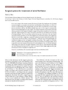
Fig. Frequency
and
characteristics
of the
symptoms
reported
by both categories.
Fig. Plantar
cases
operated
twice as often recommended upon,
unlike
as males, bilaterally simple
procedure was usually limited to one Figure 1 shows the symptoms group. Pain and local tenderness VOl..
66-B.
No.
2. MAR(H
984
of patients.
Pain
has
been
subdivided
into
four
2
of weight-hearing feet with bilateral Type II naviculars. The medial prominences are clearly seen. particularly on the right fot.
of the accessory bone and trimming of any residual medial prominence of the main navicular bone. Table shows the composition ofeach group; females presented was
groups
view
accessory
approximately procedure
I
and the Kidner in half of the
excision
where
the
or other foot. in each operative over the medial
prominence I
(Fig.
2) was
present
in
difficulty with shoes was less common chiefly in women. The pain was usually
nearly
all
cases;
and occurred described as an
ache felt over the instep or the medial prominence towards the end of the day, although occasionally rest pain
was
also
present.
cases a sprain of precipitating cause family are sometimes
In approximately
15 per
cent
of
the foot was considered to be the of symptoms. Other members of the found to have the same condition
220
M. F. MACNICOL,
(Fig. 3), and McKusick accessory navicular manifests
(1968) considers as an autosomal
S. VOUTSINAS
from
that the dominant
trait.
The
mean
duration
of symptoms
was 33 months
patients but
are shown
presented
a few
between
were
in Figures the
ages
5 years
under
4 and
in
and
some
Table
were
middle-aged.
Clinical
appearances.
Of the 26 patients treated 22 (85 per cent) presented with
Kidner procedure, flat feet, of similar classified criteria moderate
four
one
three with
presented
rigid
calcaneonavicular
clinical accessory medial
Flat
feet
clinically
flat
were
ofthe
In none
of a neuromuscular
of 21 patients
five were
classified
foot (one severe, further case had found
to
patients
tarsal
there
coalition
or
Radiographic
appearances.
radiographs
showed
admitted simple navicular
bones, excision has
of which
Type
I is small,
may
round 6, 7 and
2 of
the
in the
discrete it should
treated
The types same
from be
right feet : a mother successfully treated
(centre). her for persisting
son (left) foot pain
8
IS
I)
S
S
39
Excision
21
8
13
5
14
23
2
(Zadek and Gold 36 Type II accessory
in the Kidner group and in the group treated by excision pre-operative
and
five
II. Clinical weight-hearing
only
in the
group
grading
of
two and alone.
radiographs
standinglateral value; 17 were treated
hat
were
retrieved
toot.
by simple
tccording
excision.
to the
Moderate
Severe
Slight
Major
(ompletc
Navicular tuherositv prominence
Slight
Moderate
Scvcrc
Heel
0 10 degrees
0
>
Central
Slight
arch
21
views were considered available in the Kidner
Mild
Calcaneal
main
her daughter,
by simple
1 5 dcgrccs
tendon
lateral
appcaranccs
I 5 degrees
excision
Considerable
lateral
deviation
patient.
the
eversion
tilt
des ation
Ankle
Neutral
Mild
Forefoot
Neutral
Mild abduction
Abduction
hut
pronation
distinguished
and
Total number of operations
R
by
accessory (Dwight
Fig. Three
groups
patients
accessory
21
ossicles. into two
present
and
operation
26
two
26
Medial
the
of the
Kidner
depression
II,
traumatic or both macro-
1.
neuromuscular
had bilateral
only
8);
of
be apparent, and known to develop
Bilateral
mobile
of
may are
F
Table when
visible Type
M
one mild), and flat foot. None
time
1 3 out
procedure
whereas
both
(Figs
the
had bilateral been classified
1907) navicular
that
for the Kidner
navicular
At
1953).
Total
group
abnormalities.
is rarely
O’Rahilly
Operation
Although
of the
bilateral
and rigid
I. A comparison
forall patients, to be ofdiagnostic
had normally shaped medial prominence.
as having
which
l949
described by Geist (1925), is closely related to of the navicular but separated by an irregular dense fibrocartilage (Figs 9 and 10). Various
naviculars respectively
to
was
sesamoid
scopically (Fig. 1 1 ) and microscopically 1948). There were three Type I and
feet
secondary
disorder. treated by excision
three moderate a severe, more
have
normal
feet
navicular, the majority arches apart from the
However,
was
with
spastic
bars.
evidence
In the group
flat one
bilaterally.
by the mobile
as mild, moderate or severe according to the in Table II. Two patients had severe flat feet, 12 flat feet and 8 mild flat feet. Of the remaining
patients,
and
severity
tibial (Wood
stages of bony union degenerative features
5. Most 1 5 years
of 10 and
of. age,
posterior
originally the body plate of
each group, ranging from 3 months to 19 years for patients treated by the Kidner procedure, and from 6 months to I 5 years for the group treated by simple excision. The age at operation and the age at presentation
for the two groups
the
radiographically
no rotation
Moderate
tilt
and
3
all of whom of the ossicle
had and
bilateral surgical
THE
accessory contouring
naviculars. of the main
JOURNAL
OF BONE
The son navicular.
AND
JOINT
had
been
SURGERY
SURGICAL
TREATMENT
OF THE
SYMPTOMATIC
ACCESSORY
221
NAVICULAR
15
U)
C
0.
0 .0
E 2
of
age
1l
l6
21
26
31%
36
61.
46..
age
O
15202530351.04550
6.
5
age at operation
11
l6.
?1.
31%
Kidner
age
3
1.1
1351.045
1O52Ol5 at
presentation
Fig. 4 Age
at
operation
(left)
compared with proportion
the age at of male and
presentation of the 26 patients treated by female patients in each age group is shown.
the
Kidner
procedure.
The
15-
15
1o
U) C
0.
0 .0
E Z5
5
2
11% 16
2l
26
19191tflc5’l31
age
30 at
36
35
6.
1.0
operation
66 65
0
50
age Simple Fig.
Age
VOL.
66-B,
at operation
No. 2, MARCH
(left)
1984
compared navicular.
with the age The proportion
at presentation of male and
Excision
6
kii
11
o
15 at
20
26
25
35
30
35
[1 61
60
45
presentation
5
of the 21 patients treated by simple excision female patients in each age group is shown.
of the
accessory
222
M. F. MACNICOL,
S. VOUTSINAS
7sj
Fig.
6
Figure 6 Radiographic appearance ofa Type I accessory navicularofthe left foot and a Type II accessory navicular of the right foot. Figures 7 and 8--Two views of the Type I accessory navicular. Figures 9 and 10 Two views of the Type II accessory navicular.
Fig.
sutures
were
it inferior
inserted
when
the
decision
Fig.
into
to the main
medially
extent
9
to
necessary
severity carry
the
of out
re-routed
navicular, (Figs
the
the
tendon
which
was
13 and
14).
symptoms
Kidner
10
to secure
contoured To
some
influenced
procedure,
the
as did
the
presence of an obviously flat foot. However, the preference of the surgeon for one or other procedure appeared to be the major factor in deciding which technique should Fig.
Degenerative changes can lateral (articulating) surface navicular.
These
radiographs
standing
lateral
the medial with
arch
a normal
were radiographs
index range
be
II be seen on the of this accessory
compared using
or navicular in Caucasians
used;
contrasted, The reviewed
hence,
although
the
two
groups
can
be
they are not strictly comparable. patients treated by the Kidner procedure were 3 to 19 years after operation (mean 10 years);
to the postoperative three
index
measurements:
(Stewart
1970)
of 1 3. 1 to 17.8
(mean:
14.5); the lateral arch or cuboid index (Stewart 1970) with a normal range in Caucasians of 3.3 to 7.3 (mean: 5.6); and the calcaneometatarsal angle (Sullivan and Miller 1979) with a normal standard deviation
mean in Caucasians of7.7 (Fig. 12).
of 134.7
and
a Fig.
RESULTS Clinical tendon
data. Surgical exploration of tibialis posterior did not
abnormal operation
route to its insertion. was used (Kidner 1929,
revealed invariably
that adopt
the an
When the Kidner 1933), non-absorbable
12
Radiographic measurements of the weight-bearing foot. I. The medial arch or navicular index = (NN’ x I00)/AB: where NN’ is the vertical projection from the inferior tip of the navicular to the line AB: and AB is the distance between the most posterior projection of the calcaneum and the distal end of the longest metatarsal, regardless of its number. 2. The lateral arch or cuboid index = (CC x l00)/AB: where C(’ is the height of the cuboid above the line AB. 3. The calcaneometatarsal angle = CMT.
THE
JOURNAL
OF BONE
AND
JOINT
SURGERY
SURGICAL
TREATMENT
Fig. Figure
13
Anteroposterior
excision the main
reviewed 3 to 20 years after Table III shows the results.
Kidner Kidner
procedure. operation
presented
the Kidner navicular
procedure. bone has
of the ossicle, with or navicular bone, were
operation
(mean
cases a similar recorded. For
of patients,
12 years).
treated by the foot of varying
pain and tenderness no alteration in the
patient
with relieved
was
posture
of
combined
her with
reported Table
Ill.
were height
tarsal coalition of neither her
feet
after
excision that
The
efrect
in the
with
their
mobile
symptoms
ofthe
two
had
procedures
upon
relieved
the shape
by
ofthe
foot
Kidnerprocedure
after
operation
Number
Normal
Excision
*
Includes
t 3 severe.
: VOl..
Includes
66-8.
No.
the
evident
single
symptoms
persisted
in one
foot
would
increase and
per
hence
respond
in the
However,
of the
2. MARCH
Improved
Unchanged
15
15
6
5
984
without
tarsal
coalition
tarsal
coalition)
the
where
symptoms
0
the
feet
whose
Kidner medial
Improved
to predict
procedure arch.
The
Unchanged
14
9t
0
0
15
l
4
2
worsened
to 22
shape
were equally divided and severe flattening
3
probably
had
posterior
process. feet appeared in 14 of the
0
were
of these
pain
tibialis
Shape
Normal
(with
but
the symptoms
of patients
sutures.
navicular
in three
it was impossible
to the
height
4*
flat foot
that
was unaltered with mild, moderate
19
spastic
feet;
case
to have
wire
relieved,
suggesting
cent).
23*
of rigid
been
was still the site of an irritative shape of the mobile flat after the Kidner operation (64
feet
by broken of four
had
Flat
case
main
appears
of the accessory
on radiographs
patients
3
3 moderate. 3 mild a rigid spastic flat foot
of the
In another
tendon
recurrence
3
Flat
portion
the outcome.
posterior
Symptoms Shape
medial
radiographically
unilateral
pre-operatively;
19 (86 per
been
as shown
after operation between those
bars.
flat foot,
failed,
patients
procedures
14
the
compromised
the tibialis
The improve
and accessory naviculars symptoms nor the abnormal
Kidner
have
tendon
relieved although there of the medial arch. The
bilateral
may
re-routing
was
was invariably normally shaped
operation:
operation ; three complained that symptoms persisted. Of these three, one had a unilateral severe mobile flat foot in which the accessory navicular was bipartite and
Lastly,
than they
bilateral
of the calcaneonavicular
Of the 22 patients cent)
since
postoperative outcome the three patients with
feet, was
223
NAVICULAR
Figure 14--After been contoured.
this
Most of the patients presented with flat
in terms
ACCESSORY
Fig. before
degree as defined by the criteria in Table II. Rather reporting the results in terms of the number of feet are
SYMPTOMATIC
13
radiograph
those treated by simple without contouring of
OF THE
by surgery
which
by age
an
of the
224
M. F. MACNICOL, Table IV. Changes in the radiographic operative and post-operative values the foot alters the indices
measurements were observed,
with these
Calcaneometatarsal
Aulnt’r
foot
influences
Right
Left
17
17
15
IS
15
15
Postoperative
value
136.2±3.3
136.2±3.4
P < 0.01
P