THE SURGICAL TREATMENT OF THE MULTIPLE GINGIVAL RECESSIONS G. MAMPIERI, C. ARULLANI*, F. CECCHETTI** Research Assistant Department of Dentistry, University of Rome “Tor Vergata” * Resident Department of Dentistry, General Hospital “Tor Vergata”, Rome ** Research Assistant Department of Dentistry, University of Rome “Tor Vergata”, Rome
SUMMARY The surgical treatment of the multiple gingival recessions The growing emphasis on aesthetics puts the dentist before new requests for intervention aimed at improving the smile not only in dental component but also in the gingival. Infact, even among patients is now widespread awareness that the beauty of the smile comes from balance existing, in the relations and proportions, between the dental and gum component. The development of plastic periodontal surgery techniques increasingly sophisticated allows the dentists to correct those deteriorations of the gingival tissues, such as multiple recessions, which are responsible for troublesome increases of the dental sensitivity and aesthetic worsening of the smile. The description of a case report of multiple recessions surgically treated with Zucchelli/De Santis’ technique demonstrates that, having carefully selected patient, it’s possible to resolve with a fine but not invasive surgery these smile’s deteriorations.
RIASSUNTO Il trattamento chirurgico delle recessioni gengivali multiple La crescente attenzione per l’estetica pone l’odontoiatra davanti a nuove richieste di intervento finalizzate a migliorare il sorriso non solo nella componente dentale ma anche in quella gengivale. Infatti, anche fra i pazienti, è ormai diffusa la consapevolezza che la bellezza del sorriso derivi dall’armonia esistente nei rapporti e nelle proporzioni tra la componente dentale e quella gengivale. Lo sviluppo di tecniche di chirurgia plastica parodontale sempre più raffinate permette ai professionisti di correggere quelle alterazioni dei tessuti gengivali, come ad esempio le recessioni multiple, responsabili di fastidiosi aumenti della sensibilità dentale e di peggioramenti estetici del sorriso. La descrizione di un caso clinico di recessioni multiple trattato chirurgicamente con la tecnica di Zucchelli/De Santis dimostra come sia possibile, dopo aver selezionato con attenzione il paziente, intervenire precocemente e risolvere con una chirurgia raffinata ma non invasiva queste disarmonie del sorriso.
Key words: multiple gingival recessions, plastic gingival surgery, aesthetic.
Parole chiave: recessioni gengivali multiple, chirurgia plastica gengivale, estetica del sorriso.
The gingival recession is clinically defined as the apical movement of the gingival margin to the cementoenamel junction (CEJ) (1). Histologically it is characterised by the loss of connective fibres of the periodontal tissue and alveolar bone (2). This clinical condition is frequently noticed in the population and particularly among patients with high oral hygiene standards. In fact, in addition to the inflammation caused by the bacterial plaque, the traumatic brush is considered one of the major causes of gingival recession.
Gingival recession increases with age; the incidence varies from 8% among children to 100% in the adult population with more than 50 years of age (3). In addition to a wrong brushing technique and the gingival inflammation, the dental malpositioning can also cause recession. In fact the predisposition to the recession is linked to the position of the teeth in the dental arch, the dental-root angulation and the mesiodistal curvature of the dental surface. Teeth rotated, inclined or moved in the buccal direction show an osseous lamina thinned
ORAL & Implantology - Anno I - N. 3/2008
1
2
or reduced in height. Mastication or brushing pressure, even moderate, damage the unsustained gingiva and causes recession (4). The basis for surgical treatment of gingival recessions are codified by the American Academy of Periodontology: patients’ demand for aesthetic reasons, reduction of root sensitivity, management of caries or cervicular abrasions (5). The treatment of gingival recession focuses on covering the exposed surface of the dental root and arrest the progressive loss of tissue. Several mucogingival procedures have been used successfully including pedicle flaps procedure, implants of free gingival grafts and coronally advanced
flaps procedure (6). It has been proved that recessions treated with coronally advanced flaps recover with the formation of a long epithelial junction between the root surface and the covering tissue in humans and animals (7-10). In fact only a small fraction of the regeneration of variable extent has been observed in the most apical portion of the exposed root surface (7, 11, 12). Mucogingival surgery can be satisfactory in the coverage of the root although it doesn’t set a material increase of the attachment apparatus. The recovery takes place mainly through a epithelial junction. The strategy to increase gingival tissue dimen-
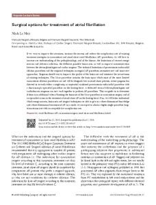
Figure 1 Multiple recessions on the elements 2.1, 2.2 respectively of mm 2, 3, ordered as I class of Miller.
Figure 3 Panoramic rx; it’s possible to see the good level of the interproximal bone around 2.1, 2.2 and 2.3.
Figure 2 Vision of the recessions by a greater magnification.
Figure 4 A partial-thickness dissection was raised starting from the line of enamel-cement of the adjacent teeth (2.1, 2.3) and convergent to the lateral incisor, drawing the new surgical papilla.
ORAL & Implantology - Anno I - N. 3/2008
sions, in width and thickness, where the keratinised tissue is considered clinically inadequate, at present is not supported by enough scientific studies which consider the dimension of the gingival tissue as important to the prevent the gingival recession and therefore this strategy as valid therapeutic option (13, 14). The clinical and histological results obtained in the coverage of the recessions through coronally advanced flaps procedure are analogous to the ones obtained through lateral sliding flap procedures and even in cases where the exposed root has been citric acid conditioned results were different (15, 16).
Figure 7 Passivating the entirely flap.
Figure 5 A full-thickness flap until the mucogingival junction without carring out released cuts
Figure 8 Radicular surfaces smoothed.
Figure 6 Interproximal papillas disepitelized.
Figure 9 Suture with Vicryl 5/0.
ORAL & Implantology - Anno I - N. 3/2008
3
Figure 10 Healing at the suture removal.
Figure 12 Control after 3 weeks.
Figure 11 Control after 2 weeks.
Figure 13 Control after 3 months; by a greater magnification it’s possible to observe the perfect healing of the periodontal tissues.
Therefore, the objective of all of these mucogingival procedures is simply to increase the quantity of keratinised tissue achieving a valid coverage of the root coverage equivalent to between 56% and 98% of the exposed root (17-22). The case report showed below proves the effectiveness of the Zucchelli/De Santis surgery technique in establishing the root coverage in patients with gingival recessions compromising the aesthetic of the smile and the dental sensitivity.
4
Case report The patient, a 37-year-old man with negative medical history and non-smoker, came for a visit suffering from gradual gingival recessions which was causing a deficit in his smile and was increasing his dental sensibility. During the clinical observation, in a context of thick gingival biotype, we discovered multiple re-
ORAL & Implantology - Anno I - N. 3/2008
cessions on the elements 2.1, 2.2, respectively of mm 2 e 3, ordered as I class of Miller. The analysis of the patient’s house dental care shows immediately a use of a wrong brush technique, which might be the cause of his recessions. Since the smile line was high, we proposed to the patient a plastic cover of the radicular exposed surface in order to improve his appearance and his dental sensibility. We decided to make a coronally advanced flaps procedure following the Zucchelli/De Santis’ technique. We realized the local infiltration anaesthesia with 2% of carbocaine, with vasoconstrictor 1:100.000 without avascularizing the papillas. Considering the measurements of the recessions, a partial-thickness dissection was raised starting from the line of enamel-cement of the adjacent teeth (2.1, 2.3) and convergent to the lateral incisor, drawing the new surgical papilla. Then we proceeded with a full-thickness flap until the mucogingival junction without carring out released cuts. After unsticking gently the mucogingival tissues we smoothed carefully the radicular surfaces by means of curets and we passivated the flap eliminating the periosteus insertions bewaring not to damage the muscle fibres. Once the interproximal papillas were disepitelized, we verified the passive positioning of the flap in order to avoid any possible muscular tension which may move up the flap. After passivating the entirely flap we proceeded to suture it using stitches distant from the base of the surgical papilla with Vicryl 5/0 threads. Finally, we provided the patient with the essential postoperative indications in order to obtain a good recovery: we prescribed to the patient a nonsteroidal anti-inflammatory, Brufen 600mg 1 every 8 hours and in case of need, we advise an appropriate home hygiene for his mouth (excluding the area surgically treated) and an antiseptic with 0,12% of clorexidina for 15 days, 2 times per day. We advised a soft post-surgical toothbrush only from the third week and then a middle bristle toothbrush from the fourth week. We removed the suture after seven days from the surgical operation, while the following check-ups
were after 2, 3, and 4 weeks and then after 3, 6 and 12 months.
Conclusions The patients’ growing attention for the appearance and the harmony of him smile, considered as the complex of teeth and gums, put the dentist in front of new problems. Today the patient detects easily the aesthetic damage, due to the presence of some gingival recessions that can modify the normal smile line. In consequence he addresses to the professional asking for an operation in order to solve such problem which may get worse if it is not blocked at the proper time. So the unskilled general dentist must have basic knowledges of the periodontology to carry out a correct diagnosis and to propose to the patient valid solutions. The case report we described shows how often the solution of such gingival problems doesn’t require big surgical operation but only very refined techniques which permit a perfect recovery of the smile in a short time and with a minimal trouble for the patient.
References 11. American Academy of Periodontology: glossary of periodontal terms. J. Periodontol 1992; 63: special issue. 12. Loe H, Anerud A, Boysen H. The natural history of periodontal disease in man: prevalence, severity and extent of gingival recession. J Periodontol 1992; 63: 489-495. 13. Glickman I. The periodontal tissues of the guinea pig in vitamin C deficiency. J Dent Res 1948; 27: 9. 14. Morris ML. The position of the margin of the gingiva. Oral Surg 1958; 11:96 15. Consensus report. Mucogingival therapy. Ann. Periodontol 1996; 1: 702-706. 16. Wennstrom JL. Mucogengival therapy. Ann Periodontol 1996; 1: 671-701. 17. Caffesse RG, Kon S, Castelli WA, Nasjleti C. Revas-
ORAL & Implantology - Anno I - N. 3/2008
5
18.
19.
10.
11.
12.
13.
14.
15.
cularisation following the lateral sliding flap procedure. J Periodontol 1984: 55: 352-358. Wilderman M, Wentz E. Repair of dentogingival defect with a pedicle flap. J Periodontol 1965; 36: 218231. Common J, McFall W. The effects of citric acid on attachment of laterally positioned flaps. J Periodontol 1983; 54: 9-18. Pfeiffer JS, Heller R. Histologic evaluation of full and partial-thickness lateral repositioned flaps: a pilot study. J Periodontol 1971: 42: 331-333. Cortellini P, De Sanctis M, Pini Prato G, Baldi C, Clauser C. Guided tissue regeneration procedure using a fibrinfibronectin system in surgically induced recessions in dogs. Int J Periodontics Restorative Dent 1991: 11: 151-163. Sugarman EE. A clinical and histological study of the attachment of grafted tissue to bone and teeth. J Periodontol 1969; 40: 381-387. Cortellini I, De Sanctis M, Pini Prato GP, Baldi C, Clauser C. Guided tissue regeneration procedure in the treatment of a bone dehiscence associated with a gingival recession: a case report. Int J Periodontics Restor Dent 1991; 11: 472-479. Cortellini P, Pini Prato GI, Tonetti M. Periodontal regeneration of human infrabony defects. I. Clinical measures. J Periodontol 1993; 64: 254-260. Gottlow J, Nyman S, Karring T, Lindhe J. Treatment
16.
17.
18.
19. 20.
21.
22.
of localized gingival recessions with coronally displaced flaps and citric acid. An experimental study in the dog. J Clin Periodontol 1986; 13: 57-43. Woodyard SG, Snyder AJ, Henley G, O’Neal RB. A histometric evaluation of the effect of citric acid preparation upon healing of coronally positioned flaps in nonhuman primates. J Periodontol 1984; 55: 203-212. Bertrand PM, Dunlap RM. Coverage of deep wide gingival clefts with free gingival autografts: root planing with and without citric acid demineralization. Int J Periodontics Restorative Dent 1988; 8: 65-77. Borghetti A, Gardella JP. Thick gingival autograft for the coverage of gingival recession: a clinical evaluation. Int J Periodontics Restorative Dent 1990; 10: 216-229. Guinard EA, Caffesse RG. Treatment of localized gingival recessions. J Periodontol 1978; 49: 351-356. Handelsman M, Davarpanah M, Celletti R. Guided tissue regeneration with and without citric acid treatment in vertical osseous defects. Int J Periodontics Restorative Dent 1991; 11: 351-363. Ibbott CG, Oles RD, Laverty WH. Effects of citric acid treatment on autogenous free graft coverage of localized recession. J Periodontol 1985; 56: 662-665. Tenenbaum H, Klewansky I, Roth JJ. Clinical evaluation of gingival recession treated by coronally repositioned flap technique. J Periodontol 1980; 51: 686690.
Correspondence to: Dott. Gianluca Mampieri Tel: +39-06-20900268 E-mail:
[email protected]
6
ORAL & Implantology - Anno I - N. 3/2008
ORAL & Implantology - Anno I - N. 3/2008
7