Original Article
71
Surgical Treatment for Giant Cell Tumor of the Thoracolumbar Spine Shih-Chieh Yang, MD; Li-Huei Chen, MD; Tsai-Sheng Fu, MD; Po-Liang Lai, MD; Chi-Chien Niu, MD; Wen-Jer Chen, MD Background: Giant cell tumor (GCT) of the bone has historically been regarded as an extremely unpredictable bone tumor. The anatomical characteristics of spinal GCT still present challenges to surgeons. Controversy remains regarding the proper treatment of patients with grade III tumors. Methods: Eleven patients with grade III GCT of the thoracolumbar spine were treated between 1992 and 2002 at a medical center by the authors. Three patients were initially treated at other institutions. Adjuvant radiotherapy was employed for local recurrence in these three patients. The other eight patients were initially treated with marginal excision. The site, size, and extent of each lesion dictated the surgical approach. Results: Five patients had tumor recurrence. One patient, who received radiotherapy, had local relapse with malignant transformation and finally died due to disease-related complications. One patient had a recurrent tumor with multiple metastases throughout the lung. Neurological status, measured using the American Spinal Injury Association scale, of one patient was worse after undergoing the procedure than preoperatively and three patients showed improvement. The other seven patients were classified as with the same grade postoperatively. Conclusion: Wide excision of GCT of the thoracolumbar spine is difficult and there is a risk of neurological deficit and spinal instability. Meticulous marginal excision with associated reconstruction may obtain good local control and preserve functional spine. Early detection of recurrent GCT during intensive follow-up can allow for treatment using en bloc excision which has achieved favorable prognoses. (Chang Gung Med J 2006;29:71-8) Key words: giant cell tumor, thoracolumbar spine, surgical treatment.
G
iant cell tumor (GCT) of the bone has historically been regarded as an extremely unpredictable bone tumor. It is a benign but potentially aggressive lesion with high rates of local recurrence and metastases.(1-4) A spinal tumor typically exhibits localized
pain or neurological disturbances as its initial symptoms. Delayed diagnosis for months or years is common; the radiographic findings of spinal GCT usually present as a grade III lesion upon the first clinical visit.(5-8) Controversy remains regarding the proper
From the Department of Orthopaedic Surgery, Chang Gung Memorial Hospital, Taipei. Received: Jun. 17, 2005; Accepted: Oct. 28, 2005 Correspondence to: Dr. Lih-Huei Chen, Department of Orthopaedic Surgery, Chang Gung Memorial Hospital. 5, Fushing Street, Gueishan Shiang, Taoyuan, Taiwan 333, R.O.C. Tel.: 886-3-3281200 ext. 2163; Fax: 886-3-3278113; E-mail:
[email protected]
Shih-Chieh Yang, et al Surgery for GCT of thoracolumbar spine
treatment of patients with grade III tumors with destruction or expansion of the cortical bone and soft tissue or natural boundary extensions.(9-11) Spinal GCT is a challenging clinicopathological entity. Treatment goals are as follows: tumor removal; reduction of the likelihood of local recurrence; pain elimination; preservation of spinal neural function; and restoration or maintenance of spinal alignment and stability. Surgical treatment consists of either curettage or en bloc resection of the lesion with subsequent reconstruction. It is generally agreed that radical resection achieves the best results; however, radical resection is often difficult in patients with spine lesions.(5,12-15) In this study we investigated surgical treatment and outcomes for 11 patients with GCT of the thoracolumbar spine. We presented the clinical experiences and offer a suitable treatment for Grade III GCT and recurrent GCT of the thoracolumbar spine.
METHODS Eleven patients with GCT of the thoracolumbar spine were treated between 1992 and 2002. The medical records, preoperative and postoperative radiographs, computed tomography (CT), or magnetic resonance imaging (MRI) scans of the patients were reviewed. All tumors were initially diagnosed using results of frozen section histological analysis. Permanent histological sections for each patient were available and reviewed. The therapy chosen was based on results of preoperative radiographic examination of the osseous
72
and extraosseous extent of the tumor, and discussion with the patients regarding oncological risks and anticipated functional outcome for various treatments and reconstruction therapies. All patients had Grade III lesions and underwent surgery for persistent symptoms of intractable back pain or progressive neurological deficit due to destructed spine with instability (Table 1). Three patients were initially treated at other institutions. They were referred to our medical center because of recurrent tumors with deteriorated symptoms. These three patients underwent surgery for tumor resection and received associated adjuvant radiotherapy. One tumor was treated as a primary GCT with marginal excision. The other two lesions broke through the cortex with multiple surrounding soft tissue extension. One of these two patients presented with progressive neurological deficit and intractable back pain at 72 months after her first operation. The exact interval between primary treatment and recurrence could not be ascertained. The other patient had involvement of three adjacent vertebrae. Intralesional curettage could only be done for these two lesions. The other eight patients were initially treated with marginal excision in our department. The surgical approach employed was based on the site and extent of the lesion. Lesions involving the anterior body necessitated an anterior approach for marginal excision and were reconstructed by bone grafting and associated anterior or posterior instrumentation. Lesions extending into the posterior elements required an additional posterior approach for as com-
Table 1. Patient Demographic Data Case
Age (years)
Gender
Site
1 2 3 4 5 6 7 8 9 10 11
18 22 24 27 26 38 29 21 66 20 34
F F F F F M F F M F M
T5 T9-11 T9 L5 T2 T9 T7 T7-8 T11 L2 L4
Involved area Body Body and posterior element Body Body and posterior element Body and posterior element Body Body Body and posterior element Body and posterior element Body Body
Clinical symptoms
Duration (months)
Weakness Weakness and radicular pain Local pain Radicular pain Weakness and radicular pain Local pain Weakness and radicular pain Weakness and radicular pain Weakness and radicular pain Local pain Local pain
2 1 5 6 3 2 1.5 18 6 3 9
Abbreviations: F: female; M: male; T: thoracic spine; L: lumbar spine.
Chang Gung Med J Vol. 29 No. 1 January-February 2006
73
Shih-Chieh Yang, et al Surgery for GCT of thoracolumbar spine
plete of a tumor removal as possible. Adequate stability indicated whether to apply fusion and/or instrumentation. All patients received follow up for at least 3 years. Patients were assessed at 3-month intervals for local recurrence and distal metastases (using plain radiographs of the spine and chest), surgical complications including neurological status, infection, implant failure, and pain and ability to work or perform daily activities.
Three of the patients who were referred for further treatment of local recurrence underwent intralesional or marginal excision combined with adjuvant radiotherapy. Of these patients, one was asymptomatic with no evidence of local recurrence during 9 years of follow up. One of the referred patients with a T2 lesion had a local relapse with malignant transformation. The preoperative neurological status staged as Frankel D progressed postoperatively to Frankel A. The patient died as a result of disease related complications after 5 years of follow up. The third patient had a recurrent tumor with multiple metastases throughout the lung. For fear of compromising lung function, lobectomy was not performed based on the recommendation of a chest surgeon. The patient’s previous spinal implants were removed. A recurrent T7-8 tumor was resected marginally, and the T6-9 region was reconstructed using an allograft and instrumentation fixation. Bone cement was incorporated to provide immediate stability. The patient did not show postoperative neurological improvement, however, her pain decreased. The other eight patients were treated with mar-
RESULTS The study population comprised of 11 patients, eight female and three male, with an average age of 30 years (range, 18-66 years). Five patients had tumor recurrence. Two of 8 patients treated primarily at our medical center had tumor recurrence. One of the three patients who had initial therapy elsewhere and presented with recurrent lesions had further recurrence with malignant transformation. Table 2 shows a summary of the surgical procedures and clinical outcomes. Table 2. Surgical Procedure and Clinical Outcome Case
Approach
Initial procedure
ASIA scale
Local recurrence site
1 2 3 4 5 6 7 8 9 10 11
A A+P A A+P P A A A+P A+P A+P A+P
Marginal excision Unknown Marginal excision Marginal excision Unknown Marginal excision Unknown Marginal excision Marginal excision Marginal excision Marginal excision
C->E D->D E->E E->E D->A E->E D->D D->E C->E E->E E->E
(-) (+) T9-11 (-) (-) (+) T2 (-) (+) T7-8 (-) (+) T11 (-) (+) L4
Case 1 2 3 4 5 6 7 8 9 10 11
Treatment for recurrence Intralesional curettage and radiotherapy
Intralesional curettage and radiotherapy Marginal excision and cementation and radiotherapy Wide excision and cementation Wide excision
Interval of recurrence (months) 3
72 2 16 11
Duration of follow-up (years)
Status
12 9 8 6 5 5 4 4 4 3 3
NED NED NED NED Malignant transformation (death) NED Pulmonary metastasis NED NED NED NED
Abbreviations: A: anterior; P: posterior; A + P: anterior and posterior; T: thoracic spine; L: lumbar spine; NED: no evidence of disease.
Chang Gung Med J Vol. 29 No. 1 January-February 2006
Shih-Chieh Yang, et al Surgery for GCT of thoracolumbar spine
ginal resection. Of these, two patients had recurrent tumors at 11 and 16 months post-resection, respectively. These two patients underwent their second operations for wide excision for recurrent tumors and subsequent reconstruction. Strut bone graft reconstruction following wide excision achieved adequate stability and acceptable alignment (Figs. 1-8). No surgical complications, such as major neurovascular injury, implant failure, or deep infection, occurred during the operations or follow-up period. The neurological status of one of the 11 patients, determined using the ASIS scale, was worse than before the operation, whereas three patients showed improved status. The other seven patients were classified postoperatively with the same grade as that preoperatively. Eight patients returned to their previ-
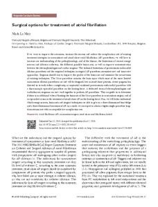
Fig. 1 Radiography showed a L4 vertebral body GCT with cortex breakthrough.
74
ous jobs without restricted daily activities.
DISCUSSION Several treatment strategies have been developed for GCT, including surgery, radiotherapy, embolization, cryosurgery, cementation, and chemical adjuvant such as phenol or liquid nitrogen. Surgical management remains the mainstay of the treatment processes. (16,17) When using either the Campanacci or Enneking grading system, the higher the radiographic grading, the more radical the required surgery. (9,10) Eckardt and Grogan recommended intralesional curettage with adjuvant therapy for stage I and II lesions and en bloc resection for most stage III lesions.(18) The anatomic characteristics of spinal GCT still present challenges for surgical excision. Surgeons and patients must choose between radical resection with potentially devastating morbidity or subtotal excision with potentially higher rates of recurrence.(5,12-15) In this study, all patients had primary stage III GCT and were treated with marginal excision without any adjuvant therapy regarding the uncertain therapeutic value and associated complications. With careful attention to the surgical margin and soft tissue and bone excision as wide as possible, the rate of recurrence may be decreased. However, complete wide excision of spinal lesions is often not possible and usually results in excision with marginal or intralesional margins. Two of the eight primarily treated tumors had local recurrence. McDonald et al.
Fig. 2 (left) and Fig. 3 (right) Sagittal T1-weighted and sagittal T2-weighted MRI revealed the L4 vertebral body GCT with retroperitoneal extension.
Chang Gung Med J Vol. 29 No. 1 January-February 2006
75
Shih-Chieh Yang, et al Surgery for GCT of thoracolumbar spine
Fig. 4 Axial MRI demonstrated the extent of the tumor.
Fig. 7 Coronal CT view revealed a reconstructed autograft and recurrent GCT at the L4 vertebral body.
Fig. 5 After marginal excision of the GCT through an anterior approach, anterior interbody fusion with autograft supplemented by posterior instrumentation was employed.
Fig. 8 Left-side anterior approach with wide excision and accompanying fibula allograft reconstruction was used for the recurrent GCT.
Fig. 6 Sagittal CT view showed a reconstructed autograft at the right L4 vertebral body and a recurrent GCT at left L4 vertebral body.
reported that recurrent lesions were probably not more biologically aggressive lesions and, in all likelihood, represented a less than complete initial curettage.(19) Under careful physical examination and use of improved imaging tools at regular follow-up intervals, recurrent tumors can be detected early. Therefore, during the earlier stages the lesions may possibly be treated using en bloc resection. Two recurrent tumors were successful diagnosed at on of the regular follow-up examinations. Wide excision combined with a reconstruction procedure for recurrent tumors achieved good results and prognoses after a minimum of 25 months of follow-up. Most patients incurring GCT of the bone are young and active with normal life expectancies. The treatment goal is not only to remove the tumor com-
Chang Gung Med J Vol. 29 No. 1 January-February 2006
Shih-Chieh Yang, et al Surgery for GCT of thoracolumbar spine
pletely, but also to preserve spinal function. After surgery, the anterior defects were reconstructed using structural autografts or allografts in most cases in this study. The advantage of the structural bone graft is that it can provide an initial load-sharing capacity for the anterior strut and aid in biological fusion. Functional spines allowed for normal daily activities in eight patients of this study. Curettage followed by bone cementing as a surgical technique for GCT of the extremities has widely been used.(20-22) Based on incomplete excision of tumors with pulmonary metastasis and old age, cement was used in two patients to supplement chemical adjuvant and offer immediate fixation and stability. Although cementation may help eradicate residual tumor tissue and produce local stability, it has no biological loading capacity. Three patients were transferred to our hospital due to tumor recurrence. Adjuvant radiotherapy was employed for incomplete excision of the tumors in these three patients. However, dose response data failed to identify optimal regimens. The risk of spinal cord myelitis, bone graft complications, and malignant transformation has been reported. (23,24) Radiotherapy of 5000 cGy in 25 fractions allowed local control in two patients. No radiation-induced malignancy or complications were noted after 4 and 9 years of follow up, respectively. The third patient, treated by laminectomy supplemented with radiotherapy, developed a sarcoma. Generally, site and extent of the lesion dictate which surgical approach is employed. In a lesion which involves the arch, a laminectomy with tumor removal and posterior fusion is performed as necessary. A lesion involving the anterior body requires an anterior approach with tumor removal and associated strut bone grafting with anterior stabilization. In common circumstances, combined anterior and posterior reconstruction is necessary for adequate stability. Radiotherapy is reserved for patients with incomplete excision and for those with local recurrence as it carries a risk of sarcomatous transformation.(23,24) The principal predictor of prognosis is the adequacy of tumor resection. However, the variability of methods and length of follow up in different series make comparison among the study results difficult. There are still no absolute clinical, radiographic, or histological parameters that indicate one correct method of treatment to reduce the likelihood of
76
recurrence. Thus, intensive clinical follow up is a critical and controllable step for the treatment of recurrent GCT. Improvements in imaging studies and surgical techniques allow physicians to improve detection of tumors during the early stages. Two patients who were initially treated at our hospital presented with localized symptoms and deterioration of daily activities during regular follow up. Early diagnosis allowed for en bloc excision and accompanying reconstruction, thereby, providing good prognosis. Nevertheless, our analytical results can only be used to generate hypotheses due to the small number of patients in this study. Further investigation with more cases with long-term follow up is required to determine the rate of late recurrence. The typical staging classifications used to evaluate and compare the treatment results for GCT were developed by Campanacci and Enneking. (9,10) However, the staging appropriate for extremities may not be suited to the spine. The definition of grade III GCT still retains some distinct variation and differentiation, especially for spinal lesions. The anatomy of the spine and the difficulty of surgical resection require specific consideration and modification.(25) Furthermore, primary tumors of the spine are rare. A single institution does not have a sufficient number of patients to assess treatment protocols. There is certainly a need for a simple, reproducible staging system which can facilitate scientific communication and carry out reliable multicenter studies on spinal lesions.
REFERENCES 1. Campanacci M, Baldini N, Boriani S, Sudanese A. Giant cell tumor of bone. J Bone Joint Surg 1987;69A:106-14. 2. Dahlin DC, Cupps RE, Johnson EW. Giant cell tumor: a study of 195 cases. Cancer 1970;25:1061-70. 3. Bertoni F, Present D, Enneking WF. Giant cell tumor of bone with pulmonary metastasis. J Bone Joint Surg 1985;67A:890-900. 4. Bertoni F, Present D, Sudanese A, Baldini N, Bacchini P, Campanacci M. Giant cell tumor of bone with pulmonary metastasis. Six case reports and a review of the literature. Clin Orthop 1988;237:275-85. 5. Dahlin DC. Giant-cell tumor of vertebrae above the sacrum: a review of 31 cases. Cancer 1977;39:1350-6. 6. Di Lorenzo N, Spallone A, Nolletti A, Nardi P. Giant cell tumors of the spine: a clinical study of six cases, with emphasis on the radiological features, treatment, and follow-up. Neurosurg 1980;6:29-34.
Chang Gung Med J Vol. 29 No. 1 January-February 2006
77
Shih-Chieh Yang, et al Surgery for GCT of thoracolumbar spine
7. Larsson SE, Lorentzon R, Boquist L. Giant-cell tumors of the spine and sacrum causing neurological symptoms. Clin Orthop 1975;111:201-11. 8. Ng ES, Saw A, Sengupta S, Nazarina AR, Path M. Giant cell tumor of bone with late presentation: review of treatment and outcome. J Orthop Surg 2002;10:120-8. 9. Campanacci M. Giant-cell tumor and chondrosarcomas: grading, treatment and results (studies of 209 and 131 cases). Recent Results Cancer Res 1976;54:257-61. 10. Enneking WF. A system of staging musculoskeletal neoplasms. Clin Orthop 1986;204:9-24. 11. Present D, Bertoni F, Hudson T, Enneking WF. The correlation between the radiologic staging studies and histopathologic findings in aggressive stage 3 giant cell tumor of bone. Cancer 1986;57:237-44. 12. Shikata J, Yamamuro T, Shimizu K, Shimizu K, Kotoura Y. Surgical treatment of giant-cell tumors. Clin Orthop 1992;278:29-36. 13. Savini R, Gherlinzoni F, Morandi M, Neff JR, Picci P. Surgical treatment of giant-cell tumor of the spine: the experience at the Istituto Ortopedico Rizzoli. J Bone Joint Surg 1983;65A:1283-9. 14. Sanjay BK, Sim FH, Unni KK, Mcleod RA, Klassen RA. Giant-cell tumors of the spine. J Bone Joint Surg 1993;75B:148-54. 15. Ozaki T, Liljenqvist U, Halm H, Hillmann A, Gosheger G, Winkelmann W. Giant cell tumor of the spine. Clin Orthop 2002;401:194-201. 16. Sung HW, Kuo DP, Shu WP, Chai YB, Liu CC, Li SM. Giant cell tumor of bone: analysis of two hundred and
Chang Gung Med J Vol. 29 No. 1 January-February 2006
17.
18. 19. 20.
21.
22.
23.
24.
25.
eight cases in Chinese patients. J Bone Joint Surg 1982;64A:755-61. Shih HN, Chen YJ, Huang TJ, Ho WP, Hsueh S, Hsu RW. Treatment of giant cell tumor of long bone. Chang Gung Med J 1996;19:16-23. Eckardt JJ, Grogan TJ. Giant cell tumor of bone. Clin Orthop 1986;204:45-58. McDonald DJ, Sim FH, McLeod RA, Dahlin DC. Giant cell tumor of bone. J Bone Joint Surg 1986;68A:235-42. Pals SD, Wilkins RM. Giant cell tumor of bone treated by curettage, cementation, and bone grafting. Orthopedics 1992;15:703-8. Persson BM, Ekelund L, Lovdahl R, Gunterberg B. Favorable results of acrylic cementation for giant cell tumors. Acta Orthop Scand 1984;55:209-14. Bini SA, Gill K, Johnston JO. Giant cell tumor of bone. Curettage and cement reconstruction. Clin Orthop 1995;321:245-50. Khan DC, Malhotra S, Stevens RE, Steinfeld AD. Radiotherapy for the treatment of giant cell tumor of the spine: a report of six cases and review of the literature. Cancer Invest 1999;17:110-3. Chen ZX, Gu DZ, Yu ZH, Qian TN, Huang YR, Hu YH, Gu XZ. Radiation therapy of giant cell tumor of bone: analysis of 35 patients. Int J Radiat Oncol Biol Phys 1986;12:329-34. Hart RA, Boriani S, Biagini R, Currier B, Weinstein JN. A system for surgical staging and management of spine tumors. A clinical outcome study of giant cell tumors of the spine. Spine 1997;22:1773-82.
78
11
1992
2002
3 8 5 ASIA scale
3 7
(
94
2006;29:71-8)
6
17
94
10
28 333
(03)3278113; E-mail:
[email protected]
5
Tel.: (03)3281200
2163; Fax: