323 Relato de caso
Giant Cervical Neurofibroma with Concomitant Deformity Surgical Strategies for treatment Neurofibroma gigante cervical com deformidade concomitante: estratégias cirúrgicas para o tratamento Gleidson C. Rodrigues1 Andrei F. Joaquim2 Enrico Ghizoni2 Luciano Queiróz3 Helder Tedeschi4
RESUMO
ABSTRACT
Apresentamos um caso de paciente de 27 anos de idade apresentando um neurofibroma cervical gigante com compressão medular e cifose. No presente artigo, discutimos detalhes técnicos de cirurgias múltiplas para ressecção tumoral completa e correção da deformidade em cifose. O algoritmo de nossa instituição utilizado para tratamento de cifose cervical é exposto em detalhes.
A 27 years-old woman presenting with a giant cervical neurofibroma with spinal cord compression and cervical kyphosis is reported. We discuss the surgical nuances of the multiple surgeries required to achieve a total tumor resection and deformity correction. An algorithm used in our institution to treat cervical kyphosis is also presented.
Palavras Chave: deformidade da neurofibroma, cifose pós laminectomia
coluna
cervical,
Keywords: cervical spine deformity, neurofibroma, postlaminectomy kyphosis
Resident of Neurosurgery. Neurosurgery Division, State University of Campinas (UNICAMP), Campinas-SP, Brazil. Assistant Neurosurgeon. Neurosurgery Division, State University of Campinas (UNICAMP), Campinas-SP, Brazil. 3 Professor, Department of Pathology, State University of Campinas (UNICAMP), Campinas-SP, Brazil. 4 Assistant Professor, Neurosurgery Division, State University of Campinas, (UNICAMP), Campinas-SP, Brazil 1 2
Recebido em 9 de julho de 2012, aceito em 5 de setembro de 2012 Rodrigues GC, Joaquim AF, Ghizoni E, Queiróz L, Tedeschi H. - Giant Cervical Neurofibroma with Concomitant Deformity - Surgical Strategies for treatment
J Bras Neurocirurg 23 (4): 323-327, 2012
324 Relato de caso
I ntroduction Neurofibromas are benign tumors originated from the Schwann cells and are characterized for their slow growth rate. Differently from schwannomas, which are capsulated tumors of the nerve wall, these are stroma-rich tumors with fibroblasts mixed with axons in a more chaotic arrangement (22). Neurofibromas can present as giant tumors, leading to severe neurological deficits, spinal deformity and cosmetic compromise (16). Kyphosis can develop secondarily due to the common association with neurofibromatosis, or after laminectomy for surgical treatment (6,14,16). Stevenson et al. reported that patients with neurofibromas can present with abnormal osseous absorption which increase the risk for deformity (3,20). In this report, we present a case of a 27 year-old woman with a giant cervical neurofibroma with spinal cord compression who developed swan neck deformity and required multiple surgeries for tumor resection and spinal realignment. We also discuss the nuances of the surgical strategy adopted for the tumor resection and deformity correction.
Case R eport A 27-year-old woman was referred to our institution five years after a posterior C3-4-5 laminectomy for the treatment of a giant right side cervical mass. The tumor, a spinal schwannoma, had been partially resected and the patient had been treated conservatively since then. On her admission, she complained of progressive tumor growth and weakness on the right side of her body and decreased tactile sensation on the left side. On her right side, Muscle strenght was grade II in the right sperior limb and grade III in the right inferiro limb, with clear pyramidal signs. Cervical spine magnetic resonance (MRI) on admission was compatible with an extensive intradural and extradural tumor at C3-4-5 level (Fig. 1).
A
B
C
D
Figure 1: Cervical MRI - T1 sequence with gadolinium (A- axial, B- sagittal, C- axial )) shows a right sided intraspinal mass, extending anteriorly through the neural foramen, with spinal cord compression C. Coronal T2 sequence. D. CT scan reconstruction showing the extension of the laminectomy.
Surgical treatment was planed with multiple step procedures, considering two main goals: total tumor resection and spinal alignment. For tumor resection, we initially proposed a posterior (intradural and extradural resection), followed by an anterior resection of the anterior cervical mass, while for spinal stabilization anterior multiple level discectomies with plating and finally a posterior C2-T1 instrumented fusion. A CT scan angiogram was also performed to evaluate the relationship of the vertebral artery with the tumor. A posterior approach was the initial procedure, using the same laminectomy performed at the first surgery, followed by dural opening and intradural resection of the residual tumor. After dural closure, resection of the extradural component was performed, visualizing the vertebral artery and the anterior extension of the tumor in the opened neural foramen ( fig. 2). The patient woke-up with no neurological worsening. After one week, an anterior cervical approach was performed, and her plexiform lesion was completely excised (Fig. 2). Histology exam was compatible with neurofibroma (Fig. 3).
Rodrigues GC, Joaquim AF, Ghizoni E, Queiróz L, Tedeschi H. - Giant Cervical Neurofibroma with Concomitant Deformity - Surgical Strategies for treatment
J Bras Neurocirurg 23 (4): 323-327, 2012
325 Relato de caso
one year of follow-up, she is doing well, ambulatory without assistance, with muscular strength grade IV in her right side and occasional neck pain , relieved with analgesics.
A
B
C
D
E
F
Figure 2: Intraoperative view of the posterior approach in A and B. In A, a large right extradural posterior mass can be clearly seen and in B, after dural opening and total intradural resection and C, after extradural resection. D. Anterior view of the right cervical mass. E. Exposure of skin incision of the mass and F after total resection.
C A
B
Figure 3: A (black and white) and B - hematoxylin-eosin staining: the tumor is constituted by bipolar cells, with long projections mixed within delicate collagen fibers. The cells are separated from each other by a highly hydrated extracellular matrix. Nuclei are elongated in a cigar shape and have a dense chromatin, without atypias. There are no mitosis or necrosis. The diagnosis corresponds to a neurofibroma.
After tumor resection, postoperative cervical spine MRI and flexion extension lateral radiographs of the cervical spine revealed a severe post-laminectomy kyphosis (Fig. 4). Once cervical lordosis could not be restored in extension, we considered it as a rigid one. Although the patient had some neurological improvement after surgery, she had also severe axial neck pain, even when wearing a cervical collar. One month after the last surgical procedure, she underwent 48 hours of cervical traction with Gardner tongs (Fig. 4), followed by a three level anterior discectomy with anterior iliac crest graft (C3-4, C4-5 and C5-6) and plating for stabilization. At the following day, she reported significant improvement in her neck pain. Three weeks later, a posterior C2-T1 fusion was performed, with autologous iliac crest bone for grafting (Fig. 8). She was discharged home two days after surgery. At
A
B
D
E
Figure 4: A. Sagittal T2 sequence cervical MRI showing cervical kyphosis and total tumor resection. B. Cervical lateral x-ray in orthostatic position showing the post-laminectomy kyphosis. C. Cervical lateral x-ray after three level discectomies. D and E. Final construction after anterior and posterior instrumented fusion.
Discussion Nerve sheath tumors are mostly represented by schwannomas (97.6%), followed by neurofibromas (2.1%) and malignant tumors (0.3%). Considering primary intraspinal tumors, 30% of them originate from the nerves, most of them represented by schwanommas (4,21). Neurofibroma can be found in the context of type 1 neurofibromatosis or in isolation (19). Although these tumors are more commonly located in the thoracic and lumbar spine, our cervical case had a disproportionate extradural portion compared with its intradural extension, probably explained by the theory of Jinnai and Koyama (9) who suggested that the intradural portion of the nerve roots enlarge craniocaudally. Surgical approach to nerve sheath tumors is based on their
Rodrigues GC, Joaquim AF, Ghizoni E, Queiróz L, Tedeschi H. - Giant Cervical Neurofibroma with Concomitant Deformity - Surgical Strategies for treatment
J Bras Neurocirurg 23 (4): 323-327, 2012
326 Relato de caso
extradural extension and intradural component (18). The most rational and logical surgical route is the posterior approach (18) , but it can be combined with an anterior approach, as in our case, when a significant extradural component is present. A lateral cervical approach has been suggested by Lot and George, in 1997, in an attempt to preserve the nerve roots (15). Microsurgical techniques combined with protection of the spinal cord and the nerve roots are the cornerstone of the treatment, resulting in cure of most of these patients when total resection is achieved (10,12,13). When associated with spinal deformities, surgery for tumor resection can be a challenge. Iatrogenic kyphosis after laminectomy for intraspinal tumor resection has multiple etiologies and risk factors (5), such as young age, preoperative spinal deformity, facet joints injuries, concomitant radiotherapy, and posterior column resection, among others (11). Straightening of the physiological cervical lordosis increases twice the risk of postlaminectomy kyphosis (11). Muscle denervation and ligaments destruction, associated with resection of the facet joints, result in weakness of the posterior tension band, leading to kyphosis with pain and neurological worsening (5, 11). In our case, the tumor was responsible for important facet destruction with foramen enlargement. We also mentioned that the patient underwent a three level cervical laminectomy, elevating three fold the risk for kyphosis (11). Many strategies had been proposed to avoid deformity after intraspinal resection of tumors. Concomitant instrumented arthrodesis was proposed at the same surgical procedure, but it increases surgical time and morbidity, especially in oncologic patients that will require adjuvant therapy or are in growth age (7,8). In our case, although a high risk for deformity was presented, our primary intention was complete tumor resection. We postponed spinal stabilization in the same sitting and waited for the patient’s recovery after two large surgical procedures. Moreover, as we did not have neurophysiological monitoring, we performed all operations separately, in order to avoid misjudgment of the potential causes for neurological deterioration. Cervical kyphosis should be treated in symptomatic patients. Sagittal plan of the cervical spine deformity can be treated using anterior, posterior or combined approaches, according to reducibility and the degree of the kyphosis (1,2,23).
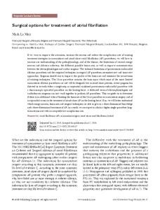
In general, dynamic x-rays are the main determinants of the approach to be chosen (6). Rigid deformity (non reducible in extension) without ankylosed facet joints can be treated using an anterior approach first to restore lordosis. If the facet joint is ankylosed, posterior osteotomies can be required prior to an anterior approach, commonly followed by a posterior approach. Traction can help decrease the stiffness of the deformity, also promoting muscle adaptation before cervical alignment after multilevel discectomies or corpectomies. Anterior approaches can restore the anterior column height thus restoring cervical lordosis, as shown in our case. After straightening her cervical spine, a posterior fusion was performed to avoid late pseudoarthrosis and maintainance of the physiological cervical lordosis. The posterior approach is generally used for instrumentation and fusion, not to restore alignment, although osteotomies can be performed in selected cases (17). Our institutional algorithm to treat cervical deformity is presented in Figure 5.
Cervical Kyphosis (dynamic x-rays/ CT to see flexibility, MRI to evaluate cord compression, fine cut CT to evaluate bone anatomy)
Symptomatic or progressive worsening
Asymptomatic
Surgical treatment
Clinical and radiological follow-up
Consider Preoperative Traction/ Evaluated dynamic x-rays
Kyphosis Reducible in extension
Kyphosis Rigid with posterior ankylosed facets
Kyphosis Rigid without posterior ankylosed facet joints
Posterior approach
Posterior release, followed by anterior approach and them a posterior instrumented fusion
Anterior approach followed by a posterior instrumented fusion
Figura 5: Institutional algorithm of the Neurosurgery Division - State University of Campinas - to treat cervical kyphosis
Rodrigues GC, Joaquim AF, Ghizoni E, Queiróz L, Tedeschi H. - Giant Cervical Neurofibroma with Concomitant Deformity - Surgical Strategies for treatment
J Bras Neurocirurg 23 (4): 323-327, 2012
327 Relato de caso
Conclusion A multiple-step surgery can be performed to treat complex cervical spine pathologies. Total tumor resection is the main goal when dealing with benign nerve sheath tumors, but complications like post-laminectomy kyphosis can be found. An early intervention in deformity correction can allow surgeons to achieve better results, improving patient outcomes.
Disclaimer The authors report no conflict of interest concerning the materials or methods used in this study or the findings specified in this paper.
R eferences 1. Butler JC, Whitecloud TS 3rd. Postlaminectomy kyphosis. Causes and surgical management. Orthop Clin North Am. 1992: 23:505–11. 2. Cattell HS, Clark GL Jr. Cervical kyphosis and instability following multiple laminectomies in children. J Bone Joint Surg Am. 1967: 49:713–20. 3.
Chaglassian JH, Riseborough EJ, Hall JE. Neurofibromatous scoliosis. Natural history and results of treatment in thirty-seven cases. J Bone Joint Surg Am 1976; 58:695–702.
4.
Darwish B, Balakrishnan V, Maltra R. Intramedullary ancient schwannoma of the cervical spinal cord: Case report and review of literature. J Clin Neurosci 2002; 9: 321-3.
5. Deutsch H, Haid RW, Rodts GE, Mummaneni PV. Postlaminectomy cervical deformity. Neurosurg Focus. 2003, 15 (3): Article 5. 6.
Fassett DR, Clark R,Brockmeyer DL, Schmidt MH. Cervical spine deformity associated with resection of spinal cord tumors. Neurosurg Focus. 2006; 20(2):E2.
7. Heller JG, Edwards CC Jr, Murakami H, Rodts GE. Laminoplasty versus laminectomy and fusion for multilevel cervical myelopathy: an independent matched cohort analysis. Spine. 2001; 26:1330–6. 8.
Herman JM, Sonntag VK. Cervical corpectomy and plate fixation for postlaminectomy kyphosis. J Neurosurg. 80,1994:963-70.
9. Jinnai T, Koyama T. Clinical characteristics of spinal nerve sheath tumors: analysis of 149 cases. Neurosurgery. 2005;56(3): 510-5. 10. Joaquim AF, Almeida JP, Ghizoni E, Santos MJ, Tedeschi H, de Oliveira E. Surgical Management of Intradural Extramedullary Tumors Located Anteriorly to the Spinal Cord. J Clin Neurosci. 2012; 19(8):1150-3. 11. Joaquim AF, Cheng I, Patel AA. Post-operative spinal deformity after treatment of intracanal spine lesions. Spinal J. 2012; 12(11):1067-74. 12. Joaquim AF. Santos MJ, Tedeschi H. Manejo cirúrgico dos hemangioblastomas. Coluna/ Columna. 2009; 8(3):274-8. 13. Joaquim AF. Santos MJ, Tedeschi H. Surgical Management of Intramedullary Ependymomas. Arq Neuro-Psiquiatr. 2009; 67(2A):284-9. 14. Katsumi Y, Honma T, Nakamura T. Analysis of cervical instability resulting from laminectomies for removal of spinal cord tumor. Spine. 1989; 14:1171–6. 15. Lot G, George B. Cervical neuromas with extradural components: surgical management in a series of 57 patients. Neurosurgery. 1997; 41(4):813-20. 16. Ma J, Wu Z, Yang X, Xiao J. Surgical treatment of severe cervical dystrophic kyphosis due to neurofibromatosis Type 1: a review of 8 cases. J Neurosurg: Spine. 2011; 14(1): 93-8. 17. McMaster MJ. Osteotomy of the cervical spine in ankylosing spondylitis. J Bone Joint Surg Br. 1997;79:197–203. 18. Salcman M, editor. Tumor Intra e Extradural: Schwannoma em Ampulheta. - Neurocirurgia operatoria de Kempe, Santos Editora; 2006 19. Seppälä MT, Haltia MJ, Sankila RJ, Jääskeläinen JE, Heiskanen O. Long-term outcome after removal of spinal neurofibroma. J Neurosurg.1995; 82(4): 572-7. 20. Stevenson DA, Schwarz EL, Viskochil DH, Moyer-Mileur LJ, Murray M, Firth SD, et al. Evidence of increased bone resorption in neurofibromatosis type 1 using urinary pyridinium crosslink analysis. Pediatr Res. 2008; 63(6):697-701. 21. Suh YL, Koo H, Kim TS, Chi JG, Park SH, Khang SK. Tumors of the central nervous system in Korea. J Neurooncol. 2002; 56: 251-9. 22. Wippold FJ 2nd, Lubner M, Perrin RJ, Lämmle M, Perry A. Neuropathology for the neuroradiologist: Antoni A and Antoni B tissue patterns. AJNR Am J Neuroradiol. 2007; 28(9):1633-8. 23. Zdeblick TA, Bohlman HH. Cervical kyphosis and myelopathy. Treatment by anterior corpectomy and strut-grafting. J Bone Joint Surg Am. 1989:71:170–82.
Corresponding author Dr. Andrei F. Joaquim e-mail:
[email protected]
Rodrigues GC, Joaquim AF, Ghizoni E, Queiróz L, Tedeschi H. - Giant Cervical Neurofibroma with Concomitant Deformity - Surgical Strategies for treatment
J Bras Neurocirurg 23 (4): 323-327, 2012