Sequence polymorphism of the COBRA gene of Kelampayan (Neolamarckia cadamba)
Aaron Bong Sing Woei (20575)
This project is submitted in partial fulfilment of the requirements for the degree of Bachelor of Science with Honours
Resource Biotechnology Department of Molecular Biology Faculty of Resource Science and Technology Universiti Malaysia Sarawak
2011
ACKNOWLEDGEMENT
First of all, I would like to thank God for His blessing upon the completion of this project. My deepest gratitude to my supervisor, Dr. Ho Wei Seng, for his encouragement, guidance and support in making this project possible. Besides that, my grateful thanks to my cosupervisor, Dr. Pang Shek Ling for guiding me in experimental processes and providing me information on this project. I would like to extend my gratitude and appreciation to my colleagues, Mr. Phui Seng Loi (Msc. Student), Mr. Liew Kit Siong (Msc. Student), Ms. Tchin Boon Ling (Msc. Student) and all my laboratory members for guiding me patiently and providing a supportive environment throughout the whole project. Lastly, I would like to thank my friends and family for giving me encouragements, supports and tolerances through this project.
i
DECLARATION
I hereby declare that this thesis is based on my original work except for quotations and citation, which have been duty acknowledged. I also declare that it has not been previously or concurrently submitted for any other degree at UNIMAS or other institutions.
___________________________________ Aaron Bong Sing Woei Resource Biotechnology Programme Department of Molecular Biology Faculty of Resource Science and Technology Universiti Malaysia Sarawak
ii
TABLE OF CONTENT ACKNOWLEDGEMENT
I
DECLARATION
II
TABLE OF CONTENTS
III
LIST OF ABBREVIATIONS
V
LIST OF TABLES AND FIGURES
VI
ABSTRACT/ABSTRAK
1
INTRODUCTION
2
LITERATURE REVIEW
4
2.1 Neolamarckia cadamba (Kelampayan)
4
2.2 Wood Formation
5
2.3 COBRA Gene
7
2.4 Single Nucleotide Polymorphism (SNP)
9
2.5 Application of Single Nucleotide Polymorphisms (SNPs)
10
MATERIALS AND METHOD
12
3.1 Plant Materials
12
3.2 DNA Extraction and Purification
12
3.2.1 Chemicals and Reagents
12
3.2.2 Total Genomic DNA Isolation
13
3.2.3 DNA Purification
14
3.3 DNA Quantification
14
3.3.1 Agarose Gel Electrophoresis
14
3.3.2 Spectrophotometric Quantification of DNA
15
3.4 Data Mining and Primer Design
16
3.5 Polymerase Chain Reaction (PCR)
17
3.6 PCR Optimization with MgCl2
18
3.7 PCR Product Purification
18
3.8 Cloning of PCR Products
19
3.8.1 Ligation
19
iii
3.8.2 Transformation and Blue/White Screening
19
3.9 Confirmation of the Desired Insert
20
3.10 DNA sequencing and analysis
20
22
RESULTS AND DISCUSSION 4.1 Collection of Plant Samples
22
4.2 DNA Extraction and Purification
23
4.3 DNA Quantification
24
4.4 Primer Design
26
4.5 Polymerase Chain Reaction (PCR)
27
4.6 PCR Optimization
28
4.7 PCR Product Purification
29
4.8 Cloning of PCR Products
30
4.9 Confirmation for Desired Insert
31
4.9.1 Colony PCR
31
4.10 Plasmid Purification
32
4.11 DNA Sequencing and Data Analysis
33
4.11.1 Evaluation of sequence variation in COBRA gene
33
among N. cadamba 4.11.2 Single nucleotide polymorphism discovery
37
4.11.3 Determination of non-synonymous mutation and
39
synonymous mutation 4.11.4 Identification of possible restriction enzymes for
41
SNP site
CONCLUSIONS
43
REFERENCES
45
iv
LIST OF ABBREVIATIONS [K+]
Concentration of salt
AFLPs
Amplified fragment length polymorphisms
Bp
Base pair(s)
CTAB
Cetyltrimethylammonium bromide
ddH2O
Double distilled water
DNA
Deoxyribonucleic acid
EDTA
Ethylenediamine tetraacetic acid
EST
Expressed Sequence Tag
GAP
Glycosylphosphatidylinositol (GPI)-anchored protein
GPI
Glycosylphosphatidylinositol
IPTG
Isopropyl β-D-1-thiogalactopyranoside
LB
Luria broth
LD
Linkage disequilibrium
MAS
Molecular marker-assisted selection
NaCl
Sodium chloride
ORF
Open reading frames
PCR
Polymerase chain reaction
PVP
Polyvinylpyrrolidone
QTL
Quantitative trait locus
RFLP
Restriction fragment length polymorphism
SFC
SARAWAK FORESTRY Cooperation
SNP
Single nucleotide polymorphism
TE
Tris-EDTA
Tm
Melting temperature
X-gal
5-bromo-4-chrolo-3-indolyl-β-galactopyranoside
ΔH
Enthalpy for helix formation
ΔS
Entropy for helix formation
v
LIST OF TABLES AND FIGURES Figures 2.1
Page N. cadamba (a) The tree of N. cadamba, and (b) Cylindrical
5
trunk with coarse surface 2.2
Model of wood formation
6
2.3
Structure of glycosylphosphatidylinositol (GPI) anchor
7
3.1
Description of plots at Landeh Reserve Nature
12
4.1
Gel electrophoresis of purified genomic DNA samples on a 0.8%
24
agarose gel 4.2
Gel electrophoresis of PCR products on a 1.5% agarose gel
27
4.3
Gel electrophoresis of PCR products on a 1.5% agarose gel.
29
4.4
Gel electrophoresis of purified PCR product on a 1.5% agarose
30
gel 4.5
Gel electrophoresis of colony PCR products on a 1.5% agarose
31
gel 4.6
Gel electrophoresis of purified plasmid on a 1.0% agarose gel
32
4.7
Alignment result of Populus Tomentosa DNA (GU053565) and
34
sample COBRA A 4.8
Alignment result of Populus Tomentosa mRNA (GQ410777)
35
and sample COBRA A 4.9
Comparison of sequence structure of Populus Tomentosa
36
genotype 38 COBRA-like 4 protein gene (GU053565), Populus Tomentosa COBRA-like 4 protein mRNA (GQ410777) and sequence of COBRA gene from sample A 4.10
Alignment result of six partial sequences of COBRA gene using
39
ClustalW software 4.11
Alignment of the protein sequences using ClustalW software
40
4.12
Possible restriction enzymes for SNP site from COBRA W using
42
NEBcutter V2.0 4.13
A close up of possible restriction enzymes for SNP site from COBRA W using NEBcutter V2.0
vi
42
Tables
Page
3.1
Ligation reaction mixture and volume
19
4.1
Plot of the collected trees
22
4.2
Characteristics of collected Kelampayan trees
23
4.3
Estimated purified DNA concentration of a N.cadamba based on
25
band intensity of samples and marker on a 0.8% agarose gel 4.4
Estimated DNA concentration and DNA purity of the six
25
purified N.cadamba samples 4.5
Primer pairs designed to amplify the COBRA gene
26
4.6
PCR reaction mixture used to amplify the COBRA gene
27
4.7
PCR reaction mixture in PCR optimization used to amplify the
28
COBRA gene 4.8
BLASTn output for amplified partial 517 bp COBRA gene
33
4.9
BLASTp output for COBRA gene sequence with intron removed
37
4.10
Position of sequence variations within the sequenced samples
39
4.11
The non-synonymous and synonymous mutation occurred in the
40
samples
vii
SEQUENCE POLYMORPHISM OF THE COBRA GENE OF KELAMPAYAN (Neolamarckia cadamba) AARON BONG SING WOEI Resource Biotechnology Faculty of Resource Science and Technology Universiti Malaysia Sarawak
ABSTRACT Neolamarckia cadamba or locally known as Kelampayan is a fast growing deciduous tree with great commercial values. COBRA gene is involved in cellulose deposition and orientation of cell expansion. Any single nucleotide differences may influence the functions of the gene. In this study, the DNA sequence polymorphisms in COBRA gene from N. cadamba were detected. The targeted DNA sequence of COBRA gene was amplified with the designed primer by using Polymerase Chain Reaction (PCR) technique. The 517 bp of COBRA gene amplicons were subjected to BLASTn analysis to search for homology sequence and validate the identity of the obtained sequences through NCBI. Multiple alignment was carried out by ClustalW for manual detection of Single Nucleotide Polymorphisms (SNPs). Five SNPs were detected in the exon region and two SNPs in the intron region of COBRA gene amplicons. The sequence of six partial COBRA gene was then subjected to in silico restriction analysis. One possible restriction enzyme, HpyCH4III was detected for SNP at nucleotide 384 bp of a sample which could be useful for genetic marker development. Key words: Neolamarckia cadamba, polymerase chain reaction (PCR), COBRA gene, single nucleotide polymorphisms (SNPs), molecular markers
ABSTRAK Neolamarckia cadamba atau lebih dikenali sebagai Kelampayan merupakan species pokok kayu yang cepat membesar dan mempunyai nilai komersial yang tinggi. Gen COBRA memainkan peranan dalam deposisi selulosa and orientasi dalam pengembangan sel. Sebarangan pembezaan nukleotida akan mempengaruhi fungsi gen. Dalam kajian ini, polimorfisme nukleotida tunggal (SNP) dalam gen COBRA DNA dari N. cadamba telah ditemui. Jujukan DNA bagi gen COBRA diamplifikasi oleh penanda molekul dengan menggunakan teknik tindakbalas berantai polimerase (PCR). Produk PCR bersaiz 517 bp gen COBRA dianalisa dengan BLASTn untuk mencari urutan homologi dan mengesan identiti untuk jujukan yang diperolehi dalam pangkalan data NCBI. Penjajaran jujukan oleh ClustalW dilakukan untuk mengesan SNP secara manual. Lima SNP telah dikesan dalam bahagian ekson dan dua dalam bahagian intron bagi gen COBRA. Enam jujukan gen COBRA dianalisa oleh enzim penyekatan secara in silico. Penemuan satu pengkhususan enzim, HpyCH4III bagi SNP pada nukleotida 384 bp adalah amat berguna dalam penghasilan penanda molekul. Kata kunci: Neolamarckia cadamba, tindakbalas berantai polimerase (PCR), COBRA gene, polimorfisme nukleotida tunggal (SNP), penanda molekul
1
1.0
INTRODUCTION
Neolamarckia cadamba, also locally known as Kelampayan is one of the fast growing deciduous trees. It is an indigenous species for areas from India through Southeast Asia to Papua New Guinea. Due to the fast growth rate and great economic value of its timber, plantations of N. cadamba have been raised in India, Sri Lanka, Myanmar, Indonesia, Malaysia and the Philippines. It grows well in freshwater swamps and it is commonly found in logging area of lowland dipterocarp forests (Nair, 2007). The timber is generally used for light-weight purposes like moulding, skirting, frames and so on (Lim et al., 2005). Besides that, N. cadamba is one of the selected plant species for planted forestry in Sarawak. The increase of global demand for wood requires an increase in forest production. It is essential for research and development (R&D) of faster growth, high-yield and short rotation plantation forests as compared to the conventional breeding which is timeconsuming and costly. The role of forest biotechnology to provide adequate tools for tree breeders and production of high quality plantation are required to augment traditional tree improvement activities. In Sarawak, SARAWAK FORESTRY Corporation (SFC) in collaboration with UNIMAS has ventured in the development of forest genomics, biotechnology and tree improvement research for development of planted forest and conservation of forest resources. The forest biotechnology researches are to develop adequate tools to produce trees that adapt to the country’s conditions and significantly achieve economic benefits. Other than that, application of forest genomics will enhance tree productivity and wood quality. Development of highly informative and polymorphic genetic markers such as single nucleotide polymorphisms (SNPs) can be used in the selection of better trees for plantation and genetic improvement activities. According to
2
Gupta et al. (2001), the application of SNPs at molecular level can be used in selective breeding with desired traits by plant breeders. Plant cell wall plays a vital role in human life as it provides large scale of dietary fibers and raw materials for textile, lumber, pulping, and potentially for biofuels. In economic point of view, understanding the regulatory mechanisms involved for biosynthesis of cell wall and identifying regulatory genes such as cellulose, hemicelluloses and lignin are of obvious important (Zhang et al., 2009). Cellulose that composes 40% to 50% of wood is an essential component of the load-bearing network by providing strength required in cell walls through cellulose deposition. The physical properties of cellulose also determine the orientation of cell expansion (Brown, 2003). The presence of COBRA gene in the woody plant species is essential as it plays the role in the regulation of cellulose deposition and orientation of cell enlargement. Identification of COBRA gene and sequence polymorphism controlling wood quality traits are vital especially in forest tree breeding programs. An improvement of percentages in traits such as pulp yield can produce large gains for a pulp and paper mill industry. Therefore, the objective of this study was to design primers for the amplification of the partial COBRA genomic fragment in the six samples of different N. cadamba trees through PCR. Moreover, single nucleotide polymorphisms (SNPs) were detected in this study by comparing the genotype of the samples. Non-synonymous or synonymous mutations due to single base differences in partial COBRA genomic sequence which may result in changes of phenotypic trait or encoding gene were identified as well. Restriction enzyme that recognizes and restricts at the identified SNP site in the COBRA gene was identified which is necessary in developing marker for selection of planting material at the seedling stage.
3
2.0
LITERATURE REVIEW
2.1
Neolamarckia cadamba (Kelampayan)
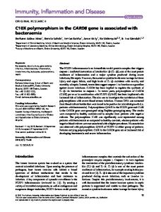
Neolamarckia cadamba or locally known as Kelampayan is under the family of Rubiaceae. N. cadamba is a medium to tall growing deciduous tree that grows up to 40 m to 45 m tall. Its bole is straight and cylindrical. This species usually starts branching after 25 m and a diameter of up to 100 cm with buttresses to 2 m tall. It is widely dispersed in lowland to mountain forests of up to 1000 m altitude where it is often found by streams, rivers and in open sites of the forests (Lim et al., 2005). According to a study done by Lim et al. (2005), N. cadamba is classified as one of the light-coloured timbers with low density. Due to the presence of large vessels, the timber of N. cadamba is white with coarse surfaces. Light and soft properties of the timber promote the usage for light-weight purposes such as moulding, picture frames, skirting, disposable chopstick, wooden sandals, veneer, plywood and general utility furniture. Besides that, the seeds have great medicinal value as it cures astringent, ulcer, digestive, diarrhoea, expectorant, fever and vomiting (Peter, 2007). N. cadamba leaves is largely ovate, whole margin of elliptic-oblong with smooth surface and pinnate venation. The length of the leaves varies from 7.5 to 18 cm with 4.5 to 16 cm breadth. Thick prominent midrib and uniform thin lamina is observed in the leaf through microscope. Short and thick wall of unicellular trichomes are observed on the epidermis layer of the leaves. The presence of unicellular, lignified trichomes, paracytic stomata, simple starch grains and sandy balls of calcium oxalate crystals are observed through the microscopic study of N. cadamba leaf powder (Patel and Kumar, 2008).
4
(a)
(b)
Figure 2.1 N. cadamba (a) The tree of N. cadamba, and (b) Cylindrical trunk with coarse surface. (Adapted from (a) http://www.flickriver.com/photos/tags/anthocephaluscadamba/interesting/ (b) http://www.flickr.com/photos/dinesh_valke/2065669915/)
2.2
Wood Formation
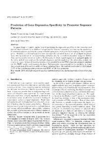
Wood is also known as secondary xylem which is constructed by four major ecological sequences, including cell division, cell expansion, cell wall thickening, and programmed cell death (Demura and Fukuda, 2006). Primary wall formation and modification are involved in the expansion of procambial cells and the cambial daughter cells. These two cells produced by cambial initials will differentiate into phloem cells and xylem cells (tracheary elements and fibre). Components of tracheary elements are tracheids and vessels. Vessels such as protoxylem vessels have annular and spiral secondary wall thickenings while metaxylem vessels have reticulate and pitted thickening (Demura and Fukuda, 2006). The expansion of cell is followed by the thickening of cell wall that forms the secondary wall. Formation of secondary wall is driven by the coordination of numerous genes that involved in the biosynthesis of cell wall proteins and polysaccharides (cellulose,
5
hemicelluloses), and formation of lignin. Lastly, an empty tube is formed with secondary walls when programmed cell death happens (Demura and Fukuda, 2006).
(a)
(b)
Figure 2.2 Model of wood formation. Adapted from (a) Demura and Fukuda, 2006) (b) http://www.bcb.uwc.ac.za/ecotree/trunk/Formationwood1.htm
According to Plomion et al. (2001), wood is made up of between 40% and 50% of cellulose. Cellulose is composed of 1,4-β-glucan chains that are synthesized by a cellulose synthase complex. This multi-protein complex simultaneously synthesizes 18 to 36 glucan chains that bond together in a single microfibril (Liepman et al., 2010). This basic structural crystallized microfibril formed as the result of a strong association of intrachain and interchain hydrogen bonds between the different chains of β-linked glucose residues (Plomion et al., 2001). In order to provide strength to cell walls, cellulose microfibrils have to be deposited in a specific orientation. Cellulose microfibrils are usually deposited in analog arrays transverse of growing cells to the direction of growth.
6
2.3
COBRA Gene
The COBRA gene in the woody species is responsible for the regulation of cellulose deposition and orientation of cell enlargement. This gene encodes for putative glycosylphosphatidylinositol (GPI)-anchored protein (GAP) targets the cell surface of the plant (Zhang et al., 2009). COBRA protein is fixed by GPI to the external plasma membrane leaflet and inserted into the endoplasmic reticulum. The addition of GPI anchor involved the cleavage of a hydrophobic C-terminal peptide and followed by linkage of a preassembled GPI anchor through an amide bond onto the last amino acid residue which is the remains of the cleavage. This attachment site is called ω-site. Specific phospholipases cleave GPI anchor and causes the release of the protein from the membrane (Roudier et al., 2002). The modified protein is transported to the cell surface and attached to the plasma membrane aided by the fatty acid chains of the GPI anchor (Cooper, 2000).
Protein C terminus Ethanolamine
Mannose
N-acetylgalactosamine Glucosamine Inositol
+
NH3-CH2-CH2
Figure 2.3 Structure of glycosylphosphatidylinositol (GPI) anchor (Cooper, 2000).
7
In a study done by Zhang et al. (2009), a COBRA-like protein was successfully isolated and identified as PtCOBL4 when comparison of nucleotide sequence was compared with the known full-length of Arabidopsis COB gene sequences. It is confirmed as one of the members of COBL family, PtCOBL4 shared 72.7%, 64.8%, and 65.3% similarity with Arabidopsis COBL members, rice BC1 members and maize BK2 proteins respectively. Besides that, PtCOBL4 was examined for these characteristics: CCVS motif, an N-terminal signal peptide sequence for secretion, a highly hydrophobic C terminus, and specific features around the ω-site. With all these criteria, PtCOLB4 is classified as COBL protein. In Arabidopsis, AtCOBL4 is identified to be implicated in the cellulose biosynthesis in the secondary wall through mutant analysis (Brown et al., 2005). The expression of AtCOBL4 is found to be similar with Arabidopsis cellulose synthase (AtCesA) genes that are involved in secondary cell wall synthesis (Persson et al., 2005). According to Zhang et al. (2009), PtCOBL4 is suggested to be an ortholog of AtCOBL4 as they shared high nucleotide homology (70.9%) and amino acid identity (72.7%). Other than that, PtCOBL4 gene is found to be expressed dominantly in the mature xylem fiber cells during the late stage of cell wall thickening. This showed that PtCOBL4 is involved in the secondary cell wall deposition in the stem of trees (Zhang et al., 2009). Studies by Roudier et al. (2005) proved that COBRA gene is found to be distributed in a banded pattern that is perpendicular to the longitudinal axis via a microtubuledependent mechanism. Mutants of COBRA gene are defective in elongation of cell growth that occurs after the initial cell expansion and this indicates the involvement of COBRA gene in morphogenesis (Roudier et al., 2005). According to Roudier et al. (2005), Arabidopsis mutants of COBRA gene has shown to reduce cellulose content and cause the cells to swell due to disorganize cellulose microfibril deposition. However, the 8
disorganization of cellulose occurs before the depletion of cellulose content in the cell wall. This shows that COBRA is involved in the orientation of deposition of cellulose microfibrils while the reduction in cellulose content is likely caused by a feedback mechanism that reflects the already disordered deposition of cellulose microfibrils (Roudier et al., 2005).
2.4
Single Nucleotide Polymorphism (SNP)
Before the emergence of molecular marker as an indicator of sequence polymorphism, plant breeders select the plant based on the phenotypic agronomic traits. With the development of fingerprinting,
technologies and genetic markers, approaches such as DNA mitochondrial
DNA
sequencing
or
restriction
fragment
length
polymorphism (RFLP) analysis, amplified fragment length polymorphisms (AFLPs), nuclear gene sequencing and genotyping of various types of nuclear loci are adopted (Aitken et al., 2004). However, some of these approaches are time-consuming and labour intensive in large-scale genome analysis (Edwards and Mogg, 2001). Approaches like RFLPs are laboured intensive and costly. Larger quantities of DNA are needed yet it produces only low quantity of polymorphism (Williams et al., 1991). Besides that, these approaches are confronted with technical and analytical issues that limit their application. For example, the highly variable mutation rate of gene limits the analysis of genome (Aitken et al., 2004). According to Aitken et al. (2004), an ideal genetic marker for population and evolutionary studies should possess at least three properties. Firstly, biases associated with single locus analysis can be avoided by using markers that are abundant and distributed widely across the genome. To facilitate analysis and interpretation, well-understood and 9
well-characterized models of evolution should be used. Lastly, technical applications must allow data acquisition from numerous loci marked in large population samples, while the data have to be comparable in laboratories using different genotype scoring procedures or technologies (Sunnucks, 2000). All these ideal marker characteristics are fulfilled by single nucleotide polymorphisms (SNPs). Single Nucleotide Polymorphisms (SNPs) are referred to single DNA base differences between homologous DNA fragments including small insertions and deletions of multiple bases. Polymorphisms of single base are usually biallelic (Dietrich et al., 1999). These variations in DNA bases can be used as genetic markers that can be identified in the neighboring of virtually every gene (Rafalski, 2002). These molecular markers are essential in genetic localization, map-based positional cloning, detection of marker-trait gene relationship through linkage and linkage disequilibrium (LD) mapping and the assessment of genetic association between organisms.
2.5
Application of Single Nucleotide Polymorphisms (SNPs)
In the study of functional genomics where expressed sequence tags predominate, the use of SNPs as molecular markers for individual genotyping are needed for molecular markerassisted selection (MAS) (Gupta et al., 2001). In the genome, SNPs may occur in the coding, non-coding and intergenic region. SNP in plant increase our knowledge to understand the genetic basis of important agronomic traits. The presence of SNPs in the coding region will demonstrate the association with the trait even though the mutant phenotype may or may not able to be determined. However, recognition of SNPs that associate with trait is useful for MAS and for gene isolation. In DNA, SNPs are recognized as the most frequent type of variation found in DNA. Recent estimation has shown that one
10
SNP occur every 100-300 bp in any genome. This shows that SNPs are recognized as the most abundant molecular markers in the present time (Gupta et al., 2001). Detection of associations between allelic forms of a gene and phenotypes especially in common diseases with multifactorial genetics can be done with SNPs too. The study of linkage disequilibrium (LD) able to provide information regarding positional cloning, linkage and evolution by constructing LD map. Positional cloning of genes for disease susceptibility is dependent on linkage and allelic association (Maniatis et al., 2002). This showed that LD is a promising tool for fine mapping of gene diseases in a population. According to Moon et al. (2007), this molecular marker, SNPs are the most essential forms of DNA variation for quantitative trait loci (QTL) mapping. QTL is defined as the genes influencing heritable quantitative trait, a phenotypic characteristic that is attributed by two or more genes (Grisel, 2000). By implementing QTL mapping, the underlying genes in a quantitative trait can be discovered with the utilization of molecular markers like SNPs. The greater number of polymorphic DNA markers used, the greater chances of discovering closely linked genes or the relevant genes (Grisel, 2000). The association analysis between the markers and the quantitative trait contributes to the identification of phenotypic variation as it helps researcher to derive a better insights into the genetic basis of a complex trait (Wu et al., 2007). QTL mapping can be carried out by comparing same genotype strains of various inbred and the interactions of two or more genes with their environment (Grisel, 2000). Hence, the utilization of SNPs markers enables the identification of heritable beneficial quantitative trait which can be used in selective breeding programme.
11
3.0
MATERIALS AND METHODS
3.1
Plant Materials
Fresh young leaves were collected from six randomly selected Neolamarckia cadamba trees for total genomic DNA extraction from the Kelampayan Trial Plot at Landeh Nature
Tree 1 – Tree 9
Reserve, Semengok, Sarawak (Figure 3.1).
Tree 1 – Tree 16 Figure 3.1: Description of plot at Landeh Nature Reserve
3.2
DNA Extraction and Purification
3.2.1 Chemicals and Reagents The reagents used in this study are liquid nitrogen, CTAB extraction buffer (100 mM TrisCL pH 8.0; 1.4 M NaCl; 20 mM EDTA; 2% CTAB; 1% polyvinylpyrrolidone (PVP); 2% (v/v) β-mercaptoethanol), isopropanol, TE buffer, chloroform:isoamy alcohol (24:1) and sterile distilled deionized water (ddH2O).
12
3.2.2 Total Genomic DNA Isolation Total genomic DNA of N. cadamba was extracted by using the modified CTAB method from Doyle and Doyle (1990). Six thousand micro liters of CTAB extraction buffer was added with 2% of βmercaptoethanol in a 15 ml snap cap Falcon tube. The tube was then preheated for 30 minutes at 65°C. The sterilized mortar, pestle and spatula were pre-chilled in liquid nitrogen. Plant material of approximately 1.0 g was ground to powder form in liquid nitrogen. During the grinding process, the tissue should be kept frozen. It is essential to grind the tissue to a very fine powder in order to obtain high yield of molecular weight DNA. The ground tissue powder was then scraped and placed into the preheated buffer and swirled gently to mix. The sample was then incubated at 65°C for 1 hour with shaking. After incubation, the sample was cooled down for 5 minutes. Then, 750 µl of the mixture in the Falcon tube was transferred into 1.5 ml microcentrifuge tubes with excised wide-bored tips. Equal volume of chloroform/isoamyl alcohol (CIA) (24:1; v/v) was added and mixed gently. The sample was centrifuged at 13,000 rpm for 10 minutes at room temperature. Later, the aqueous phase was transferred to new 1.5 ml microcentrifuge tubes. Equal volume of CIA was added to the tubes and the tubes were centrifuged again. This process is to ensure any carbohydrates impurities were removed. Following centrifugation, the aqueous phase was transferred to clean microcentrifuge tubes. About 2/3 volume of cold isopropanol was added to the tubes and mixed gently. Precipitation of nucleic acids was observed. The sample was then stored overnight at -20°C. The next day, the sample was thawed and centrifuged for 15 minutes at 13,000 rpm. Pellet was formed and the supernatant was carefully poured off. Five hundred micro liters of 70% cold ethanol was added and centrifuged for 10 minutes at 13,000 rpm. The 13
supernatant was decanted and air dry for 10 – 15 minutes. The pellet was resuspended by adding 80 µl of ultra pure water. The tubes were flicked gently to dissolve the pellet and later on stored for an hour at -20°C.
3.2.3 DNA Purification The isolated DNA was purified by using Wizard® Genomic DNA Purification Kit (Promega, USA) based on the manufacture’s protocol. The sample was top up with ultra pure water to 600 µl. RNase of 3 µl was extracted from the kit and added into the sample. The sample was incubated for an hour at 36.5°C. An hour later, 200 µl of Protein Precipitation Solution was then added. The tube was flicked gently and centrifuged for 3 minutes at 13,000 rpm. Six hundred micro litres of supernatant was transferred to a new tube containing 600 µl isopropanol. The tube was mixed gently and centrifuged for 10 minutes at 13,000 rpm. The pellet was formed and the supernatant was carefully poured off. Later, 70% of ethanol was added and centrifuged for 5 minutes at 13,000 rpm. The ethanol was then discarded and the tube was left to air-dry for 15 minutes. The pellet of DNA was resuspended in 30 µl of ultra pure water. It was then incubated for an hour at 36.5°C and stored at -20°C for further analysis.
3.3
DNA Quantification
3.3.1 Agarose Gel Electrophoresis The presence and concentration of isolated genomic DNA from the leaf was determined by running a 0.8% agarose gel. Agarose powder of 0.32g was added into a conical flask
14
containing 40ml 1X TAE Buffer and the mixture was heated at 300°Cfor 2 minutes. Later, the mixture was cooled while swirled gently and poured into gel casing. Bubble was poked and comb of 16 wells was inserted. The gel was left to cool and solidify for 45 minutes at room temperature. Five micro liters of genomic DNA was mixed with 2 µl of 1X loading dye. Three micro liters of Lambda-HindIII (Promega, USA) was used as DNA marker and mixed with 1 µl of 1X loading dye. The gel was run under 50V, 40A for 90 minutes. Later, the gel was stained with ethidium bromide for 10 seconds. It was then followed by de-staining with distilled water for 30 minutes. Presence of any bands were detected by visualized the gel under a UV transilluminator. The concentrations of the genomic DNA samples were checked by comparing the intensity of samples’ band with standard DNA of known concentration. An estimation of the DNA concentration was made by comparing the brightness of the known band with the sample bands. The concentration of DNA was calculated based on the formula below: DNA Conc. (ng µl) =
Marker frag. size Marker vol. (µl) X Marker Conc. (µg µl) X X 10³ Marker total size DNA vol. (µl)
3.3.2 Spectrophotometric Quantification of DNA The concentration of purified DNA was quantified using Lambda 25 UV/VIS Spectrophotometer (Perkin Elmer, USA). Three micro liters of DNA sample was mixed with 2,997 µl of ultra pure water in a quartz cuvette. The absorbance reading was taken at wavelength of 230nm, 260nm, and 280nm. The absorbance ratio of A260/A280 and A260/A230 15
were calculated to account the purity of the DNA samples. The concentration of each DNA sample was calculated based on the formula below: [DNA] (ng/µl) = A260 x OD260 The OD260 unit for dsDNA correspond to a concentration of 50 µg/ml. The dilution factor has automatically counted by the programme while giving the value of A260.
3.4
Data Mining and Primer Design
Primer was designed using Primer Premier 6.0 (PREMIER Biosoft International, USA). The forward and reverse primer pairs were designed based on the EST of Neolamarckia cadamba with the nucleotide sequence length of 536 bp that is obtained by Forest Genomics and Informatics (fGiL). In PCR, primers should be designed so that there are no inverted repeats or homopolymeric regions. There should be approximately 50% of GC contents in the base composition. At the 3’ end of primers, there should be more than 1 G or C residues. Any shortage of above criteria will affect the stability of primer-template interaction (Middendorf et al., 2005). In designing primers, it is important to have optimal length of the primer as it controls the specificity. Longer primers are prone to form secondary structures like hairpins, dimmers, and self-complementation. It is vital to avoid the forming of secondary structure as it would limit the number of primers available for bonding. It is best to use shorter primer that would provide the best balance between specificity and efficiency. Optimally, primers length should be between 18 and 24 bases. Besides that, melting temperature (Tm) can be calculated based on the following formula: 16