JOURNAL OF VIROLOGY, Mar. 1989, p. 1123-1133 0022-538X/89/031123-11$02.00/0 Copyright © 1989, American Society for Microbiology
Vol. 63, No. 3
Identification and Nucleotide Sequence of the Glycoprotein gB Gene of Equine Herpesvirus 4 DAVID E. ONIONS'* Department of Veterinary Pathology, University of Glasgow Veterinary School, Bearsden Road, Bearsden, Glasgow MARCELLO P.
G61
RIGGIO,1 ANN A. CULLINANE,2
AND
IQH, Scotland,' and Irish Equine Research Centre, Johnstown, Naas, County Kildare, Ireland? Received 26 July 1988/Accepted 8 November 1988
The nucleotide sequence of the glycoprotein gB gene of equine herpesvirus 4 (EHV-4) was determined. The located within a BamHI genomic library by a combination of Southern and dot-blot hybridization with probes derived from the herpes simplex virus type 1 (HSV-1) gB DNA sequence. The predominant portion of the coding sequences was mapped to a 2.95-kilobase BamHI-EcoRI subfragment at the left-hand end of BamHI-C. Potential TATA box, CAT box, and mRNA start site sequences and the translational initiation codon were located in the BamHI M fragment of the virus, which is located immediately to the left of BamHI-C. A polyadenylation signal, AATAAA, occurs nine nucleotides past the chain termination codon. Translation of these sequences would give a 110-kilodalton protein possessing a 5' hydrophobic signal sequence, a hydrophiic surface domain containing 11 potential N-linked glycosylation sites, a hydrophobic transmembrane domain, and a 3' highly charged cytoplasmic domain. A potential internal proteolytic cleavage site, Arg-Arg/Ser, was identified at residues 459 to 461. Analysis of this protein revealed amino acid sequence homologies of 47% with HSV-1 gB, 54% with pseudorabies virus gpII, 51% with varicella-zoster virus gpII, 29% with human cytomegalovirus gB, and 30% with Epstein-Barr virus gB. Alignment of EHV-4 gB with HSV-1 (KOS) gB further revealed that four potential N-linked glycosylation sites and all 10 cysteine residues on the external surface of the molecules are perfectly conserved, suggesting that the proteins possess similar secondary and tertiary structures. Thus, we showed that EHV-4 gB is highly conserved with the gB and gpII glycoproteins of other herpesviruses, suggesting that this glycoprotein has a similar overall function in each virus. gene was
Equine herpesvirus 1 (EHV-1) and EHV-4 are alphaherpesviruses which cause serious disease in the horse, EHV-1 being predominantly associated with abortion and neurological disease and EHV-4 being a major cause of respiratory disease (28). The envelope glycoproteins are important immunogens of herpesviruses involved in producing a protective immune response (40, 41). EHV-1 and EHV-4 are known to contain at least eight highly abundant envelope glycoproteins (1). However, little is known about the structure of these glycoproteins, although the six major glycoproteins of EHV-1 have recently been mapped (2). The major glycoproteins gB and gC of herpes simplex virus type 1 (HSV-1) have been sequenced and characterized (8, 16, 31), and they have been shown to possess determinants generating cytolytic and virus-neutralizing antibody (17, 41). Glycoprotein gB is essential for the production of infectious virus, since temperature-sensitive mutants have been mapped and isolated in gB (8, 25, 27), and it is thought to have a role in cell fusion and viral penetration of infected cells (25, 38). Syncytial (27) and fast-entry (13) phenotypes have been mapped within HSV-1 gB and have been shown to be the result of single amino acid substitutions-a Val-to-Ala substitution at residue 552 and an Arg-to-His substitution at residue 857 account for the fast-entry and syncytial phenotypes, respectively (9). Wild-type gB exists as a dimer (38) and appears on the surface of virions and virus-infected cells. In localizing EHV-4 gB, we took advantage of the known colinearity of the EHV-4 genome with the ISL-IL arrangement of the HSV-1 genome (12). The available map position for HSV-1 gB enabled us to locate the region of the genome containing the EHV-4 gB gene, and the 5' and 3' boundaries *
were determined with DNA probes derived from the HSV-1 gB gene. In this report, we present the nucleotide sequence of the EHV-4 gB gene and report that this protein shares amino acid homology with the gB glycoproteins of HSV-1 (8, 31), Epstein-Barr virus (29), and human cytomegalovirus (11) and the gpII glycoproteins of pseudorabies virus (PRV) (34) and varicella-zoster virus (VZV) (21).
MATERIALS AND METHODS Recombinant DNA methods. The EcoRI F restriction fragment of HSV-1 (19) cloned in plasmid pACYC184 (26) was kindly provided by J. B. Clements (MRC Institute of Virology, Glasgow), and this recombinant plasmid was designated pACYC-EcoRI(F). The entire HSV-1 gB gene was excised from plasmid pACYC-EcoRI(F) as a 3.3-kilobase (kb) XhoIKpnI fragment, which was subsequently directionally cloned into the XhoI and KpnI sites of pIC20R to generate the plasmid pICgB. Digestion of pICgB with NarI yielded a 650-base-pair fragment (corresponding to amino acids 121 to 336 of HSV-1 gB), and digestion of pICgB with PstI yielded an 810base-pair fragment (corresponding to amino acids 530 to 799 of HSV-1 gB), which were used as 5' and 3' HSV-1 gB DNA hybridization probes, respectively, in dot-blot and Southern blot analyses (see Fig. 2). The genomic library of EHV-4 strain 1942 (12) contained the 13.6-kb BamHI C fragment in the BamHI site of pUC9. The 2.95-kb BamHI-EcoRI fragment at the left end of BamHI-C was excised from pUC9 as a 2.95-kb EcoRI fragment and cloned into the EcoRI site of Bluescript M13+ vector. This recombinant plasmid was designated pBSgB. Bacterial strains. Escherichia coli JM83 and JM101 were used for recombinant DNA experiments and were grown in L broth medium (L broth contains 10 g of tryptone, 5 g of
Corresponding author. 1123
1124
J. VIROL.
RIGGIO ET AL. C
M
Eco RI
Bam HI
i
m
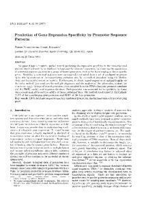
FIG. 1. Diagram showing strategy for sequence determination of the EHV-4 gB gene. Sequence data were generated with synthetic deoxyoligonucleotide primers. Dots represent the 5' end and arrowheads show the 3' end of each portion of the sequence. The EHV-4 gB ORF is denoted by a thick bar.
yeast extract, and 10 g of NaCl per liter). Bluescript M13+ recombinant DNA was propagated in JM101 cells, and all other recombinant plasmids were propagated in JM83 cells. Cells containing recombinant plasmids were selected on L broth agar plates (1.5% agar in L broth) containing ampicillin at 100 ,ug/ml or tetracycline at 12.5 ,ug/ml. Plasmid DNA isolation. Plasmid DNA was isolated from bacteria by the boiling method (5) for small-scale preparations. For large-scale isolations, plasmid DNA was extracted from bacteria by the alkaline lysis method (26) and further purified by banding on cesium chloride gradients containing 50% (wt/vol) CsCl and 200 ,ug of ethidium bromide per ml. Ethidium bromide was removed by multiple extractions with isopropanol, and the DNA was dialyzed against 0.1 x TE (1 mM Tris hydrochloride, 0.1 mM EDTA [pH 8.0]), followed by ethanol precipitation. Dot-blot analysis. The C, F, and M viral restriction fragments cloned in the BamHI site of pUC9 were excised with BamHI, purified through low-melting-point agarose gels, and spotted onto Gene-Screen hybridization membranes in 100-, 200-, and 400-ng amounts. In a later experiment, the four subfragments of BamHI-C obtained by digestion with SmaI were spotted onto membranes as above. Samples (200 ng) of vector DNAs pUC8 and pBR322 were used as negative hybridization controls and 200 ng of the appropriate probe DNA was used as a positive hybridization control in all experiments. Membranes were prehybridized in 10 ml of hybridization buffer (40% formamide, 5x SSC [lx SSC is 0.15 M NaCl plus 0.015 M sodium citrate], 5x Denhardt solution [0.1% (wt/vol) Ficoll, 0.1% (wt/vol) polyvinylpyrrolidone, 0.1% (wt/vol) bovine serum albumin], 10% dextran sulfate, 50 mM sodium PPi [pH 6.5], 100 p.g of denatured salmon sperm DNA per ml) at 42°C for 16 to 20 h. HSV-1 gB 5' and 3' probe DNA was prepared by nick translation (33) to a specific activity of 108 cpm/l,g of DNA and was added at a concentration of 106 cpm/ml of hybridization buffer (10 ng/ml) to the prehybridized membranes. Hybridization was carried out at 42°C for 16 to 20 h. Membranes were prewashed in 2x SSC for 15 min at room temperature and then washed in 2 x SSC-0. 1% sodium dodecyl sulfate (SDS), lx SSC-0.1% SDS, or O.lx SSC0.1% SDS for three 20-min washes at 65°C. Finally, membranes were rinsed in O.1x SSC, air dried, and exposed to Fuji-RX X-ray film at -70°C for 24 h. Southern blot analysis. The BamHI-C viral restriction fragment contained in pUC9 was digested with BamHI and then double digested with BamHI and each of the restriction enzymes BglI, EcoRI, PstI, PvuII, and SmaI to release viral DNA sequences from vector sequences. Digested DNA was run on 0.8% agarose gels in 1 x TEA buffer (50x TEA is 2 M Tris base, 0.1 M EDTA, 1 M sodium chloride, 1 M sodium acetate, pH 8.1), denatured, neutralized, and transferred to Gene-Screen hybridization membranes (39). Membranes
were hybridized to HSV-1 gB 5' and 3' DNA probes as outlined above. DNA sequencing. The 2.95-kb BamHI-EcoRI EHV-4 DNA fragment contained in plasmid pBSgB was sequenced by the dideoxy chain termination method (37) with [a-35S]dATP as a label (4). Extensive use was made of synthetic deoxyoligonucleotide primers to rapidly generate sequencing data, in addition to Bluescript M13+ specific sequencing primers, and the sequencing strategy is shown in Fig. 1. Since the BamHI-M viral restriction fragment contains the start of the gB gene, the right-hand end of this fragment was sequenced in pUC9 with the M13 reverse primer and synthetic deoxyoligonucleotide primers.
- cc -
a
m v co
m w a.
EE
E EE E
E E
Kb
m m mm
co
23.6-
p
b
enb
C
0
oE o
CD
co3 >
a mXmcn
cn
E E E :E E E mn co m m Cn m
Kb 23.6-
9.66..64.
to
=3 >
a
3O.
9.66.6-
a
4.3-
*
v....
-
as
2. .32. *0-
TM
a
2.32.0-
0,16-
40
Probe: 650bp Nar I HSV-1 gB
0.6-
Probe: 81Obp Pst I HSV- 1 gB
FIG. 2. Southern blot analysis of EHV-4 DNA. (a) Southern blot analysis of BamHI-C DNA with the 650-base-pair (bp) NarI HSV-1 gB (5') DNA as a hybridization probe. (b) As in panel a, but with the 810-base-pair PstI HSV-1 gB (3') DNA as a hybridization probe. All procedures were done as described in the text. Hybridization was only observed when membranes were washed at low stringency (2x SSC-0.1% SDS at 650C).
VOL. 63, 1989
EQUINE HERPESVIRUS 4 GLYCOPROTEIN gB GENE
Single-stranded template for sequencing was produced by alkaline denaturation of 1 pmol of plasmid DNA in 0.2 M NaOH-0.2 mM EDTA in a volume of 20 ,u for 5 min at room temperature, followed by neutralization with 2 ,ul of 2 M ammonium acetate (pH 5.3) and ethanol precipitation. The resulting single-stranded DNA template was suspended in 8 ,ul of 0.1 x TE (pH 8.0) and mixed with 2 pmol of sequencing primer, 3 ,lI (30 ttCi) of [ox-35S]dATP, and 1.5 pul of 1Ox Klenow annealing buffer (100 mM Tris hydrochloride [pH 8.0], 50 mM MgCl2) in a total volume of 15 ,ul. The mixture was annealed for 15 min at 37°C and then used in standard sequencing reactions at 37°C. Samples were run on 6% polyacrylamide wedge-shaped gels containing 7 M urea at 60°C. Gels were bonded to glass plates by treatment of the plates with 2% dimethylchlorosilane in 1,1,1-trichloroethane, fixed in 10% acetic acid-10% methanol, and dried in an 80°C oven before being exposed to X-ray film at -70°C for 18 to 24 h. RESULTS Identification of DNA fragment containing the EHV-4 gB gene. Since the EHV-4 genome is colinear with the 'L arrangement of the HSV-1 genome (12) and the exact location and sequence of the HSV-1 gB gene was known (8, 31), we were able to predict the region of the EHV-4 genome which would probably contain the gB gene.
Hybridization of DNA fragments from this region to both 5' and 3' HSV-1 gB DNA probes in dot-blot analyses localized the gB gene to a 6.5-kb subfragment of BamHI-C (data not shown). Southern blot analysis of BamHI-C more precisely located the gene to the left terminal 2.95-kb BamHI-EcoRI subfragment of BamHI-C (Fig. 2a and b). The 5' probe hybridized to 2.95-kb BamHI-EcoRI, 0.56-kb BamHI-PvuII, 6.5-kb BamHI-SmaI, 3-kb BamHI-PstI, and 0.42-kb BamHI-BglI fragments; weak hybridization was seen to 4.2-kb BglI and 0.14-kb PvuII fragments. The 3' probe hybridized to 2.95-kb BamHI-EcoRI, 2.7-kb PvuII, 6.5-kb BamHI-SmaI, 3-kb BamHI-PstI, and 4.2-kb BglI fragments. Some hybridization of probes to vector DNA (pUC9) was observed which was probably due to cross hybridization of G+C-rich regions of pUC9 to G+C-rich regions of HSV-1 DNA. These observations strongly suggested that the gene is located predominantly within a 2.95-kb BamHI-EcoRI fragment at the left terminus of BamHI-C with a left-to-right transcription orientation (Fig. 3d and e). Since the 5' HSV-1 gB probe does not contain the first 357 nucleotides of the coding region of the HSV-1 gB gene, it was predicted that a corresponding portion of the EHV-4 gB gene and its transcriptional control domains reside within the BamHI M fragment, which is immediately to the left of BamHI-C.
a.
TRS
IRS L.191bl
Bam Hi
b.
r
b
I
11111
~d
i
a
'h
1125
S135KbI
c I 'a k n dn I h I cdqlml
kop-.q
.
Ib
~
i
n
Eco RI
BamHI ,, -.-', -7 7-9 65 IMI ,,-
C.
2-!5,,3-6, r-_r-__
Al
10
BanmHI
d. IMI
./
*
0
Eco RI
Eco _-_ RI '-_
~~~II
Sin e
Sma I
BamHI
10-3
IF
I
Sma
134
I
\BamHI 024 0-33 0-42 I
I
Cis Sst I
I
I
Bol I
056 0-70 2-95-. 3.4 3.6 486 1*7 I~~ I ~ ~~~~~~~~~~~~~~ I --I-I I gI1 Hind III Pvu Pvu Eco Pvu Eco II II It RI I RI t
0.
650bp HSV-1 Nar I 0
15'I
065
1.38
810bp HSV-I Pst 1 2-19
t
65 Sme
\\
IFI 13
I t
13'I FIG. 3. Map of the EHV-4 genome showing restriction enzyme sites and DNA fragments used in studying the glycoprotein gB gene. (a) Structure of EHV-4 genome. This consists of a unique long region (L) and a unique short region (S) bounded by inverted repeats (IRS, TRS). (b) Arrangement of BamHI and EcoRI restriction enzyme sites along the genome. (c) Location of EcoRI and SmaI restriction enzyme sites in the 13.6-kb BamHI C fragment of the genome. (d) Detailed restriction enzyme mapping of the 2.95-kb BamHI-EcoRI subfragment of BamHI-C, which contains the predominant portion of the gB gene. The first PvuII, BgIl, and SmaI restriction enzyme sites to the right of the 2.95-kb BamHI-EcoRI subfragment are indicated by arrows. (e) Arrows show the regions of the 2.95-kb BamHI-EcoRI subfragment of BamHI-C to which the 5' and 3' HSV-1 gB DNA probes hybridized. bp, Base pairs.
1126
J. VIROL.
RIGGIO ET AL. EHV-4 gB HSV- 1 gB GA- -ACT -------- AGCT --------------------- CGGTTTAT ------------------- GGTTACTGCGG- - -CTAAA----------------
1
111 11111 I1111
111I li1
11 III
GTCAACGGGCCCCTCTTTGATCACTCCACCCACAGCTTCGCCCAGCCCCCCAACACCGCGCTGTATTACAGCGTCGAGAACGTGGGGCTCCTGCCGCACCTGAAGGAGGAGCTCGCCCGG TATA
CAT
34
------------------------ GGTG- - -ATTGGTCAATTAGCGAGTTTCAAAGGTTTTATTGCTTTGAGGGTGTGACAGGTGTGACGGCCACGCAACGGCTGGCGTGGAAATATATC
121
i IIII1 1 11 111 11 III iii li1 TTCATCATGGGGGCGGGGGGCTCGGGTGCTCATTGGGCCGTCAGCGAATTTCAGAGGTTTTACTGTTTTGACGGCATTTCCGGAATAACGCCCACTCAGCGCGCCGCCTGGCGATATATT
127
mRNA init GGGGAGCTCATTCTAGCTGCCGCAGTATTCTCTTCGGTTTTCCACTGCGGAGAGGTGCGCCTTCTGCGCGCAGATCGTACATATCCAAACACCAACGGCGCACAGCGCTGCGCTAGCGGC
241
CGCGAGCTGATTATCGCCACCsCACTCTTTGCCTCGGTCTACCGGTGCGGGGAGCTCGAGTTGCGCCGCCCGGACTGCAGCCGCCCGACCTCCGAGGTCGTTACCGTTACCCGCCCGGC
247
ATTTACATAACATACGAGACGTCATG7'CCACTTGTTGCCGTGCTATTTGTGGCCCCCAACGGTGTTATTGGCGAAGAGACTGTGGTATTTACGACAGCGACGTGTTCTCGCTTCTATAC
361
GTATATCTCACGTACGACTCCGACTGTCCGCTGGTGGCCATCGTCGAGAGCGCCCCCGACGGCTGTATCGGCCCCCGGTCGGTCGTGGTCTACGACGCCGACGTTTTCTCGATCCTCTAC
367
ACCGTACTCCAGCAGCT-GGCTCCTGGCT- -CT - --GGAGCCA- -ATTAGGAAATGTAAACTTGCCA-GCTAC - - -CTCCCCCATGT ------- CTMAGACTCGAC- -AT ----- CT
481
TCGGTCCTCCAGCACCTCGCCCCCAGGCTACCTGACGGGGGGCACGACGGGCCCCCGTAGTCCCGCCATGCACCAGGGCGCCCCCTCGTGGGGGCGCCGGTGGTTCGTCGTATGGGCGCT
i*iit
111111111
1 111111 11111 11111111 11 11111
I111111
111 1 111I 11
11 I I 11 111111111 111111 1ii I1111miIIII
11111 II
1111
start
i IIIIii 111111 1111111 H1il I iii I 11 I1 MI III III
I 1111111111111 II 111111 11
I
11
1 1 1111
1 11111 11
III11111 1
111 11
1111 1111111 1111111111111 11111
start
459
C-TGGGGGTGA------ GAACAATAGT- -CAT-TGCGTGTTTG-GTTCTCTTGGGA- - - -TGTTGTATTGTG ------------- GAA-GCTGTACCAACCACGCCAAGTTCTCAGCCCA
601
CTTGGGGTTGACGCTGGGGGTCCTGGTGGCGTCGGCGGCTCCGAGTTCCCCCGGCACGCCTGGGGTCGCGCGCGACCCAGGCGGCGAACGGGGGCCCTGCCACTCCGGCGCCGCCGCCCT
550
GTACTCC ------- CGCGTCAACCCAGTCCG -------- C-TAAAACCG-TTGACCAAACGCTTCTA- --- -CCAACTGAAAC-ACCAGACC- -CG-CTC-AGACTGG- --CTGTACGCGA
721
TGGCGCCGCCCCAACGGGGGACCCGAAACCGAAGAAGAACAAAAAACCGAAAACCCAACGCCACCACGCCCCGCCGGCGACAACGCGACCGTCGCCGCGGGCCACGCCACCCTGCGCGA
641
G- - - -TCCGG- -TAT- -ACTCGCAGA- - - -GGATGGAGACTTTTACACCTGCCCGCCGCCTACTGGATCCACAGTTGTACGCATTGAACCCCCACGGTCATGTCCCAAGTTTGATCTGGG
841
GCACCTGCGGGACATCAGGCGGAGAACACCGATGCAAACTTTTACGTGTGCCCACCCCCCACGGGCGCCACGGTGGTGCAGTTCGAGCAGCCGCGCCGCTGCCCGACCCGGCCCGAGGG
749
GAGGAACT TCACGGAGGGCATTGCTGTTATTTTCAAGGAAACATAGCCCCGTACATTTAGAGCAACGTCTACTAcl AGACATTGTAGTGACAAAGGT T TGGA GI ACAGCCA
961
TCAGAACTACAACGGAGGGCATCGCGGTGGTCTTCAAGGAGAACATCGCCCCGTACAAGTTCAAGGCCACCATGTACTACAAAGACGTCACCGTTTCGCAGGTGT GGTTCGGCCACCGCTA
869
CACCTCTTTATCCGATAGATACAATGACAGAGTGCCAGTTTCAGTGGAGGAGATATTCACTCTCATCGATAGCMAGGAAATGTTCTTCTAGGCAGAGTACCTCCGAGATMCATTAT
1081
CTCCCAGTTTATGGGGATCTTTGAGGACCGCGCCCCCGTCCCCTTCGAGGAGGTGATCGACAAGATCAACGCCAAGGGGGTCTGTCGGTCCACGGCCAAGTACGTGCGCAACAACCTGGA
Il
I
989
ULiA IIAU
A
I 11 I I III I I MII
11111111
1111111 'III 1111
111 1 111 111
1 11
11 1 11111
III
I1 IIII 111111
11 11
111
1111 111MI
11
A IUAIA UAIALIAAWIII
IIHIM I'IIIIIIII 11111II'11 I1111
LUAU1
I
11L. 1 IA I
1 111AtI
11.
1
111
III
1111
11 I 1111111
11111111111111III111111 'I 1111111111111111111111111111 11 I I1111111111111 IIIII IIIIIIII
11111
1 1 Hi111 111111 IIII11 11 I 11I1111
MII1llIll
111 1111
11 III IIIII,I 11
Ii*WW%Ai
11 11 11
III 1
II 11111111 111 *_
1201
11 I I III 111 11 11111 I I1111 111111111111 11 1 11111III 11 I 1111 1111I1III GACCACCGCGTTTCACCGGGACGACCACGAGACCGACATGGAGCTGAAACCGGCCAACGCCGCGACCCGCACGAGCCGGGGC -TGGCACACCACCGACTCAAiiuGTACAACCCCTCGCGGG
1107
TGGATGCCATGGAGGCACTACACATCAACCTCTGTCAACTGCATTGTCGAAGAGGTAGAAGCGCGGTCTGTT TACCCATACGACTCCTTTGCCCTATCGACCGGTGATATTGTGTACACC
1320
TGGAGGCGTTCCA-CCGGTAC- --- GGGACGACGGTAMCTGCATCGTCGAGGAGGTGGACGCGCGCTCGGTGTACCCGTACGACGAGTTTGTGCTGGCGACTGGCGACTTTGTGTACATG
1227
TCACCGTT TTACGGCCTTCGGTCAGCTGCTCAGTTAGAACACAATAGCTACGCACAGGAGCGCTTTAGACAAGTTGAGGATACCAACCAAGAGACTTGGACAGTAMTTACAGGCCG1A
1436
TCCCCGTTTTACGGCTACCGGGAGGGGTCGCACACCGAACACACCAC,GTACGCCGCCGACCGCTTCAAGCAGGTCGACGGCTTCTACGCGCGCGACCTCACCACCAAGGCCCGGGCCACG
1347
GAGCCAGTTACCAAACTTTATTACTACACCTCATGTTACAG-TCAGCCTGGACTGGACTGAAAAAAGATAG- AGGCGTGTACACTAACTAATGGAAGGAGGTTGACGAACTTGTCAG
1556
GCGCCGACCACCCGGAACCTGCTCACGACCCCCAAGTTCACCGTGGCCTGGGACTGG-GCGCCCMGCGCCCGTCGGTCTGCACCATGACCAAGTGGCAGGAAGTGGACGAGATGCTGCG
1466
AGATGAGT TTCGGGGGTCCTACAGGTT TACTATTCGA- TCCATTTCGTCCACGTTTATTAGCAACACTACTCAAT TTAAGCTAGAAGATGCCCCACTCACCGACTGTGTGTCAAAAGAAG
1675
CTCCGAGTACGGCGGCTCCTTCCGATTCTC-CTCCGACGCCATATCCACCACCTTCACCACCAACCTGACCGAGTACCCGCT-----CTCGCGCGTGGAC-- -CTGGGGGACTGCA-TCG
1585
CCAAAGATGCCATAGACTCTATATACCGAAAACAGTATGAGTCTACACACGT TT TTAGTCGGGGATGTGGAATTTTACTTGGCACGTwGAGGGTTCT TAATCGCATT TAGACCGATGAT TT
1785
GCAAGGACGCCCGCGACGCCATGGACCG- - -- CA ------ TCTTCGCCC --------- GCAGGTACAACGCGAC- --GCACATCAAGGTGGGCCAGCCGCAGT --------------
1705
CTACGACTTGCCAGCTGTACCTCGAGCTTGTGAGATCTACCGCACCTATGACCTTCTGTTAAACCCCACGCCCATATACCAATCGACACGCAGGTCGCTAC
1111 11. I I I III
11 I 11 11111111 11111'11111 11 11111 11 11 11111 111111
IIII II I I 11 II IIII 11
I III
H 1 11 11 11 11 I I I I I III I IIII I I I III11
III 1111I1 11 11 11 I I 111 1111ll11111 11 III I
MI I 11 I IIII111111 1 I*1
I 111'1 11 1111
1 11111 1 11 1111 11111111I1111I11
111111111 1I
1 11111
1111 11 1111 11 11 111111111
1111
I
11 1111
I 1111
I 11 11 11 I 11 11111111ll11111111 I I 1
II
11 I I
111111
III
11 11'
11
I II
11111 111
III11
1
1 1 1 111111
1 11111
1111111 III
11
CA- -GC-A----------------AACACGCTCG-CGGAGC -.-.-.TCT GGGGCTT-TCTGATC- - -GCGTACC-AGCCCCTFIG. 4. Alignment of EHV-4 gB and HSV-1 gB (KOS) DNA sequences. The top line is the DNA sequence of EHV-4 gB, and the bottom line is the HSV-1 gB DNA sequence. Identical residues are indicated with a vertical bar, and dashes mark spaces introduced to maximize homology between the two sequences. The salient features of the sequences (CAT box, TATA box, mRNA cap site, termination codon [ter], and polyadenylation signal [polyA]) are indicated for each sequence. 1866
-- -ACTACCTGGCCA- -ATG-
Consequently, the 2.95-kb BamHI-EcoRI subfragment of BamHI-C and the right-hand end of BamHI-M were sequenced and analyzed for an open reading frame (ORF) which contains the gB gene. Analysis of DNA and amino acid sequences. (i) Nucleotide sequence of the EHV-4 gB gene showing homology to HSV-1 gB. The DNA insert of pBSgB and the right-hand end of
-
BamHI-M were sequenced as described above. An ORF of 2,925 nucleotides was found in the sequence with an ATG initiation codon at position 270 and a chain termination codon at position 3195. The predicted translation product of this ORF would be a protein of 975 amino acids, which is larger than the 903 amino acids predicted for HSV-1 gB and the 913 amino acids predicted for PRV gpII.
EQUINE HERPESVIRUS 4 GLYCOPROTEIN gB GENE
63,
1127
TATCAATACCAGAAMCTACTCCAACCCMAAGAAGCCTCCACAGAGAACAAATACTACATCGCCTACACAAACGAGCAGTGGAGGCTGCGAATAGTACAMCTCTTCCAACGTCACCGCCA
1825
I I I I IIII IIII I I I11 111111 I I 11 11 III II 11 iI Iiii 111 ---CGCCC---GGGGCCAGCG TGTACGTGCGGGAA-CACCTCCGA--GAGCAGAG---CCGCA-AGCCCCCAAACCCCA-CGCCCCCGC-
1932
-------------
111111 111
CCAACGCGTCCG---
2133
AACAACTAGAGCTAATCACAAACCGTCCTCTATTGAGTTTGCTATGCTACAGTTTGCATACGATCACATCCAATCCCACGTTAATGAGATGCTAAGTAGGAkTAGCAACTJGCGTGGTGTA 11111 1111111 III IIIIII I IIIIIIIII 11 11 1111111 1111111 111111111 I II'l TGGAGCGCATCAAGACCACCTCCTCCATCGAGTTCGCCCGGCTGCAGTTTACGTACcAACCAATAcAGCGCCATGTCAACGATATGTTGGGCCGCGTTGCCATCGCGTGGTGCG CACTACMAACAAAGAGCGGACCCTCTGGAATGAGATGGTAAAGGTTAACCCAAGCGCTATTGTTTCCGCCACTCTTGACGAGCGAGTTGCGGCAAGGGTTTTGGGAGACGTTATAG;CCA 1111111 I IIIIIIIIIIiIIII 111111II 11 1111111 ill I 1 Hi111111I1II11 I 11 111 I I III I AGCTACAGMTCACGAGCTGACCCTGTGGAACGAGGCCCGCAAGCTGAACCCCAACGCCATCGCCTCGGTCACCGTGGGCCGGCGGGTGAGCGCGCGGATGCTCGGCGACGTGATGGCCG
2185
TAACACATTGTGTAAAAAT- --AGAGGGCAATGTGTACTTACAAACTCTATGCG-TC- -CTCGGACAGCAACACGTGCTACTCCCGcCCACCTGTAACGTTTACCATTAcTAAAAATG
1945 2019
-- - --
2065
1 11111111 il 111111111 1111 11 MI I I 1111 111111 1111111 1I Ill 11111 1 rTCCACGTGCGTGCCGGTGGCCGCGGACAACGTGATCGTCCAAAACTCGATGCGCATCAGCTCGCGGCCCGGGGCCTGCTACAGCCGCCCCCTGGTCAGCTTT--cGGTAcGAA - -G CAAACAGCAGAGGGACGATAGAGGGCCAGTTGGGAGMAAAACGAGGTTTATACGGAGCGCAAGCTTATCGAGCCGTGCGCTATCAATCAAAAACGrATCTTTMGTTTrGGCAACAGTG IIl I I 11 11111111111111 1 illl I 11111 1111111111111 1 11 I 111111 1111 1 - -ACCAG- GGCCCGTTGGTCGAGGGGCAGCTGGGGGAGAACAACGAGCTGCGGCTGACGCGCGATGCGATCGAGCCGTGCACCGTGGGACACCGGCGCTACTTCACCTTCGGTGGGGGCT ATGTTTACTATGAGAACTACACGTACGTTCGCAAAGTGCCCCCGACTGAAATCGAAGTGATCAGCACCTACGTTGAACTAAACTTAACTCTTTrGMGCACCGCGCAGTTTCTACCCCTGG I 11 1 1111 111 11111 1 1 1 11111 11111111111 111111 H 1111111 i1111111 I1111111 111Ill I ACGTGTACTTCGAGGAGTACGCGTACTCCCACCAGCTGAGCCGCGCCGACATCACCACCGTCAGCACCTTCATCGACCTCAACATCACCATGCTGGAGGATCACGAGTTTGTCCCCCTGG
2253
-
2299 2367
2419 2484 2539
AGGTTTACACGCGAGCTGAGCTTGAAGACACGGGGCTATTGGATTACAGCGAGATACAGCGCCGTAACCAGCTTCACGCCCTCCGATTCTACGATATAGACAGCGTTGTCMCGTrGACA MI 1111111 llII 11. 1111 11 111M M IlIll Ill111111111111111I 1 I III I 1 II II 1 Il Ill
2604
AGGTGTACACCCGCCACGAGATCAAGGACAGCGGCCTGCTGGACTACACGGAGGTCCAGCGCCGCAACCAGCTGCACGACCTGCGCTTCGCCGACATCGACACGGTCATCCACGCCG,ALu
2659
2844
ACACTGCTGTCATTATGCAGGGMTTGCCACCJTTTTTAAAGGCCTTGGTAAGGTGGGAGAGGCAGTTGGGACGCTTGTACTTGGAGCGGCTGGCGCGGTTGTTTJCTACAGTATCGGGTA 1 111 1 11 I 11 11 Hill CCAACGCCGCCATGTTCGCGGGCCTGGGCGCGTTCTTCGAGGGGATGGGCGACCTGGGGCGCGCGGTCGGCAAGGTGGTGATGGGACTCGTGGGCGGCGTGGTATCGGCCGTGTCGGGCG TAGCCTCATTTATAAACAACCCATTTGGGGGGCTCGCAATAGGCCTGTTGGTAATTGCGGGCTTAGTGGCTGCGTrTTTTGCCTACCGGTATGTAATGCAACTGCGCAGCAACCCCATGA I l11111111 111111 111111 11111 1 11111111 I 11llII 1I11111111111 1111111111 1111 1111111111111 TGTCCTCCTTCATGTCCAACCCCTTTGGGGCGCTGGCCGTGGGTCTGTTGGTCCTGGCCGGCCTGGCGGCGGCCTTCTTCGCCTTTrCGTTACGTCATGCGGCTGCAGAGCAACCCCATGA
2899
AAGCTCTATACCCAATAACAACCAGGAGCCTTAAAAACAAAGCCAAAGCCTCATACGGCCAAAACGACGATGATGACACTAGCGACTTCGATGAAGCCAAGCTGGAGGAGGCACCGCGAA
2724 2779
I 11111111 11 M11 11111 111111 11 11
I I
11111111111
3081
111111111 11111 1 11I AGGCCCTGTACCCTCTMCCACCAAGGAGCTCAAGAACCCCACC-AACCCGGACGCGTCC- -GGGGAGGGCGAGGAGGGCGGCGACTTTGACGAGGCCMGCTAGCCGAGGCCAGGGAGA TGATCAAATATATGTCTATGGTTTCTGCCCTGGAAAACAGGAAAGCAA TGGAAGAAAACAAGGGGrTGGAC TTATTGCCAGCAACGTTTCAAAAACTCGCACTGCG;CAGCGCG 1111 lIM 11I111111 111 I Hill1I1H11111 I 11 I I1 11111 1 111111 1111 l TGATACGGTACATGGCCCTGGTGTCGGCCATGGAGCGCACGGAACACAAGGCCAAGAAGAAGGGCA- -CGAGCCGG-CTGCTCAGCGCCAAGGTCACCGACATGGTCATGCGCAAGCGcC
3139
ter poly A G- -TC-CGAAATATACCC- -GTCTTCGA-GAGACGATCCATGGAAGCGAAAAAATG-GTTTAAAAATTAAAr-ATTTGACACGTACTTGTGGGTTGAcTCATATTGCAT
2964
3019
| | 111111111 11 1111111
3198
111 1 111_ I
11
111111
11 11 1
1 1111111 1
GCAACACCAACTACACCCAAGTTCCCAACAAAGACGGTGACGCCGACGAGGACGACCTGTGACGGGGGGTTTGTTGTAAATAAAAACCACGGGTGTTAAACCGCATGCGCATCrTTTGGT ter poly A AACATCTTTJCTAGTTCCGGCTATAAGCCTATTTAAGCCTAGTAJTTTTGCCAAAAGTTTATCATCCTCTACAAGCGCACATCCTCTCAAAAGAGTTG.AAJTTTGCTGTTTATTACGCTAT
3251
11 I I ill
111
I I I 11
11'
11illill 11
1111
1111 11
111 111 M 11
3432
CCCCTTGGTCAGTGCCGATTTCCTCCCCCCCACGC-CTTCCTCCACGTCAAGGCTTTTGCATTGTAAAGCTACCCGCCTACCCGCMCTCCYCAT_GAACATACACCAAT
3489
GATTCAGACACGCCCGCTGCCACGGGGTGT- -CAAGA-CCACACCTACGC-TCGCCGGCTCACCGAGAATGGTGC-AATCGAAGAGATMACACGGCTGATcTACTGGAAATGTGcTr
l 11 1
1
1
1111111 111 1 1 111 1 1 1 11111111I1
1 1
I MI1
3551
111GAACAAACGGCGu II 11 GGGTCTTATTTGGTAlTACrttuuuiTATTTAAAGATATACAGTAAGACATCCCATGGTACCAAAGACCGGGGCGAATCAGCGGGCCCCCATCATCTGAGAGAC-
3604
CTTCTGAAAACGCTCAAAGCGAACCCGGAATTC
3670
C-GCGGGCCGTG-TCAACGTCCACGTGTGCTGCGCTGCTGGCGTTGACMGGCCCCGGCCTCCGCGTTGGATrGCTCCGGTTGGGATCC FIG. 4-Continued.
I II
111111
11 1
1
EHV-4 ICP 18.5 HSV-1 ICP 18.5
1
638 39
E L A R f
I
L
W K Y
I
II Y I
11111111 I
I II E L I
I A
T T L
I F
A
II S V
Y R
I
I
I IIR
T P T
A A
iIII C G E
L E
I L
R
I R
P
I D
C S R
I P
1N
G A 0 R
I
T S E G f
Y R
Yt I T Y E T S C P L V A V L F V A P N G V I G E E T V V I Y D S D V F
I
I IIII1I1 I
II
I
II
II
II
I II
V Y L T Y D S D C P L V A I V E S A P D G C I G P R S V V V Y D A D V F
119
S L L Y T V L Q Q L A P G S G A N(TERM)
758
S
I
I
I
C A S G I Y P P
I
R
79
I G
I
W A V S E F Q R F Y C F D GIS G I
L A A A V F S S V F H C G E V R L L R A D R T Y P N T
678
718
N V T A A IC G -D W S I S E F 0 R F Y C F E G V T G V T A T 0 R L A
G E L I
I W
R
-
I II111I A R F I G A G G S
L Y S V L Q H L A P R L P D G G H D G P P
(TERM)
FIG. 5. Alignment of the predicted carboxy-terminal amino acids of the ICP18.5 protein of EHV-4 and HSV-1 (KOS). The top line is the EHV-4 sequence, and the bottom line is the HSV-1 sequence, shown in the single-letter amino acid code. Residue 1 in the EHV-4 sequence aligns with residue 638 in the HSV-1 sequence. Identical residues are indicated with a vertical bar, and dashes have been introduced to maximize homology between the two sequences.
1128
J. VIROL.
RIGGIO ET AL. (-56 NSTCCRAICGPQRCYWRRDCGNLRQRRVLASI RTPAAGSWLWSOLGNVNLPATSP -1)
EHV-4 pB HSV- 1 9B
1
Signal sequence/ 1N S K D S T S L G V R- T I V I A C L V L - L G C C I V E A
I
II
R
I R
II
W f V V W A L L G L T L G V L V A S S T P A S T Q S A K T V D - -
-
I A
V P T T p.
A P S S P G T
-
I
- Q T L L P T E T P DP L
34
SSQaP
41
A R D P G G E R G
63
R L A -
----R E --S G I L A E --D G D F Y t
PT G
81
R P A G D N A T V A A G H A T L R E H L R D I K A E N T D A N F Y V
P T G
87
S T V V R I E P P R S C P K F D L G R N F T E G I A V I
121
A T V V Q F E Q P R R C
127
N V Y Y K D I V V T K V W K G Y S H T S L S D R Y
161
T N
167 201
D S K G K C S S K A E Y L R D N I N H N A Y H D D E D E V E L D L V P S K FA T I IIIi I I II I I N A K G V C R S T A K Y V R N N L E T T A F H R D u n E T D N E L K r A N AA T
207
P G A R A UW
241
R T S R G W H T T
247
V Y P Y D S F A L S T G D
280
V Y P Y D E F V
287
Q V E G Y Q P R D L D S K L a A G E P V T K N F I
T T P H V T V S W N W T EK K
T T K A R A T A P T T R N L L
T T P K f T V A W D W V PK R
320
-
-
-
-
I I I I Y Y K D
V T
D G F Y A
K
P E G Q
I N
Y
I T
E
I
W F G H R Y S Q F N G
I G
I
F K E N I A P Y K FR A P Y K FK A
I
E N D R V P V P V E
F
I
f TL I
E V I DK I
T T -N D T T S Y V G W N P W R H Y T S T S V N C I V E E V E AR S
II
I II I I
II Q V
s a
I v
r
II
I
II
I
1 V
I
I
S G A A A L G A A P T G D P K P K K N K K P K N P
V
I
III
I
I
I
1 L
I
II I
I
I
I
V D A R S -D L K Y N P S R V E A F H R Y G T T V N C I V E E PGT
IT
II
I V Y T S P F Y G L R S A A Q L E N N S Y
I
1I 1I1I1 AI H T T Y
A T G D F V Y N S P F Y G Y R E G S H T E
II I R D L
II I
E RF R
A
A D R F K
I
I
360
I E A C T L T K U K E V D E L V R D E F R G S Y R f T I R S I S S T F I S NT T TTPKFQYEST II HAU I I I III II P S V C T M T K W Q E V D E M L R S E Y G G S F R F S S D A I S T T F T T NL T
367
Q F K L E D A P L T D C V S K E A K D A I D S I Y
400
E Y P L S R V D L G D C I G K D A R D A M D R
407
F Y L A R G G F L
440
Y Y L
447
L N P N A N H N T N R T R R S L L S I P E P T P T Q E S L H R E aI L N R L H K
469
-
487
R A V E A A N S T N S S NI V T A K Q L E L
327
484
I
I I IA
G
-
-
-
I I
N
-
-
Y 0 P L L S N T L A E L Y-
-R- E 0 S
I
F S G DV E
II
I
F A R R Y N A T H I
I A f R P M I S N E L A R L Y L N E L V R S N R T Y D L KN L
I I I I I I G G F L I A
-
I
S A N A
-
R K
r
r
N
-
E H L
R
P T P-
---
I K T T SS II
I E F I I
I
-V -----E R I K T T S S I E F
A
-
-
rP
rP
rP
M L a f A Y D H I
I II I RLQ0F
A
-
T
I I YNNH I
FIG. 6. Alignment of the predicted amino acid sequences of the gB proteins of EHV-4 and HSV-1 (KOS). The top line is the EHV-4 and the bottom line is the HSV-1 sequence, shown in the single-letter amino acid code. Identical residues are indicated with a vertical bar, and dashes show spaces introduced to maximize homology between the two sequences. The predicted signal peptidase cleavage site is indicated for both proteins, as are the hydrophilic surface, hydrophobic transmembrane, and cytoplasmic domains of the proteins. Potential N-linked glycosylation sites are underlined. The additional 56 amino acids that appear to form the start of an uncharacteristically long 84-amino-acid signal sequence for EHV-4 gB, as discussed in the text, are indicated in parentheses above the alignment (residues -56 to -1). sequence,
The aligned DNA sequences of EHV-4 gB and HSV-1 gB shown in Fig. 4. The two genes are well conserved and show DNA homology of 52%. The main features of the transcriptional and translational
are
control signals of the EHV-4 gB gene are as follows. (a) A TATA box, AATATAT, is found at nucleotides 119 to 125. This aligns well with the HSV-1 gB TATA box (ATATATT) at nucleotides 234 to 240 in the HSV-1 gB sequence. The first
VOL. 1989 VOL. 1989 63,63, ~~~~EQUINE HERPESVIRUS 4 GLYCOPROTEIN gB GENE 527
S N V NE NL SRRIA TA WC TLON K
ERkTL W NE NV KV NP SA IV SA
513
II II I II II I I11 II II II I I III III Q R NVND NLGCR V A IA WC ELQNHNEI L TLWN E A RK L NPN AI
567
TL D ER VA A RVL G DVIARIT N CV -K IE G NV YL0N SN R
553
TVG R RV SA R NL G DVN A V STCV PVA A DN V IV Q NSNR I S SRP
605
N T C Y S R P P V T F T I T K N A N S ft G T I E G 0 L G E E N E V Y T E R K L I
593
G A C Y S ft P L V S F
645
E P C A I
III I III1I1I II
I
I1 II
I
I
-
-
-
I111
I
I
IIIY Y
I ****I***I E E Y A VYS
E P C T V
685
V E LN L
670
I
725
1 N A L ft
710
L N 0 L ft
765
T L VLG A A GAVV ST VSG I A SFI N NPF
II1
N ft ft Y F T F G G G
V
F
N 0
II
1LS
ft A D
II 1
1 111 1 11
1
I
11ILL1 11 D Y
K D S G
SSD S
Rt
D A
I
I E V I
S T
Y
I
I 1
I
T T V S T
T L L E D ft E F L P L E V Y T ft A E L E D T G L L D Y S E T N L E D N E F V P 1 E V Y T ft N E
A SV
II1
ft Y E D 0 G P L V E G 0 L G EI N N E L ft L T
630
D ILN I
-
IIII 11 I11
I
N 0 K ft Y F K F G K E Y V Y Y E N Y T Y V ft K V P P T E
II III IIIII G
12 1129
I
0 R
Rt
F
N 0
1 111 1 Rt N Q
T E V 0 R
/T ransmewbrare domaI n
I IIF
II
I V
I G
G I AT F FKGILG KV GE AV G
F YD ID SV V NV DN TA V I N
I II
I
I
II
III I V V vu G
I A
GG1LA
K V
805
/Cytoplasmic doamin A FF A YR YVVNQL R S NP NK AILYP IT T R SILKN
L.
II I
S
V S G V S
I GL LVI AG LV A
I Ii I II II I IiIiII S F N S N P F G A L A V G L L V
750
N
I
AD I DT V IHA DA NA ANF A G LIGA F FE G NGDLGuR AV G
--
I II
1
L A G L A A
KA K A SYG 0N
790
I I I III I I II IIII I I I I I AF FA F RY V NRIL0 S NP NK ALVYPILT T
843
D D D D T S D
F D E A K L E E A ft E N
I K Y N S N V S A L E K 0 E K K A N K K N
827
E G E E G G D F D E A K L A E A ft E N
I ft Y N A L V S A N E ft T E H K A K K K G
883
KCGV GL IA SN V SK L AILfR-fRtRtGP K YT RILRED D P N E S E K N
867
T SRILL
I 1II II I1I1I 1I I 11I
I
I SA
I
-K V TD N V N RK
II RR
II
II
I II K ELK NP T
I II
I
I
II
I
NPD A S-Gu-
II
-
II1
V (TERN)
N TN YT0V P NK D G D A D E D D L (TERN)
FIG. 6-Continued.
transcribed base of eucaryotic mRNA is usually an A residue surrounded by pyrimidine residues (7). The sequence CAGT was observed at nucleotides 149 to 152. Since the A residue at position 150 is 25 nucleotides downstream from the TATA box, it is likely that this is the first transcribed base of EHV-4 gB mRNA. This would be in keeping with the typical 24- to 32-nucleotide spacing between the TATA box and mRNA cap site (7). Furthermore, this presumptive mRNA cap site aligns well with the HSV-1 gB mRNA cap site. (b) The sequence ATTG at nucleotides 38 to 41 reads CAAT on the other strand and is likely to function as a CAT box, since it is about 100 nucleotides upstream from the mRNA cap site (6). This CAT box is identical to, and aligns perfectly with, the HSV-1 gB CAT box. (c) The modified scanning hypothesis of translation predicts that the first AUG in a mRNA serves as the initiation codon, assuming that the local environment will allow this to occur (23). The first AUG in the mRNA occurs at nucleotides 270 to 272 and is in frame with two nearby AUGs in the sequence. Use of this first ATG as the initiation codon would give rise to an unusually long signal sequence of 84 amino acids, and similarly long signal sequences of 53 and 67 amino acids have also been reported for the gB homologs of PRV and bovine herpesvirus 1, respectively (34, 44). The second available ATG is at nucleotides 412 to 414 but is immediately followed by an in-frame chain termination codon at nucleotides 415 to 417. The next available ATG is at nucleotides 438 to 440, and the DNA sequences around
both this ATG and the ATG at nucleotides 270 to 272 suggest that either ATG could function as an efficient initiation site (22). Thus, two initiation codons can be predicted for EHV-4 gB, although we consider the first ATG as the most likely initiation codon for this gene. (d) The stop codon, TAA, occurs at nucleotides 3195 to 3197. The first polyadenylation signal, AATAAA (3), occurs 9 nucleotides past the termination codon and occurs about 20 nucleotides before the poly(A) addition site (15). A second polyadenylation signal occurs at nucleotides 3470 to 3475, suggesting that other mRNA(s) which may be present in this region would terminate at this point. (e) An ORF encoding the protein ICP18.5 in HSV-1 has been shown to terminate 10 nucleotides before the HSV-1 gB ATG initiation codon. ICP18.5 has been reported to have a role in the transport of viral glycoproteins (30). On examining the EHV-4 sequence, we located the termination codon (TAG) of an upstream ORF at nucleotides 406 to 408, occurring 136 nucleotides after the predicted EHV-4 gB ATG initiation codon. Analysis of this ORF back to nucleotide 1 revealed that this region encodes a protein showing 48% homology to HSV-1 ICP18.5 (Fig. 5). A similar protein has also been reported for PRV (34) and bovine herpesvirus 1 (44), in which the ICP18.5 protein-coding sequence actually overlaps the gB homolog-coding sequence by 44 and 47 codons, respectively, which compares with the 45-codon overlap predicted here for EHV-4. Thus, in all four viruses, there is a great constraint on this region of the genome since it probably serves as the tran-
1130
J. VIROL.
RIGGIO ET AL.
t
a
J
~1 I-
3-
of I-
m
co 0 0. 0 s
CL
0.
1
2
0
2
3
=
3
i
Residue Number Residue Number FIG. 7. Hydropathic analysis of the transmembrane region of the gB proteins of EHV-4 (residues 734 to 812) (a) and HSV-1 (KOS) (residues 720 to 799) (b), as determined by the method of Kyte and Doolittle (24). The area above the x axis denotes hydrophilicity and that below the x axis denotes hydrophobicity. The predicted three membrane-spanning segments of this domain, which are indicated by hydrophobic peaks, are numbered for both proteins. These segments are perfectly aligned between the two proteins and are highly conserved regions of the molecules (see Fig. 6 and text).
scriptional control domain of the gB gene and also encodes the carboxy-terminal amino acids of the protein ICP18.5. Further constraints are imposed on this region in EHV-4, PRV, and bovine herpesvirus 1 as it must also encode the amino-terminal amino acids of the unusually long signal sequence of the gB homolog of these viruses. (ii) Primary structure of the EHV-4 gB protein which shows homology to HSV-1 gB. The alignment of the EHV-4 and HSV-1 gB proteins is shown in Fig. 6. The EHV-4 protein is predicted to have a molecular mass of 110 kilodaltons (kDa), which is larger than the 100.3 kDa reported for HSV-1 gB (8, 31). In this alignment, 47% of the amino acids are perfectly matched between the two proteins. The codon usage and amino acid composition of the EHV-4 gB protein are shown in Table 1. The G+C content of nucleotides 1, 2, and 3 of the codons is 51, 41, and 50%, respectively. Unlike HSV-1 gB, there is no strong preference for a G or C in the third base of the codons in EHV-4 gB (8), which is a reflection of the lower G+C content of the coding region of EHV-4 gB (47%), as opposed to 66% for the HSV-1 gB-coding region. The EHV-4 gB protein contains features characteristic of all envelope glycoproteins, namely, a 5' hydrophobic signal sequence, an external hydrophilic surface domain, a hydrophobic membrane-spanning domain, and a basic, highly charged cytoplasmic anchor domain, as predicted by the hydropathic analysis of Kyte and Doolittle (24) and the secondary structure analyses of Chou and Fasman (10). (a) Signal sequence domain. Features characteristic of membrane insertion (signal) sequences are a hydrophobic core preceded by positively charged residues (32), immediately followed by a signal peptidase cleavage site near a predicted beta turn (42). A hydrophobic sequence of amino acids was found from Ile-13 to Val-26 in the EHV-4 gB protein which we believe would be of sufficient length to serve as a signal sequence hydrophobic core which spans the membrane but not to serve as an anchor sequence (32). Furthermore, the protein contains a Leu residue at core position 2, a feature which is conserved in many eucaryotic
and procaryotic signal sequences (32), although the characteristic Val-Val pair at core positions 7 and 8 is replaced by Ile-Val. The hydrophobic core sequence is immediately preceded by 12 amino acids, of which 2 are positively charged, and by a further 56 amino acids which are predominantly hydrophilic (residues -56 to -1, Fig. 6). A similar hydrophilic region has also been reported for the gB homolog of PRV (34) and bovine herpesvirus 1 (44), which has led Robbins et al. (34) to speculate that these unusually long leader sequences are a result of constraints imposed on this region of the viral genome to encode both an upstream protein and a functional signal sequence. Cleavage of the EHV-4 gB signal sequence is predicted to TABLE 1. Codon usage and predicted amino acid composition of EHV-4 gB (110 kDa) 2nd
1st(%)
A
C
G
T
(32%)
(25%)
(17%)
(26%)
3rd
A (30)
36 Lys 36 Asn 16 Lys 13 Asn
17 Thr 14 Arg 23 Thr 18 Ser 14 Thr 9 Arg 22 Thr 6 Ser
21 9 18 22
Ile Ile Met Ile
A C G T
C (20)
16 Gln 12 His 13 Gln 5 His
19 11 10 5
13 Arg 16 Arg 7 Arg 6 Arg
19 14 12 16
Leu Leu Leu Leu
A C G T
13 Val
A C G
G (31) 31 Glu 30 Asp 38 Glu 17 Asp T (19)
1 Stop 33 Tyr
O Stop 10 Tyr
Pro Pro Pro Pro
19 Ala 16 Gly 12 Gly 12 Gly 6 Gly
20 Ala 11 Ala 21 Ala 11 17 6 21
Ser Ser Ser Ser
0 Stop
S Cys 12 Trp 13 Cys
10 Val 17 Val 33 Val 10 Leu 6 Phe 10 Leu 27 Phe
T A
C G T
Residue
No.
Ala Arg Asn Asp Cys Gln Glu Gly His Ile Leu
71 65 49 47 18 29 69 46 17 52 81
Lys
52 18
Met Phe Pro Ser Thr Trp Tyr Val
33 45 79 76 12 43 73
VOL. 63, 1989
EQUINE HERPESVIRUS 4 GLYCOPROTEIN gB GENE
occur after Ala-28, and this cleavage site is immediately preceded by a beta turn. That this is a probable cleavage site is also supported by the observations that a helix-breaking residue (Gly or Pro) or a large polar residue (Glu) usually occurs four to eight residues before the cleavage site (43); in the EHV-4 gB protein, a helix-breaking Gly residue occurs at position 22, six residues prior to the cleavage site, and a polar residue (Glu) is found at position 27. Most signal sequences contain either an Ala or Gly at their carboxy terminus (43). The EHV-4 gB signal sequence carries an Ala at its carboxy terminus, and the cleavage site is perfectly aligned with that of HSV-1 gB, suggesting that the cleavage site predicted for EHV-4 gB is likely to be accurate (Fig. 6). (b) Hydrophobic transmembrane domain. The hydropathic profile of the EHV-4 gB protein suggests the presence of a transmembrane domain (amino acids 741 to 809) containing three antiparallel segments connected to each other by a very short turn region which traverse the membrane (Fig. 7), as has previously been predicted for HSV-1 gB (31). These membrane-traversing segments are thought to assume a helical conformation (14). The Chou and Fasman (10) analysis of these segments suggests that the first segment (Asn-741 to Lys-754) has the potential to adopt both alpha-helical and beta-sheet conformations, as can the second segment (Leu-766 to Asn-786), while the third segment (Phe-789 to Tyr-809) has an alphahelical structure. This transmembrane region is highly conserved between EHV-4 gB and HSV-1 gB, with 39 of the 69 amino acids composing this domain being conserved. The first segment of this domain is slightly shorter in the EHV-4 protein (14 amino acids) than in the HSV-1 protein (21 amino acids), while the second and third segments are predicted to be 21 amino acids long in both proteins. A short turn followed by a random coil is predicted to occur between segments 1 and 2 of the EHV-4 protein, although this random coiling does not appear to occur in the corresponding region of the HSV-1 protein. However, the triple traverse of the membrane by gB in this way has yet to be confirmed experimentally and is predicted solely from analysis of sequence data obtained for both HSV-1 and EHV-4 gB proteins. (c) Hydrophilic surface domain. The hydrophilic surface domain of EHV-4 gB is predicted to extend from Val-29 to Asp-740. This domain is thought to reside on the outer surface of the viral envelope and to contain the antigenic determinants against which virus-neutralizing antibody is directed (20, 31). This surface domain contains 11 potential N-linked glycosylation sites, Asn-X-Ser, in which X is not proline (35). Four of the six potential N-linked glycosylation sites in HSV-1 gB are perfectly conserved with EHV-4 gB, and all 10 cysteine residues outside the signal sequence are also conserved, suggesting that the proteins possess similar secondary and tertiary structures. As predicted for HSV-1 gB, most of the glycosylation sites are located at the junction of alpha-helical and beta-sheet domains, suggesting that these sites are exposed to the outer surface of the molecule (31). Eighteen major alpha-helical domains, nine major betasheet domains, and five major turns containing at least seven amino acids are predicted for the amino acid sequence of EHV-4 gB. (d) Cytoplasmic domain. The remaining 110 amino acids of EHV-4 gB constitute the cytoplasmic domain, which is predicted to be hydrophilic and to adopt an alpha-helical conformation. This carboxy-terminal segment is thought to function as a cytoplasmic anchor domain.
1131
This domain shares a primary amino acid homology of 49% with the cytoplasmic domain of HSV-1 gB, with 54 of the 110 amino acids constituting this domain being conserved. Of the 110 amino acids in the cytoplasmic domain of EHV-4 gB, 24 are positively charged and 17 are negatively charged, giving an overall positive charge (the HSV-1 gB cytoplasmic domain is also positively charged and contains 109 amino acids). DISCUSSION We identified the glycoprotein gB gene within a genomic library of EHV-4 using both 5' and 3' HSV-1 gB DNA probes in dot-blot and Southern blot analyses. The gene was localized to a 2.95-kb BamHI-EcoRI subfragment of the BamHI C viral restriction fragment and the right-hand end of the adjacent BamHI M fragment. We sequenced this region and identified an ORF of 2,925 nucleotides, the primary translation product of which would be a 975-amino-acid protein with a molecular mass of 110 kDa, slightly larger than the 903-amino-acid (100.3-kDa) protein predicted for HSV-1 gB. The genes for these proteins show a DNA homology of 52%, and the transcriptional control signals of the two genes are highly conserved. This transcriptional control domain also encodes the carboxy-terminal amino acids of the protein ICP18.5, with EHV-4 ICP18.5 showing 48% homology at the amino acid level to HSV-1 ICP18.5 in this region. EHV-4 gB and HSV-1 gB proteins are 47% homologous and possess highly conserved hydrophilic surface, hydrophobic transmembrane, and cytoplasmic anchor domains, while the signal sequence domains show the least homology. Two signal sequences may be predicted from the data. A sequence of 28 amino acids would be similar to the 30amino-acid sequence of HSV-1 (KOS) gB and would be in keeping with the length and structure of other glycoprotein signal sequences. However, an alternative signal sequence of 84 amino acids is likely given the precedent of an 85-amino-acid signal sequence predicted by M. Whalley for EHV-1 gB (personal communication) and signal sequences of 53 and 67 amino acids which have been predicted for the PRV and bovine herpesvirus 1 gB homologs, respectively (34, 44). Whichever sequence is adopted, the mature form of EHV-4 gB is predicted to be 891 amino acids (100.8 kDa) in size. The hydrophobic domain of both proteins is predicted to be 69 amino acids long and to contain three antiparallel segments passing through the membrane (31). From these predictions, segments 2 and 3 appear to be identical in size (21 amino acids), whereas segment 1 is thought to be somewhat shorter in EHV-4 gB (14 amino acids compared with 21 amino acids for HSV-1 gB). Furthermore, the two proteins possess similar-sized hydrophilic surface domains which contain epitopes for virus-neutralizing antibody (20, 31), and all 10 cysteine residues and four of six potential N-linked glycosylation sites in this domain are perfectly conserved. These observations suggest that the two proteins adopt similar secondary and tertiary structures. EHV-4 gB contains five more potential N-linked glycosylation sites than HSV-1 gB, and it is interesting to note the presence of three such sites immediately adjacent to each other (residues 493 to 501), with the central site being conserved with one such site in HSV-1 gB. It is possible that oligosaccharides are not added at all of these three sites, and no direct evidence exists that fully processed EHV-4 gB contains 11 oligosaccharide chains.
1132
RIGGIO ET AL.
The cytoplasmic anchor domain of EHV-4 gB is composed of 110 amino acids (compared with 109 amino acids for HSV-1 gB) and serves to anchor the protein to the membrane. It has been proposed that gB is part of a multiprotein complex which determines the social behavior of infected cells and the structure of infected cell membranes (36). The cytoplasmic domain is thought to interact with virion tegument proteins and with other membrane proteins to affect the social behavior of infected cells (29), since syn mutations have been mapped to this domain in HSV-1 gB (9, 31) (a single amino acid substitution at position 857 [Arg to His] gives rise to a syncytial phenotype [9]). It has been predicted that a stretch of amino acids before this Arg residue may adopt both a hydrophobic and hydrophilic character and that this region interacts with other proteins (29). The gB counterpart in VZV, gpII, is known to exist as a disulfide-linked dimer (18). The mature species of VZV gpII has further been suggested to exist as a disulfide-linked
heterodimer generated by an in vivo proteolytic cleavage between Arg-431 and Ser-432 of gpII (21). We observed that the residues Asn-426-Thr-427-Arg-428-Ser-429-Arg-430Arg-431-Ser-432 immediately before and including this cleavage site are aligned with the residues Asn-454-Thr455-Asn-456-Arg-457-Thr-458-Arg-459-Arg-460-Ser-461 of EHV-4 gB. Six of these residues are perfectly matched, including the cleavage site. It would therefore be interesting to speculate whether EHV-4 gB would also exist as a disulfide-linked dimer as a result of proteolytic cleavage between Arg-460 and Ser-461. This would bisect the molecule, as is the case with VZV gpII proteolytic cleavage. We compared the predicted amino acid sequence of EHV4 gB with the VZV gpII, PRV gpII, Epstein-Barr virus gB, and human cytomegalovirus gB amino acid sequences and report homologies of 51, 54, 30, and 29%, respectively. The gB glycoproteins of the alphaherpesviruses show a similar degree of homology to each other (47 to 54%) and significantly less homology to the gB protein of the betaherpesvirus human cytomegalovirus (29%) and the gammaherpesvirus Epstein-Barr virus (30%). Furthermore, Epstein-Barr virus gB and human cytomegalovirus gB are only 30% homologous, suggesting that each family of herpesviruses has separate ancestral origins. We also report that the EHV-4 gB glycoprotein shows 88% homology to the gB glycoprotein of EHV-1 (M. Whal-
ley, personal communication).
It would now be of interest to produce EHV-4 gB protein in a suitable E. coli expression vector system and to assess the efficacy of the purified protein as a subunit vaccine. The prediction of the structure of potentially immunogenic viral glycoproteins by DNA sequence analysis and the subsequent expression, purification, and testing of such proteins as potential vaccines against the virus will, it is hoped, lead us to a better understanding of viral infection. ACKNOWLEDGMENTS We thank Alan May for skilled photographic work and Maisie Riddell for typing the manuscript. We are grateful to the Horserace Betting Levy Board for financial assistance during the course of this work. LITERATURE CITED 1. Allen, G. P., and J. T. Bryans. 1986. Molecular epizootiology, pathogenesis and prophylaxis of equine herpesvirus-1 infections, p. 78-144. In R. Pandey (ed.), Progress in veterinary microbiology and immunology, vol. 2. S. Karger, Basel.
J. VIROL. 2. Allen, G. P., and M. R. Yeargan. 1987. Use of Agtll and monoclonal antibodies to map the genes for the six major glycoproteins of equine herpesvirus 1. J. Virol. 61:2454-2461. 3. Berget, S. M. 1984. Are U4 small nuclear ribonucleoproteins involved in polyadenylation? Nature (London) 309:179-182. 4. Biggin, M. D., T. J. Gibson, and C. F. Hong. 1983. Buffer gradient gels and 35S label as an aid to rapid DNA sequence determination. Proc. Natl. Acad. Sci. USA 80:3963-3965. 5. Birnboim, H. C., and J. Doly. 1979. A rapid alkaline extraction procedure for screening recombinant plasmid DNA. Nucleic Acids Res. 7:1513-1523. 6. Breathnach, R., and P. Chambon. 1981. Organisation and expression of eucaryotic split genes coding for proteins. Annu. Rev. Biochem. 50:349-383. 7. Busslinger, M., R. Portman, J. Irminger, and M. Birnsteil. 1980. Ubiquitous and gene-specific regulatory 5' sequences in a sea urchin histone DNA clone coding for histone protein variants. Nucleic Acids Res. 8:957-978. 8. Bzik, D. J., B. A. Fox, N. A. DeLuca, and S. Person. 1984. Nucleotide sequence specifying the glycoprotein gene, gB, of herpes simplex virus type 1. Virology 133:301-314. 9. Bzik, D. J., B. A. Fox, N. A. DeLuca, and S. Person. 1984. Nucleotide sequence of a region of the herpes simplex virus type 1 gB glycoprotein gene: mutations affecting rate of virus entry and cell fusion. Virology 137:185-190. 10. Chou, P. Y., and G. D. Fasman. 1978. Prediction of the secondary structure of proteins from their amino acid sequence. Adv. Protein Chem. 47:45-148. 11. Cranage, M. P., T. Kouzarides, A. T. Bankier, S. Satchwell, K. Weston, P. Tomlinson, B. Barrell, H. Hart, S. E. Bell, A. C. Minson, and G. L. Smith. 1986. Identification of the human cytomegalovirus glycoprotein B gene and induction of neutralizing antibodies via its expression in recombinant vaccinia virus. EMBO J. 5:3057-3063. 12. Cullinane, A. A., F. J. Rixon, and A. J. Davison. 1988. Characterization of the genome of equine herpesvirus 1 subtype 2. J. Gen. Virol. 69:1575-1590. 13. DeLuca, N., D. Bzik, V. C. Bond, S. Person, and W. Snipes. 1982. Nucleotide sequences of herpes simplex virus type 1 (HSV-1) affecting virus entry, cell fusion, and production of glycoprotein gB (VP7). Virology 122:411-423. 14. Engelman, D. M., and T. A. Steitz. 1981. The spontaneous insertion of proteins into and across membranes: the helical hairpin hypothesis. Cell 23:411-422. 15. Fitzgerald, M., and T. Shenk. 1981. The sequence 5'-AAUAAA3' forms part of the recognition site for polyadenylation of late SV40 mRNAs. Cell 24:251-260. 16. Frink, R. J., R. Eisenberg, G. Cohen, and E. K. Wagner. 1983. Detailed analysis of the portion of the herpes simplex virus type 1 genome encoding glycoprotein C. J. Virol. 45:634 647. 17. Glorioso, J., C. H. Schroder, G. Kurnel, M. Szczesiul, and M. Levine. 1984. Immunogenicity of herpes simplex virus glycoproteins gC and gB and their role in protective immunity. J. Virol. 50:805-812. 18. Grose, C., D. P. Edwards, K. A. Weigle, W. E. Friedrichs, and W. L. McGuire. 1984. Varicella-zoster virus specific gpl40: a highly immunogenic and disulfide-linked structural glycoprotein. Virology 132:138-146. 19. Holland, L. E., R. M. Sandri-Goldin, A. L. Goldin, J. C. Glorioso, and M. Levine. 1984. Transcriptional and genetic analyses of the herpes simplex virus type 1 genome: coordinates 0.29 to 0.45. J. Virol. 49:947-959. 20. Hopp, T. P., and K. R. Woods. 1981. Prediction of protein antigenic determinants from amino acid sequences. Proc. Natl. Acad. Sci. USA 78:3824-3828. 21. Keller, P. M., A. J. Davison, R. S. Lowe, C. D. Bennett, and R. W. Ellis. 1986. Identification and structure of the gene encoding gpII, a major glycoprotein of varicella-zoster virus. Virology 152:181-191. 22. Kozak, M. 1984. Comparison of initiation of protein synthesis in procaryotes, eucaryotes, and organelles. Microbiol. Rev. 47: 1-45. 23. Kozak, M. 1984. Point mutations close to the AUG initiator
VOL. 63, 1989
24. 25.
26. 27.
28. 29.
EQUINE HERPESVIRUS 4 GLYCOPROTEIN gB GENE
codon affect the efficiency of translation of rat preproinsulin in vivo. Nature (London) 308:241-246. Kyte, J., and R. F. Doolittle. 1982. A simple method for displaying the hydropathic character of a protein. J. Mol. Biol. 157:105-132. Little, S. P., J. T. Jofre, R. J. Courtney, and P. A. Schaffer. 1981. A virion-associated glycoprotein essential for infectivity of herpes simplex virus type 1. Virology 115:149-160. Maniatis, T., E. F. Fritsch, and J. Sambrook. 1982. Molecular cloning, a laboratory manual. Cold Spring Harbor Laboratory, Cold Spring Harbor, N.Y. Manservigi, R., P. G. Spear, and A. Buchan. 1977. Cell fusion induced by herpes simplex virus is promoted and suppressed by different viral glycoproteins. Proc. Natl. Acad. Sci. USA 74: 3913-3917. O'Callaghan, D. J., G. A. Gentry, and C. C. Randall. 1983. The equine herpesviruses, p. 215-318. In B. Roizman (ed.), The herpesviruses, vol. 2. Plenum Publishing Corp., New York. Pellett, P. E., M. D. Biggin, B. Barrell, and B. Roizman. 1985. Epstein-Barr virus genome may encode a protein showing significant amino acid and predicted secondary structure homology with glycoprotein B of herpes simplex virus 1. J. Virol.
56:807-813. 30. Pellett, P. E., F. J. Jenkins, M. Ackermann, M. Sarmiento, and B. Roizman. 1986. Transcription initiation sites and nucleotide sequence of a herpes simplex virus 1 gene conserved in EpsteinBarr virus genome and reported to affect the transport of viral glycoproteins. J. Virol. 60:1134-1140. 31. Pellett, P. E., K. G. Kousoulas, L. Pereira, and B. Roizman. 1985. Anatomy of the herpes simplex virus 1 strain F glycoprotein B gene: primary sequence and predicted protein structure of the wild type and of monoclonal antibody-resistant mutants. J. Virol. 53:243-253. 32. Perlman, D., and H. 0. Halvorson. 1983. A putative signal peptidase recognition site and sequence in eukaryotic and prokaryotic signal peptides. J. Mol. Biol. 167:391-409. 33. Rigby, P. W. J., M. Dieckmann, C. Rhodes, and P. Berg. 1977.
34.
35. 36.
37.
38.
39. 40. 41.
42. 43. 44.
1133
Labelling deoxyribonucleic acid to high specific activity in vitro by nick-translation with DNA polymerase I. J. Mol. Biol. 113:237-251. Robbins, A. K., D. J. Dorney, M. W. Wathen, M. E. Whealy, C. Gold, R. J. Watson, L. E. Holland, S. D. Weed, M. Levine, J. C. Glorioso, and L. W. Enquist. 1987. The pseudorabies virus gll gene is closely related to the gB glycoprotein gene of herpes simplex virus. J. Virol. 61:2691-2701. Rose, J. K., R. F. Doolittle, A. Anilionis, P. J. Curtis, and W. H. Wunner. 1982. Homology between the glycoproteins of vesicular stomatitis virus and rabies virus. J. Virol. 43:361-364. Ruyechan, W. T., L. S. Morse, D. M. Knipe, and B. Roizman. 1979. Molecular genetics of herpes simplex virus. II. Mapping of the major viral glycoproteins and of the genetic loci specifying the social behavior of infected cells. J. Virol. 29:677-697. Sanger, F., S. Nicklen, and A. R. Coulson. 1977. DNA sequencing with chain-terminating inhibitors. Proc. Natl. Acad. Sci. USA 74:5463-5467. Sarmiento, M., M. Haffey, and P. G. Spear. 1979. Membrane proteins specified by herpes simplex viruses. III. Role of glycoprotein VP7 (B2) in virion infectivity. J. Virol. 29:11491158. Southern, E. M. 1975. Detection of specific sequences among DNA fragments separated by gel electrophoresis. J. Mol. Biol. 98:503-517. Spear, P. G. 1985. Glycoproteins specified by herpes simplex viruses, p. 315-356. In B. Roizman (ed.), The herpesviruses, vol. 3. Plenum Publishing Corp., New York. Vestergaard, B. F. 1980. Herpes simplex virus antigens and antibodies. A survey of studies based on quantitative immunoelectrophoresis. Rev. Infect. Dis. 2:899-913. von Heqne, G. 1984. How signal sequences maintain sequence specificity. J. Mol. Biol. 173:243-351. Watson, M. E. E. 1984. Compilation of published signal sequences. Nucleic Acids Res. 12:5144-5164. Whitbeck, J. C., L. J. Bello, and W. C. Lawrence. 1988. Comparison of the bovine herpesvirus 1 gI gene and the herpes simplex virus type 1 gB gene. J. Virol. 62:3319-3327.