Indian J. Anim. Res., 50 (3) 2016:357-365
AGRICULTURAL RESEARCH COMMUNICATION CENTRE
Print ISSN:0367-6722 / Online ISSN:0976-0555
www.arccjournals.com/www.ijaronline.in
Nucleotide sequence analysis of Mx1 gene in Japanese quail Diwesh Kumar Niraj, Pushpendra Kumar*, Chinmoy Mishra, Neeraj Kashyap, Raj Narayan1, T.K. Bhattacharya2, Bharat Bhushan, A.K. Tiwari, V.K. Saxena1, N.R. Sahoo and Deepak Sharma Animal Genetics Division, Indian Veterinary Research Institute, Izatnagar Bareilly-243 122, India. Received: 05-01-2015 Accepted: 08-07-2015
DOI:10.18805/ijar.7091
ABSTRACT During the process of increasing production potential of poultry, other economically important traits like disease resistance are neglected and ultimately leading to decrease in quality food production. The poultry health management is important due to emergence of highly pathogenic diseases like Avian Influenza. Mx1 gene has been reported to provide resistance against avian influenza virus in chicken. Therefore, present research work was focused to identify the genetic variation of Mx1 gene in Japanese quail. Total three fragments viz. Frag-I of 185 bp (Exon 3 region), Frag-II of 161 bp (Exon 7 region) and Frag-III of 176 bp (Exon 13 region) of Mx1 gene in 170 Japanese quail birds were amplified and screened for polymorphism by PCR-SSCP technique. All the three fragments were found to be monomorphic which was confirmed by dideoxy nucleotide sequencing. Comparative analysis of Mx1 sequence of Japanese quail by MegAlign programme of DNASTAR showed 100% homology with common quail and more than 80% homology with Rhode Island Red and Silki chicken breeds. Key words: Allele, Genotype, Mx1, PCR-SSCP, Quail, Virus. INTRODUCTION The poultry production is a profitable business due to its low input and high output. The increasing demand for eggs and poultry meat to meet the recommended nutritional requirement paves the way for rearing of alternate poultry species like ducks and quail which are known for their ability to produce more eggs and better meat in comparison to chicken. Japanese quail (Coturnix coturnix japonica) is a migratory bird of Japan, now domesticated throughout the world as a good source of egg and meat. In India also it has gained popularity among poultry breeders. For economic and sustainable production, good health and survival of birds along with optimum productive performance are fundamental factors. But, the new emerging diseases and some important zoonotic diseases are posing serious threats to the poultry industry, out of them avian influenza is the most important one being its widespread occurrence (de Jong and Hien, 2006). Current methods to control diseases like antibiotic treatment and vaccination are not much effective due to rapid changes in the pathogenicity, vaccination failure and problem of drug residues in poultry products. So the future research activities shall mostly focus on utilization of diversified approaches such as development of disease resistant lines, well suited to prevailing climatic conditions and having better adaptability and production capacity. The genetic markers can provide useful information to identify genetic basis of disease resistance (Orita et al., 1989). Myxovirus resistance (Mx1) gene is an interferon induced gene that inhibits single
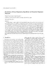
stranded negative sense RNA viruses’ proliferation including avian influenza virus (Ko et al., 2002; Sasaki et al., 2013; Wang et al., 2014). The chicken Mx1 gene consists of 21289 bp linear DNA, 14 exons and 2118 bp long coding sequence. The Mx protein of chicken (Gallus gallus) is predominantly cytoplasmic consisting of 705 amino acids (Bernasconi et al., 1995). However, study on quail Mx1 gene was known very little globally. Therefore, the present investigation was undertaken to find out the DNA polymorphism in Mx1 gene of Japanese quail using PCR-SSCP and nucleotide sequencing techniques. MATERIALS AND METHODS Isolation of DNA: The blood samples were collected from 170 quail birds maintained at Central Avian Research Institute (CARI), Izatnagar, Bareilly. The genomic DNA was isolated (Sambrook and Russell, 2001).The quality and purity were assessed by agarose gel electrophoresis and spectrophotometer. Design of PCR Primers: Three different regions (Fragment I of 185bp, Fragment II of 161 bp and fragment III of 176bp) of quail Mx1 gene were selected for amplification (Figure 1). The PCR primers were designed on the basis of available sequences of chicken and common quail (wild) using Primer3 and OligoAnalyzer softwares (Table 1). Setting up of PCR reactions: The PCR master mix, containing all the reaction components except genomic DNA, was prepared (Table 2). Care was taken to add Taq DNA
*Corresponding author’s e-mail:
[email protected]. 1CARI, Izatnagar, Bareilly- 243 122, U.P, 2-PDP, Rajendra Nagar, Hyderabad.
358
INDIAN JOURNAL OF ANIMAL RESEARCH
Table 1: Primer sequences used to amplify Mx1 gene in Japanese quail Fragments
Primer
Primer Sequence
I
Forward Reverse Forward Reverse Forward Reverse
5’- GCAGCAGAACACAGCTTTCA -3’ 5’- CTAGGAAGAGCAACACCAGAC -3’ 5’- TCCTCACTAAACCAGATCTGGTG - 3’ 5’-TTGCTGGATTACAGAGGCCAAGGA- 3’ 5’- GCAAGCAACAGCTGCGAAAA - 3’ 5’-AAACCATTTCCAGGGCAAAGCTGG -3’
II III
polymerase at the end. After all the components were added, the master mix was mixed gently, followed by spinning and dispensed (24 l) to each labeled PCR tube kept in ice. Finally, 1 l (80-100 ng) of good quality diluted genomic DNA was added to each tube. The PCR tubes were kept in a preprogrammed thermocycler (Eppendorf gradient, Germany). The PCR programme were as, initial denaturation (940C for 4min), denaturation (940C for 1 min), the annealing (610C, 59.20C and 61.20C for 1 min for fragment I, II and III, respectively), extension (720C for 1 min) and the final extension (720C for 10 min) Checking of the amplified product: Horizontal submarine agarose gel electrophoresis was carried out to check the amplified products. A 1.5% w/v agarose (Low EEO) gel was prepared in 0.5 X TBE and subsequently 5 l PCR product mixed with 1 l of 6 X gel loading dye (Bromophenol blue and xylene cyanol) was loaded along with 100 bp ladder (Gene RulerTM Plus) as a marker in a separate lane. The electrophoresis was done at 60 volt for 2 hr. The amplified product in the gel was observed under UV transilluminator and documented by photography. PCR-SSCP analysis: The PCR products were resolved on 15% polyacrylamide gel. About 6 µl of PCR product and 12 µl of denaturing formamide dye (Formamide, 95%; Xylene cyanol, 0.025%; Bromophenol blue, 0.025%; 0.5 M EDTA, 4%) were taken in a 0.2 ml PCR tube and mixed properly. The mixture of PCR product and formamide dye were denatured at 95°C for 10 min (by keeping on 95°C hot Table 2: PCR reaction components for fragment- I, II and III Reaction components dNTPs mix (10 mM) Primers Forward Primer (E3 QF) Reverse primer (E3 QR) MgCl2 (25 mM) 10 x PCR assay buffer Taq DNA polymerase (1U/ml) Genomic DNA Nuclease free water Total
Amount 1X (25 l)
Final concentration
1 l
100 M
1l 1l 2l 2.5 l 1 l 1 l 15.5 l 25 l
10 pmole 10 pmole 2mM 1X 1 unit 80-100 ng
Length (Base) 20 21 23 24 20 24
Amplification length 185 161 176
Fig 1: Amplified regions of Mx 1 gene in quail
water) and snap chilled on ice for 15 min. The product was loaded in gel carefully. The electrophoresis was performed at 4°C for 13-16 hr at 130 constant volts with subsequent silver staining for visualization of bands under gel documentation system (Bassam et al., 1991). The genotypes were identified by visualizing SSCP patterns of each sample in the gel. The gene and genotype frequencies were estimated by applying standard procedure (Falconer and Mackey, 1996). DNA sequencing: Different genotypes of amplified fragment of Mx1 gene were selected for sequencing and subjected to BLAST (www.ncbi.nlm.nih.gov/BLAST) analysis to ascertain that sequences were of Mx1 gene. Nucleotide sequences were then aligned with the corresponding common quail (NCBI Acc. No. EF575605) and different breeds of chicken viz. RIR (NCBI Acc. No. DQ788613), SILKIE (NCBI Acc. No. DQ788614) and WLH (NCBI Acc. No. DQ788615) Mx1 gene sequences using the ClustalW method of MegAlign Programme of Lasergene Software (DNASTAR). The amino acid sequences were deduced from different alleles of Mx1 gene in quail and compared with the corresponding deduced chicken amino acid sequences. The nucleotide and amino acid substitutions and diversity analyses (by DNASTAR) were reported. Percentage similarity and phylogenetic analysis were also performed to determine the evolutionary relationship. RESULTS AND DISCUSSION DNA Polymorphism of Mx1 gene: The 185 bp fragment in 3rd exon, 161 bp fragment of 7th exon and 176 bp fragment of 13 th exon of Mx1 gene of quail were amplified (Figure 2a, 2b and 2c). The SSCP analysis revealed
Volume 50 Issue 3 (2016) monomorphic pattern in all the three fragments (Figure 3a, 3b and 3c). The PCR-SSCP of all the three fragments of Mx1 gene has not been studied so far in quail or any other breeds of chicken. Therefore, present findings could not be compared.
359
Nucleotide sequence analysis: Different amplified fragments of Mx1 gene of Japanese quail were sequenced by Sanger’s dideoxy chain termination sequencing method
Fig 3a: PCR-SSCP genotypes of 185 bp fragment of Mx1 gene Fig 2a: Specific 185 bp amplified PCR product of Mx1 gene in Japanese quail
Fig 3b: PCR-SSCP genotypes of 161 bp fragment of Mx1 gene Fig 2b: Specific 161 bp amplified PCR product of Mx1 gene in quail
Fig 2c: Specific 176 bp amplified PCR product of Mx1 gene in Japanese quail
Fig 3c: PCR-SSCP genotypes of 176 bp fragment of Mx1 gene
360
INDIAN JOURNAL OF ANIMAL RESEARCH
(Sanger et al., 1997) and were submitted to NCBI GenBank data with Acc. Nos. KC571220, KC571225 and KC571226 for 185,161 and 176 alleles, respectively. All the three sequences and the corresponding reported sequences of different breeds of chicken viz. RIR (NCBI Acc. No. DQ788613), SILKIE (NCBI Acc. No. DQ788614) and WLH (NCBI Acc. No. DQ788615) were aligned using MegAlign programme of DNASTAR software (Figure 4, 5 and 6). The nucleotide substitutions were observed at 21 different positions in fragment I, 27 different positions in fragment II and 58 different positions in fragment III of quail Mx1 gene in comparison to chicken Mx1 gene. However, when sequences of Japanese quail and common quail were compared, nucleotide variation was observed at 180 th position (Cytosine was present in Japanese quail and
Thymine was present in common quail) in fragment III. The fragment I and II showed 100% similarity in Japanese quail and common quail. Diversity analysis among alleles: Percentage similarity/ dissimilarity was done for three fragments of Japanese quail Mx1 gene and common quail along with other breeds of chicken (RIR, Silki and WLH). The 100% similarity was observed between Japanese quail and common quail for fragment I and fragment II of Mx1 gene (Figure 7a and 7b). However, 99.4% similarity was observed for fragment III between Japanese quail and common quail (Figure 7c). In contrast to this Japanese quail was more distinct from RIR, Silki and WLH breed of chicken (Range 83.2% to 89.2%).
Fig 4: Nucleotide sequence alignment of 185 bp fragment of Mx 1 gene of Japanese quail with Common quail and different breeds of chicken
Volume 50 Issue 3 (2016)
361
Fig 5: Nucleotide sequence alignment of 161 bp fragment of Mx 1 gene of Japanese quail with Common quail and different breeds of chicken
Phylogenetic analysis : The phylogenetic analysis showed that Japanese quail and common quail formed single cluster while all chicken breeds remain in another cluster for all the three fragments (Figure 8a, 8b and 8c). Comparison of amino acid sequences: The deduced amino acid sequences of 185, 161 and 176 bp fragments of Mx1 gene quail was compared with those of the published sequences of chicken breeds viz. RIR, Silki and WLH (Figure 9a, 9b and 9c). Total forty six (Twelve in fragment I, fourteen in fragment II and twenty in fragment III) amino acid substitutions were observed in quail Mx1
gene as compared to chicken (Table 3). The highest similarity of amino acid composition was observed between Japanese quail and common quail and more dissimilarity was observed between quail and different breeds of chicken. However, when amino acid sequences of Japanese quail and common quail were compared, amino acid variation was observed at 60 th position (Alanine was present in Japanese quail and Valine was present in common quail) in fragment III. The fragment I and II showed 100% identity in Japanese quail and common quail.
362
INDIAN JOURNAL OF ANIMAL RESEARCH
Fig 6: Nucleotide sequence alignment of 176 bp fragment of Mx 1 gene of Japanese quail with Common quail and different breeds of chicken
Table 3: Amino acid substitutions at different positions of three fragments of Japanese quail Mx1 gene Fragment
I II III
Positions
3 8 4
4 9 10
6 10 15
7 13 18
9 17 19
12 22 19 24 24 27
26 36 28
32 39 32
37 41 34
39 42 37
44 47 39
48 46
50 48
49
50
51
54
56
60
Volume 50 Issue 3 (2016)
Fig 7a: Similarity and divergence between Japanese quail, Common quail and different breeds of chicken on the basis of 185 bp Mx1 gene fragment
363
Fig 7b: Similarity and divergence between Japanese quail, Common quail and different breeds of chicken on the basis of 161 bp fragment of Mx 1 gene
Fig 7c: Similarity and divergence between Japanese quail, Common quail and different breeds of chicken on the basis of 176 bp fragment of Mx 1 gene
Fig 8a: Phylogenetic tree based on 185 bp fragment of Mx 1 gene in Japanese quail
Fig 8b: Phylogenetic tree based on 161 bp fragment of Mx 1 gene in Japanese quail
Fig 8c: Phylogenetic tree based on 176 bp fragment of Mx 1 gene in Japanese quail
364
INDIAN JOURNAL OF ANIMAL RESEARCH
Fig 9a : Amino acid sequence alignment of 185 bp fragment of Mx 1 gene in Japanese quail
Fig 9b: Amino acid sequence alignment of 161 bp fragment of Mx 1 gene in Japanese quail
Fig 9c: Amino acid sequence alignment of 176 bp fragment of Mx 1 gene in Japanese quail
CONCLUSION The Mx1 gene was found to be conserved in quail. The other regions of this gene need to be sequenced for the complete characterization of this gene in quail.
ACKNOWLEDGMENTS The authors are thankful to the Director, Indian Veterinary Research Institute (IVRI), Izatnagar, Bareilly, India for providing necessary facilities to carry out this work.
Volume 50 Issue 3 (2016)
365
REFERENCES Bassam, B.J., Caetano-Anolles, G. and Gresshoff, P. M. (1991). Fast and sensitive silver staining of DNA in polyacrylamide gels. Anal Biochem., 196:80-83. Bernasconi, D., Schwartz, A. and Staeheli, P. (1995). The interferon-induced Mx protein of chickens lacks antiviral activity. J. Int. and Cyt. Res., 15:47-53. de Jong, M.D. and Hien, T.T. (2006). Avian influenza A (H5N1). J. Cli. Vir., 35:2-13. Falconer, D.S. and Mackay, T.F.C. (1996). Introduction to Quantitative Genetics. 4th Ed. Addision Wesley Longmann Ltd. Essere. England, pp1-12. Ko, J.H., Jin, H.K., Asano, A., Takada, A., Ninomiya, A., Kida, H., Hokiyama, H., Ohara, M., Tsuzuki, M., Nishibori, M., Mizutani, M. and Watanabe. T. (2002). Polymorphisms and the differential antiviral activity of the chicken Mx gene. Gen. Res., 12:595-601. Mishra, C., Das, D., Kumar, P., Khanna, K., Singh, A.P., Dayal, S., Selvaramesh, Bhattacharya, T.K., Bhushan, B. and Sharma, A. (2011). Nucleotide sequencing and PCR-SSCP of Mx1 gene in chicken. Ind. J. Ani. Res., 45:276-282. Orita, M., Iwahana, H., Kanazawa, H., Hayashi, K. and Sekiya, T. (1989). Detection of polymorphisms of human DNA by gel electrophoresis as single strand conformation polymorphisms. Pro. Nat. Acad. Sci. USA, 86:2766-2770. Sasaki, K., Yoneda, A., Ninomiya, A., Kawahara, M. and Watanabe, T. (2013). Both antiviral activity and intracellular localization of chicken Mx protein depend on a polymorphism at amino acid position 631. Biochem. Biophys. Res. Commun., 430:161-166. Sambrook, J. and Russell, D.W. (2001). Molecular Cloning: A Laboratory Manual. 3rd Ed. Cold Spring Harbor Laboratory Press, New York, USA, pp 5.61-5.90. Sanger, F., Nicklen, S. and Coulson, A.R. (1997). DNA sequencing with chain terminating inhibitors. Pro. Nat. Acad. Sci. USA, USA, 74: 5463-5467. Wang, Y., Lupiani, B., Reddy, S.M., Lamont, S.J. and Zhou, H. (2014). RNA-seq analysis revealed novel genes and signaling pathway associated with disease resistance to avian influenza virus infection in chickens. Pol. Sci., 93: 485-493.