KU
LAK
BURUN
BO
ĞA
HA LARI
B EH B UT C
AL IK ST
EV AN
R
Z
Şİ
.
BO
R
N
BA
Ş
EĞ
VE
İ
.
YU
N C E R R A Hİ S
E İD
Kulak Burun Bogaz Ihtis Derg 2015;25(3):144-151
doi: 10.5606/kbbihtisas.2015.68878
144
Original Article / Çalışma - Araştırma
Retrospective analysis of surgical treatment of choanal polyps Koanal poliplerin cerrahi tedavisinin retrospektif analizi Kadri İla, MD., Murat Topdağ, MD., Murat Öztürk, MD., Mete İşeri, MD., Ömer Aydın, MD., Gürkan Keskin, MD. Department of Otolaryngology, Medical Faculty of Kocaeli University, Kocaeli, Turkey
ABSTRACT Objectives: This study aims to discuss the efficacy of the surgical method performed in patients with choanal polyp in the light of the literature. Patients and Methods: The study included 76 patients (42 males, 34 females; mean age 25.36 years; range 7 to 73 years) diagnosed with choanal polyp in the sinonasal region between January 2005 and December 2013 in the otorhinolaryngology clinic of Kocaeli University. Age, sex, and presenting complaints of the patients, as well as the characteristics of the polyps (localization, direction) were retrospectively investigated. Results: The majority (98.68%) of the patients with choanal polyp presented with nasal obstruction, followed by snoring, sleeping with the mouth open (25.0%), and nasal discharge (21.05%). According to their localizations, the origin of the polyps was the maxillary sinus in 65 patients, sphenoid sinus in six patients, middle turbinate in two patients, septum in two patients, and ethmoid sinus in one patient. Conclusion: Endoscopic approach is a safe and effective procedure for choanal polyp treatment. There was no significant difference between the success rates of the endoscopic approach and combined approaches. Keywords: Caldwell-Luc procedure; choanal polyp; endoscopic surgery; paranasal sinus.
ÖZ Amaç: Bu çalışmada, koanal polibi olan hastalarda uygulanan cerrahi yöntemin etkinliği literatür ışığında tartışıldı. Hastalar ve Yöntemler: Çalışmaya Kocaeli Üniversitesi Kulak Burun Boğaz kliniğinde Ocak 2005 - Aralık 2013 tarihleri arasında sinonazal bölgede koanal polip tanısı konulmuş 76 hasta (42 erkek, 34 kadın; ort. yaş 25.36 yıl; dağılım 7-73 yıl) dahil edildi. Hastaların yaş, cinsiyet ve kliniğe başvuru yakınmaları ile beraber poliplerin özellikleri (yerleşim yeri, yönü) retrospektif olarak araştırıldı. Bulgular: Koanal polibi olan hastaların büyük çoğunluğu (%98.68) burun tıkanıklığı ile başvurdu; bunu horlama, ağzı açık uyuma (%25.0) ve burun akıntısı (%21.05) izledi. Yerleşim yerlerine göre polipler 65 hastada maksiller sinüs, altı hastada sfenoid sinüs, iki hastada orta konka, iki hastada septum ve bir hastada etmoid sinüs kaynaklıydı. Sonuç: Endoskopik yaklaşım koanal polip tedavisinde güvenilir ve etkili bir işlemdir. Endoskopik yaklaşım ve kombine yaklaşımların başarı oranları arasında anlamlı farklılık yoktu. Anahtar Sözcükler: Caldwell-Luc işlemi; koanal polip; endoskopik cerrahi; paranazal sinüs.
Choanal polyps occur due to inflammation or edema in mucosa of paranasal sinuses or other nasal configurations. They usually originate
Available online at www.kbbihtisas.org doi: 10.5606/kbbihtisas.2015.68878 QR (Quick Response) Code
from the maxillary sinus and these are termed antrochoanal polyps.[1] The etiology remains unclear although chronic inflammation and
Received / Geliş tarihi: August 27, 2014 Accepted / Kabul tarihi: December 15, 2014
Correspondence / İletişim adresi: Kadri İla, MD. Kocaeli Üniversitesi Tıp Fakültesi Kulak Burun Boğaz Hastalıkları Anabilim Dalı, 41380 Umuttepe, Kocaeli, Turkey. Tel: +90 554 - 479 70 66 e-mail (e-posta):
[email protected]
Retrospective analysis of surgical treatment of choanal polyps
allergy are thought to play a role in the etiology of choanal polyps.[2] Recently, it has been suggested that arachidonic acid metabolism and urokinase-type plasminogen activator might play a role in the pathogenesis.[3,4] Antrochoanal polyps account for approximately 4-6% of nasal polyps in the general population. However, this ratio is about 33% in the pediatric population.[5] Treatment of choanal polyps is surgical. The present study aimed to discuss the effect of surgical method performed in the patients with choanal polyps on success rates in the light of the literature. PATIENTS AND METHODS
The present study comprised 76 patients (42 males and 34 females; mean age 25.36 years; range 7-73 years) diagnosed with choanal polyps in the sinonasal region between January 2005 and December 2013 in the Ear, Nose and Throat Department of Kocaeli University. Age, gender, and presenting complaints of the patients, as well as characteristics of the polyp (such as localization and direction), were retrospectively investigated. Nasal pathologies accompanying the choanal polyps were also recorded. Treatment modalities selected according to the characteristics of the choanal polyp and patients, postoperative complications, and the consequences of treatment were analyzed. All patients underwent paranasal sinus computed tomography (CT). Additionally, some cases underwent maxillofacial magnetic resonance imaging (MRI) for differential diagnosis. Endoscopic sinus surgery was performed for the treatment of patients with antrochoanal polyps. When the origin of the polyp in the maxillary sinus could not be observed, the operation was completed by performing additional Caldwell-Luc surgery or by using a trans-canine trocar method. Transnasal endoscopic method was used for sphenochoanal polyps. In patients that underwent surgery for septochoanal polyp, the polyp was endoscopically excised from the septum mucosa including 2 mm of intact margins. Endoscopic ethmoidectomy was performed for ethmoidochoanal polyps. Endoscopic excision or additional ethmoidectomy was performed for the middle turbinate choanal polyps and the surgery was completed. The mean postoperative follow-up period was 50.74 months.
145
Data were analyzed using the SPSS for Windows version 16.0 software program (SPSS Inc., Chicago, IL, USA). The results of the surgical methods used for the treatment of choanal polyps were compared using chi-square test. A p value less than 0.05 was considered statistically significant. RESULTS
With regard to age distribution, 47.36% of the patients were under the age of 16 years. The majority (98.68%) of the patients with choanal polyp presented to our clinic with nasal obstruction followed by snoring and sleeping with the mouth open (25.0%), nasal discharge (21.05%), allergic complaints (18.42%), headache (17.10%), postnasal discharge (17.10%), epistaxis (5.26%), sleep apnea syndrome (3.94%), facial pain (2.63%), anosmia (1.31%), and tinnitus (1.31%). While 70 patients underwent surgery for choanal polyp under general anesthesia, six patients underwent surgery under local anesthesia. Choanal polyps were located in the right side in 43 patients (56.57%), in the left side in 31 patients (40.78%), and were bilateral in two patients (2.63%). With regard to the nasal pathologies accompanying choanal polyp, 30 patients (39.47%) had nasal septum deviation, 19 (25.0%) had chronic sinusitis, 18 (23.68%) had inferior concha hypertrophy, 15 (19.73%) had concha bullosa, and 14 (18.42%) had allergic rhinitis. According to localization, the origin of polyps was the maxillary sinus in 65 patients, sphenoid sinus in six patients (Figure 1, 2), middle turbinate in two patients (Figure 3), septum in two patients, and ethmoid sinus in one patient (Figure 4). The origin of antrochoanal polyps was determined in 47 patients. Among these polyps, the most common origin in the maxillary sinus was the medial wall (19.14%) followed by posterior wall (12.76%), posterosuperior wall (8.51%), and anteromedial wall (8.51%) (Table 1). With regard to the relation of the polyp with the wall, the polyp was in contact with the medial wall in 42.55%, the posterior wall in 38.9%, the inferior wall in 23.4%, and the lateral wall in 19.14% of the patients.
146
Kulak Burun Bogaz Ihtis Derg
(a)
(b)
(c)
(d)
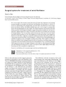
Figure 1. (a) Paranasal sinus computed tomography imaging of the polyp that arose from the right sphenoid sinus. (b, c) Endoscopic view of sphenochoanal polyp. (d) Postoperative view of sphenoid sinus. MT: Middle turbinate; S: Nasal septum; SC: Sphenochoanal polyp; SO: Sphenoid sinus ostium.
Of the patients with antrochoanal polyp, 50 (76.92%) had natural maxillary ostia, 11 (16.92%) had accessory maxillary ostia, and four (6.15%) had both accessory and natural maxillary ostia.
(a)
(b)
Among the patients who underwent surgery for choanal polyp, one had inverted papilloma, one had juvenile angiofibroma, two had inferior concha hypertrophy on postoperative
(c)
Figure 2. A 53x22 mm mass extending from the septum of the right sphenoid sinus to the nasopharynx on T1 coronal (a) and T2 sagittal (b) maxillofacial magnetic resonance imaging sections. (c) The place where the sphenochoanal polyp arose from the right sphenoidal septum (black arrow).
Retrospective analysis of surgical treatment of choanal polyps
147
(a)
(b)
(c)
(d)
Figure 3. (a, b) A mass lesion that almost completely obliterates the left choana, largely fills the nasopharynx and shows heterogeneous contrast agent uptake on the cranial magnetic resonance imaging of the choanal polyp arising from the middle turbinate in the left nasal cavity. (c) Endoscopic view of the polyp. (d) The attachment point of the choanal polyp in the posterior of the left middle turbinate. MT: Middle turbinate; P: Choanal polyp.
histopathology and they were excluded from the study. While endoscopic sinus surgery was performed in 49 (75.38%) of the patients who underwent surgery for antrochoanal polyp, combined approach (trans-canine trocar in 14 patients, Caldwell-Luc in one patient, and mini Caldwell-Luc in one patient) was performed in 16 patients (24.61%) in whom the mass could not be observed entirely during endoscopic examination. Relapse was observed in three (6.12%) of the patients who underwent endoscopic surgery alone and in one (6.25%) of the patients who underwent combined approach. No significant difference was observed between combined approach and endoscopic surgery (p=0.971) in terms of the relapse rate. Of the four
patients with relapse, one underwent endoscopic surgery, whereas three underwent combined surgery (trans-canine trocar in one and CaldwellLuc in two patients). No relapse was observed over the course of follow-up period. All the patients with choanal polyp, except for the patients with antrochoanal polyp, underwent endoscopic surgery. None of the patients developed relapse. Concomitant concha bullosa excision was performed in seven, septoplasty was performed in seven, adenoidectomy was performed in three, and tonsillectomy was performed in one of the patients who underwent surgery for choanal polyp. None of the patients who underwent surgery for choanal polyp developed postoperative complications.
148
Kulak Burun Bogaz Ihtis Derg
(a)
(b)
(c)
(d)
Figure 4. Soft tissue mass arising from the left ethmoidal sinus extending to the choana on coronal (a) and axial (b) computed tomography sections of the paranasal sinus. (c) Endoscopic view of the choanal polyp of anterior ethmoid origin. (d) Attachment of the mass to the anterior nasal ethmoid while the mass was being excised by microdebrider.
DISCUSSION
The first detailed description of choanal polyp, which was first defined by Palfyn in 1753, was Table 1. The origins of antrochoanal polyps in maxillary sinus Origins of polyps
n
%
Medial Posterior Posterosuperior Lateral Anteromedial Inferomedial Posterolateral Inferior Posteromedial Anterior Posteroinferior Anteroinferior Anterolateral Inferolateral
9 6 4 4 4 4 3 3 3 2 2 1 1 1
19.14 12.76 8.51 8.51 8.51 8.51 6.38 6.38 6.38 4.25 4.25 2.12 2.12 2.12
made by Killian in 1906.[6] It is more prevalent in males than females.[7] In the present study, male/female ratio was 1.23. Patients with choanal polyp most often present with unilateral nasal obstruction. Nevertheless, anosmia, nasal discharge, snoring, headache, obstructive sleep apnea, and epistaxis are other likely symptoms.[8] Likewise, in the present study, patients most frequently presented with nasal obstruction followed in turn by snoring and sleeping with the mouth open, nasal discharge, and allergy. Antrochoanal polyps are almost always unilateral. Only a few cases of bilateral antrochoanal polyp have been reported previously.[9-12] In the present study, bilateral antrochoanal polyp arising from maxillary sinus was detected in two patients. In a 23-case series of antrochoanal polyp, Tatlıpınar et al.[13] investigated the patients in terms of concomitant nasal pathologies and detected septum deviation in 60.86%, concha
Retrospective analysis of surgical treatment of choanal polyps
bullosa in 13.04%. Adenoid hypertrophy in 4.34%, and inferior concha hypertrophy in 4.34%. Balıkçı et al.[7] detected septum deviation in 50%, inferior concha hypertrophy in 32.5%, and concha bullosa in 17.6% of the patients in their 34-case series. In the present study, choanal polyp was accompanied by nasal septum deviation in 39.47% of patients, whereas it was accompanied by chronic sinusitis in 25%, by inferior concha hypertrophy in 23.68%, and by concha bullosa in 19.73%. Antrochoanal polyps may usually arise from the posterior, inferior, lateral, and medial walls of the maxillary sinus and may rarely arise from the anterior wall.[9] In their 34-case series of antrochoanal polyps, Balıkçı et al.[7] reported that 85.3% of the polyps arose from the posterior wall, 5.8% of the polyps arose from the lateral wall, and 5.8% of the polyps arose from the medial wall. Lee and Huang[14] performed a study in a 26-case series of antrochoanal polyps and reported that 92.3% of the polyps arose from the posterior wall, 61.5% of the polyps arose from the lateral wall, and 38.5% of the polyps arose from the inferior wall. In a 23-case series of antrochoanal polyps, Bozzo et al.[15] found that 56% of the polyps arose from the lateral wall and 17% of the polyps arose from the inferior wall. In the present study, the origin of the antrochonal polyp could be detected in 47 of 65 patients. Accordingly, the polyp was in contact with the medial wall in 42.55%, the posterior wall in 38.9%, the inferior wall in 23.4%, and the lateral wall in 19.14% of patients. Unlike the literature, in the present study, the polyp was most often in contact with the medial wall followed by the posterior wall. It has been reported that choanal polyps not only arise from the maxillary sinus, but may also arise from the sphenoidal sinus, middle turbinate, ethmoidal sinus, nasal septum, inferior concha, and cribriform plate.[16] Polyps that arise from the sphenoid sinus and extend to the nasopharynx are called as sphenochoanal polyps and are extremely rare.[17] In the present study, sphenochoanal polyps were observed in six patients and all of them underwent surgery by endoscopic transnasal approach. Choanal polyps of the middle turbinate are quite rare and there have been only four cases reported in the literature, of which one arose
149
from the inferior, two arose from the medial, and one arose from the posterior region.[1,8,18] In the present study, there were two patients with choanal polyp of the middle turbinate. These patients presented to the clinic with nasal obstruction, headache, and mucoid nasal discharge. While the polyp arose from the medial of the middle turbinate in one of these patients, it arose from the posterior in the other patient. In one patient, the polyp was removed by endoscopic ethmoidectomy resecting from the region in the posterior of the middle turbinate from where it arose. In the other patient, the polyp was endoscopically excised from the medial aspect of the middle turbinate and the operation was completed. Choanal polyp of ethmoid origin is rarer than antrochoanal polyp.[13] In the present study, ethmoidochoanal polyp was detected in one patient. The patient underwent endonasal endoscopic surgery. Septochoanal polyp is another rarely encountered choanal polyp. Removal of a small amount of intact mucosa together with the peduncle of the polyp is adequate for the treatment of septochoanal polyps.[19] In the present study, septochoanal polyp was detected in two patients. These patients presented with nasal obstruction, nasal discharge, and postnasal discharge. The patients underwent surgical procedure under local anesthesia. The polyp was excised together with its peduncle including small amount of surrounding intact tissue. Differential diagnosis of antrochoanal polyp includes juvenile angiofibroma, encephalocele, malignancies of nasophraynx, nasal polyp, adenoid hypertrophy, concha hypertrophy, and inverted papilloma.[6,9] In the present study, among the patients undergoing surgery for the prediagnosis of antrochoanal polyp, one had inverted papilloma, one had angiofibroma, and two had inferior concha hypertrophy on postoperative pathology. Previously, surgical treatment of antrochoanal polyp included simple polypectomy and the Caldwell-Luc procedure.[20] Relapse rates are high with simple polypectomy alone.[20] It is necessary to remove both the choanal and antral parts of the polyp completely for treatment.[21]
150
The choanal part can be easily removed via transnasal or transoral route after dissecting the peduncle. The antral part can be removed by the Caldwell-Luc procedure or inferior meatal antrostomy or median meatal antrostomy.[21] Cleaning the antral part is important for the prevention of relapses in antrochoanal polyp surgery.[22] The Caldwell-Luc procedure enables a clear field of vision and ensures that the polyp becames completely free from the antral part.[22] Recently, it has been demonstrated that endoscopic sinus surgery is a safe and effective method in the treatment of antrochoanal polyps.[23] By this approach, the antral part is endoscopically removed through the median meatus without need for the Caldwell-Luc procedure or inferior meatal antrostomy.[21] Comparing with the Caldwell-Luc procedure, cheek swelling is less likely with endoscopic sinus surgery, unfavorable effects on tooth and face growth in children are minimized, and a shorter healing period is provided. This procedure preserves the intact antral mucosa and contributes to mucociliary clearance by epithelialization.[24] Minimal morbidity and excellent outcomes have been obtained with the use of the angled Blakesley or giraffe forceps or microdebrider together with 30°, 70° and 120° degree angled endoscopes.[21] In the literature, it has been demonstrated that the relapse rate is between 0% and 23% in endoscopic surgery and between 0% and 6.66% with combined surgery performed for choanal polyps.[7,9,13,14,25] In the present study, relapse was observed in three (6.12%) of the patients that underwent endoscopic method alone and in one (6.25%) of the patients who underwent combined approach. In conclusion, successful outcomes were obtained by endoscopic surgery in the treatment of choanal polyps except antrochoanal polyps. Success rates of endoscopic surgery in antrochoanal polyps were consistent with the literature. In contrast to the literature, success rates were lower in the patients who underwent combined surgery. This might have resulted from longer follow-up periods in the present study. There was no significant difference between the success rates of endoscopic surgery and the combined approach.
Kulak Burun Bogaz Ihtis Derg
Declaration of conflicting interests The authors declared no conflicts of interest with respect to the authorship and/or publication of this article. Funding The authors received no financial support for the research and/or authorship of this article. REFERENCES 1. Ozcan C, Duce MN, Görür K. Choanal polyp originating from the middle turbinate. Eur Arch Otorhinolaryngol 2004;261:184-6. 2. Altun H, Teker AM, Ceran M, Gedikli O. Endoscopic approach in patients with choanal polyps. [Article in Turkish] Kulak Burun Bogaz Ihtis Derg 2008;18:74-8. 3. Jang YJ, Rhee CK, Oh CH, Ryoo HG, Kim HG, Ha M. Arachidonic acid metabolites in antrochoanal polyp and nasal polyp associated with chronic paranasal sinusitis. Acta Otolaryngol 2000;120:531-4. 4. Sunagawa M, Kinjoh K, Nakamura M, Kosugi T. Urokinase-type plasminogen activator and plasminogen activator inhibitor antigen in tissue extracts of paranasal sinus mucous membranes affected by chronic sinusitis and antrochoanal polyps. Eur Arch Otorhinolaryngol 1999;256:237-41. 5. Freitas MR, Giesta RP, Pinheiro SD, Silva VC. Antrochoanal polyp: a review of sixteen cases. Braz J Otorhinolaryngol 2006;72:831-5. 6. Aydin O, Keskin G, Ustündağ E, Işeri M, Ozkarakaş H. Choanal polyps: an evaluation of 53 cases. Am J Rhinol 2007;21:164-8. 7. Balikci HH, Ozkul MH, Uvacin O, Yasar H, Karakas M, Gurdal M. Antrochoanal polyposis: analysis of 34 cases. Eur Arch Otorhinolaryngol 2013;270:1651-4. 8. Özkırış M. Choanal polyp originating from the middle turbinate: a case report. ACU Sağlık Bil Derg 2012;3:187-9. 9. Frosini P, Picarella G, De Campora E. Antrochoanal polyp: analysis of 200 cases. Acta Otorhinolaryngol Ital 2009;29:21-6. 10. Al-Qudah M. Bilateral antrochoanal polyps: possible pathogenesis. J Craniofac Surg 2011;22:1116-8. 11. Basu SK, Bandyopadhyay SN, Bora H. Bilateral antrochoanal polyps. J Laryngol Otol 2001;115:561-2. 12. Sousa DW, Pinheiro SD, Silva VC, Bastos JP. Bilateral antrochoanal polyps in an adult. Braz J Otorhinolaryngol 2011;77:539. 13. Tatlıpınar A, Gökçeer T, Köksal S, Esen E. Our clinical experience in patients with isolated choanal polyps reaching choana. KBB-Forum 2010;9:25-9. 14. Lee TJ, Huang SF. Endoscopic sinus surgery for antrochoanal polyps in children. Otolaryngol Head Neck Surg 2006;135:688-92. 15. Bozzo C, Garrel R, Meloni F, Stomeo F, Crampette L. Endoscopic treatment of antrochoanal polyps. Eur Arch Otorhinolaryngol 2007;264:145-50. 16. Ozcan C, Duce MN, Görür K. Choanal polyp originating from the cribriform plate. J Craniofac Surg 2010;21:806-7.
Retrospective analysis of surgical treatment of choanal polyps
17. Dadaş B, Yilmaz O, Vural C, Caliş AB, Turgut S. Choanal polyp of sphenoidal origin. Eur Arch Otorhinolaryngol 2000;257:379-81. 18. Kim KS. Choanal polyp originating from the middle turbinate 2013;148:525-6. 19. Birkent H, Karahatay S, Durmaz A, Kurt B, Tosun F. Choanal polyp originating from the nasal septum: septochoanal polyp. Kulak Burun Bogaz Ihtis Derg 2009;19:163-6. 20. Al-Mazrou KA, Bukhari M, Al-Fayez AI. Characteristics of antrochoanal polyps in the pediatric age group. Ann Thorac Med 2009;4:133-6. 21. Yanagisawa K, Coelho DH, Yanagisawa E. Endoscopic removal of the antral and choanal portions of an
151
antrochoanal polyp. Ear Nose Throat J 2005;84:194-5. 22. Ozdek A, Samim E, Bayiz U, Meral I, Safak MA, Oğuz H. Antrochoanal polyps in children. Int J Pediatr Otorhinolaryngol 2002;65:213-8. 23. Yaman H, Yilmaz S, Karali E, Guclu E, Ozturk O. Evaluation and management of antrochoanal polyps. Clin Exp Otorhinolaryngol 2010;3:110-4. 24. Sato K, Nakashima T. Endoscopic sinus surgery for antrochoanal polyp using CO2 laser and/or microresector: a long-term result. J Laryngol Otol 2005;119:362-5. 25. Eşki E, Imre A, Çallı Ç, Pınar E, Öncel S. Approaches to antrochoanal polyps in adults: long-term comparative results. [Article in Turkish] Kulak Burun Bogaz Ihtis Derg 2012;22:1-5.