0021-972X/06/$15.00/0 Printed in U.S.A.
The Journal of Clinical Endocrinology & Metabolism 91(3):827– 836 Copyright © 2006 by The Endocrine Society doi: 10.1210/jc.2005-1862
Clinical Presentation and Penetrance of Pheochromocytoma/Paraganglioma Syndromes Diana E. Benn,* Anne-Paule Gimenez-Roqueplo,* Jennifer R. Reilly,* Je´roˆme Bertherat, John Burgess, Karen Byth, Michael Croxson, Patricia L. M. Dahia, Marianne Elston, Oliver Gimm, David Henley, Philippe Herman, Victoria Murday, Patricia Niccoli-Sire, Janice L. Pasieka, Vincent Rohmer, Kathy Tucker, Xavier Jeunemaitre, Deborah J. Marsh, Pierre-Franc¸ois Plouin, and Bruce G. Robinson, for the International SDH Consortium Department of Cancer Genetics (D.E.B., J.R.R., D.J.M., B.G.R), Kolling Institute of Medical Research, Royal North Shore Hospital and University of Sydney, Sydney 2006, Australia; Department of Endocrinology (B.G.R.), Royal North Shore Hospital, Sydney 2065, Australia; Hereditary Cancer Clinic (K.T.), Prince of Wales Hospital and School of Medicine, University of New South Wales, Sydney, New South Wales 2052, Australia; National Health and Medical Research Council Clinical Trials Centre (K.B.), University of Sydney, Sydney 2006, Australia; Department of Endocrinology (J.Bu.), Royal Hobart Hospital, Tasmania 7001, Australia; Department of Endocrinology and Diabetes (D.H.), Sir Charles Gairdner Hospital, Nedlands, Western Australia 6009, Australia; Department of Endocrinology (M.C.), Greenlane Clinical Centre, Auckland, New Zealand; Department of Endocrinology (M.E.), Waikato Hospital, Hamilton, New Zealand; Departments of Genetics (A.-P.G.-R., X.J.) and Hypertension (P.-F.P.), Hoˆpital Europe´en Georges Pompidou, Assistance Publique des Hoˆpitaux de Paris, L’Institut National de la Sante´ et de la Recherche Me´dicale (INSERM) U36, Colle`ge de France, University Paris 5, Paris 75015, France; Department of Otorhinolaryngology and Head and Neck Surgery (P.H.), Hoˆpital Lariboisie`re, Assistance Publique des Hopitaux de Paris, Paris 75010, France; Department of Endocrinology (J.Be.), INSERM U567, Hoˆpital Cochin Assistance Publique des Hoˆpitaux de Paris, University Paris 5, Paris 75014, France; Department of Endocrinology (P.N.-S.), Hoˆpital de la Timone, Assistance Publique des Hoˆpitaux de Marseille, Marseille 13385, France; Department of Endocrinology (V.R.), Hoˆpital d’Angers, Angers 49033, France; Department of General, Visceral and Vascular Surgery (O.G.), Martin Luther University Halle-Wittenberg, Halle 06097, Germany; West of Scotland, Regional Genetics Services (V.M.), Yorkhill G3 8SJ, Scotland; Department of Surgery (J.L.P.), Faculty of Medicine, University of Calgary, Calgary, Canada T2N 1N4; and Departments of Medicine and Cellular and Structural Biology (P.L.M.D.), University of Texas Health Science Center, San Antonio, Texas 78229-3900 Context: The identification of mutations in genes encoding peptides of succinate dehydrogenase (SDH) in pheochromocytoma/paraganglioma syndromes has necessitated clear elucidation of genotype-phenotype associations.
ical features were evaluated for evidence of genotype-phenotype associations, and penetrance was determined. Results: SDHB mutation carriers were more likely than SDHD mutation carriers to develop extraadrenal pheochromocytomas and malignant disease, whereas SDHD mutation carriers had a greater propensity to develop head and neck paragangliomas and multiple tumors. For the index cases, there was no difference between 43 SDHB and 19 SDHD mutation carriers in the time to first diagnosis (34 vs. 28 yr, respectively; P ⫽ 0.3). However, when all mutation carriers were included (n ⫽ 112), the estimated agerelated penetrance was different for SDHB vs. SDHD mutation carriers (P ⫽ 0.008).
Objective: Our objective was to determine genotype-phenotype associations in a cohort of patients with pheochromocytoma/paraganglioma syndromes and succinate dehydrogenase subunit B (SDHB) or subunit D (SDHD) mutations. Design, Setting, and Participants: The International SDH Consortium studied 116 individuals (83 affected and 33 clinically unaffected) from 62 families with pheochromocytoma/paraganglioma syndromes and SDHB or SDHD mutations. Clinical data were collected between August 2003 and September 2004 from tertiary referral centers in Australia, France, New Zealand, Germany, United States, Canada, and Scotland.
Conclusions: For clinical follow-up, features of SDHB mutation-associated disease include a later age of onset, extraadrenal (abdominal or thoracic) tumors, and a higher rate of malignancy. In contrast, SDHD mutation carriers, in addition to head and neck paragangliomas, should be observed for multifocal tumors, infrequent malignancy, and the possibility of extraadrenal pheochromocytoma. (J Clin Endocrinol Metab 91: 827– 836, 2006)
Main Outcome Measures: Data were collected on patients with pheochromocytomas and/or paragangliomas with respect to onset of disease, diagnosis, genetic testing, surgery, pathology, and disease progression. Clin-
P
ARAGANGLIOMAS ARE TUMORS derived from the parasympathetic and sympathetic nervous systems (1). The parasympathetic-associated paragangliomas arising in the head and neck are usually nonfunctioning. The
sympathetic-associated paragangliomas arise from the adrenal medulla or from the sympathetic ganglias and are generally functionally active as indicated by excess catecholamine production. The term pheochromocytoma is also currently applied to the catecholamine-secreting paragangliomas that are intraabdominal (adrenal or extraadrenal) or thoracic. During the past 10 yr, it has been recognized that pheochromocytomas are component tumors of von Hippel Lindau disease, multiple endocrine neoplasia type 2, and neurofibromatosis type 1, occurring as a result of germline
First Published Online November 29, 2005 * D.E.B., A.-P.G.-R., and J.R.R. contributed equally to this work. Abbreviations: CI, Confidence interval; HR, hazard ratio; SDH, succinate dehydrogenase; SDHB, SDH subunit B; SDHD, SDH subunit D. JCEM is published monthly by The Endocrine Society (http://www. endo-society.org), the foremost professional society serving the endocrine community.
827
The Endocrine Society. Downloaded from press.endocrine.org by [${individualUser.displayName}] on 18 January 2017. at 00:53 For personal use only. No other uses without permission. . All rights reserved.
828
J Clin Endocrinol Metab, March 2006, 91(3):827– 836
Benn et al. • Pheochromocytoma/Paraganglioma Syndromes
TABLE 1. Patient population with germline SDHB mutations and pheochromocytoma and/or paraganglioma Family ID
F-45 F-44 F-55 F-60 F-50 F-36 F-27 F-42 F-51 F-30 F-28 F-29 F-33 F-34 F-20 F-11 F-07 F-46 F-41 F-56 F-39 F-59 F-53 F-61 F-35 F-54 F-02 F-47 F-57 F-62 F-13 F-19 F-40 F-25 F-23 F-24 F-16 F-43 F-22 F-21 F-26 F-37 F-49
Country
SDHB mutation
SDHB mutation (cDNA nucleotide)
Australia/UKa NZ Australia UK Australia Australia France USA Australia France France France Australia/NZ NZ France France France Australia USA Australia USA Australia Australia Australia NZ Australia France Australia Australia UK France France USA France France France France USA France France France Germany Australia
IVS1⫹1G3 T IVS1⫹1G3 T IVS1⫹1G3 T IVS1⫹1G3 T Q30fsX76b K40Ec A43Pd R46X R46Xb R46Gd R46Qd R46Qe R46Qb R46Q R46Q P56L R90X R90X G96D C98R C98Y G99D N104fsX118 R115X I127S I127S P131fsX135g IVS4⫹1G3 A IVS4-1G3 A Q168fsX177 C192Y C196Y C196fsX219 S198fsX220d L207fsX220d L207fsX220d L207fsX220 G208E R230Cd F238fsX247 L240fsX247 R242H P254fsX255
Intron Intron Intron Intron c.88delC c.118A3 G c.127G3 C c.136C3 T c.136C3 T c.136C3 G c.137G3 A c.137G3 A c.137G3 A c.137G3 A c.137G3 A c.167C3 T c.268C3 T c.268C3 T c.287G3 A c.292T3 C c.293G3 A c.296G3 A c.311delAinsGG c.343C3 T c.379T3 G c.379T3 G c.392delC Intron Intron c.502insC c.575G3 A c.587G3 A c.588delC c.591delC c.620-621delTG c.620-621delTG c.620-621delTG c.623G3 A c.688C3 T c.713delT c.718-721delCTAT c.725G3 A c.761insC
Exon
2 2 2 2 2 2 2 2 2 2 2 2 3 3 4 4 4 4 4 4 4 4
5 6 6 6 6 6 6 6 6 7 7 7 7 7
Earliest age of onset (yr)
Ages of all patients at diagnosis (yr)
Types of tumors in family
27 68
27, 32 68 53 15 12, 23 12, 40 29 35 7 30 27 54 25– 64 (n ⫺ 6)f 43 25 60 31, 55 23 43 37 18 48 27, 47 49 29, 50 18 29 21 15 35, 51 34 24 55 36 24 39 27 20 32 9 48 34 15
A, (M) A T A, (M) P, H, (M) A, T A, T, (M) A, T A P P, A, (M) P, A, T, (M) P, A, H, (M) A, H, (M) T A A, T, (M) T A, (M) A, T A A, (M) P, H, (M) A A A, (M) H H T A, H H, (M) A, (M) A A A, (M) A, (M) A, (M) A P, A, (M) P, A A A P
12 12 27 7 29 23 51 24 40 25 60 54 17
14 48
24 18 29 21 10 33 24 55 32 20 37 9 30 9 48 34
P, Pheochromocytoma of adrenal gland; A, abdominal extraadrenal paraganglioma (pheochromocytoma); T, thoracic extraadrenal paraganglioma (pheochromocytoma); H, head and neck paraganglioma; (M), metastases; NZ, New Zealand. a Family members were submitted from both countries. b Previously published case (11). c Previously published case (12). d Previously published case (13). e Previously published case (10). f Including four presumed carriers (see Statistical analysis in Patients and Methods). g Previously published case (14).
mutations in VHL, RET, or NF1, respectively. More recently, nuclear genes encoding two mitochondrial complex II subunit proteins, SDHD and SDHB, have been associated with the development of familial pheochromocytomas and paragangliomas (2, 3), and SDHC mutations have been linked to familial head and neck paragangliomas in four families worldwide (4, 5). Although genotype-phenotype associations have been established for VHL and RET, only one large cohort study
of clinical features in SDHB and SDHD mutation carriers has been reported, in a predominantly German/Polish population with pheochromocytomas and paragangliomas (6). We undertook an international study of genotypephenotype associations in a cohort of patients with SDHB or SDHD mutations and pheochromocytoma and/or paraganglioma, to identify associations that will assist clinicians in making evidence-based decisions regarding the monitoring of disease in these patients and their families.
The Endocrine Society. Downloaded from press.endocrine.org by [${individualUser.displayName}] on 18 January 2017. at 00:53 For personal use only. No other uses without permission. . All rights reserved.
Benn et al. • Pheochromocytoma/Paraganglioma Syndromes
J Clin Endocrinol Metab, March 2006, 91(3):827– 836
Patients and Methods Patients Patients were included in this retrospective study if they had been shown to carry a germline SDHB or SDHD mutation. No data were received on SDHC mutation carriers. For the purpose of this study, families analyzed ranged from single germline mutation carriers with diagnosed pheochromocytoma and/or paraganglioma to families with up to eight mutation carriers, not all of whom were symptomatic at the time of data collection. All mutation analyses had been performed before this study using previously published methods (7–11). Data were collected from tertiary referral centers in Australia, France, New Zealand, United States, United Kingdom, Germany, and Canada. Information on 119 participants from 63 families was received, but three participants did not fulfill the requirements for inclusion. Therefore, the final data set comprised 116 participants from 62 families; 43 families were mutation positive for SDHB (Table 1), and 19 families had SDHD mutations (Table 2). The study was initiated by and coordinated from the Cancer Genetics Unit, Kolling Institute of Medical Research, University of Sydney, at Royal North Shore Hospital, Sydney, Australia, and approved by the Northern Sydney Health Human Research Ethics Committee. Patients gave informed consent. French participants were collected through the PGL.NET and COMETE networks. Participating centers complied with their local ethics requirements. Data collection commenced in August 2003 and was completed in September 2004. To ensure standardization of mutation reporting, a nucleotide numbering sheet was supplied for SDHB (NCBI accession no: NM_003000) and SDHD (NCBI accession no: NM_003002).
829
dominal paraganglioma (pheochromocytoma), extraadrenal thoracic paraganglioma (pheochromocytoma), or head and neck paraganglioma. These were the four designated tumor sites used for statistical analysis. Malignancy at any stage of the course of disease was defined by the presence of distant metastases including lung, liver, bone, and lymph nodes. The International SDH Consortium Data Sheet used in the study is available on the Kolling Institute of Medical Research Website at http://www.kolling.usyd.edu.au.
Statistical analysis The statistical software package SPSS for Windows version 12 was used to perform the analyses. Two-tailed tests with a significance level of 5% were used throughout, and 2 tests were used to test for association between categorical variables except when an expected cell size was less than 5, in which case Fisher’s exact test was employed. McNemar’s test was used to compare different types of catecholamine results within patients. Kaplan-Meier survival curves were used to illustrate the relationship between age and both survival and the cumulative percentage diagnosed with disease. Estimated median ages at first diagnosis were calculated along with their 95% confidence intervals (95% CI). Hazard ratios (HR) estimated via Cox regression models were used to quantify the associations between the survival distributions and mutation type, provided the assumption of proportional hazards was valid. Schoenfeld residues were used to test for proportional hazards. Four presumed carriers from one family (F-33), all of whom died of related disease, were excluded from the Kaplan-Meier analyses of penetrance, because they had not been positively identified as SDHB mutation carriers, although three of them had mutation-positive children.
Requested data collection The International SDH Consortium Data Sheet contained seven main sections: Patient Data, Features at Clinical or Genetic Diagnosis, Past Medical History, Primary Tumor Characteristics, Treatment Options, Systems Review at Date of Diagnosis of First Pheochromocytoma or Paraganglioma, and Subsequent Surgeries. In the literature, nomenclature for extraadrenal (abdominal and thoracic) tumors is inconsistent, sometimes being referred to as extraadrenal pheochromocytomas and frequently as paragangliomas. To avoid ambiguity in the collection of data, tumors were classified as either pheochromocytoma of the adrenal gland, extraadrenal ab-
Results SDHB and SDHD mutation carrier status
The study included 116 participants from 62 families (Fig. 1), as shown in Tables 1 and 2, with 62 index cases (38 males and 24 females). There were 43 index cases with 31 different SDHB mutations spanning exons 2–7 (Fig. 2) and 19 index cases with 16 different SDHD mutations spread across all four exons (Fig. 3). With respect to mutation type (splice site,
TABLE 2. Patient population with germline SDHD mutations and pheochromocytoma and/or paraganglioma Family ID
F-32 F-01 F-04 F-10 F-14 F-12 F-05 F-31 F-52 F-15 F-63 F-17 F-06 F-08 F-18 F-58 F-38 F-03 F-09
Country
SDHD mutation
France France France France France France France France Australia France UK France France France France Canada USA France France
L7fsX14 R22Xa R22X W43X H50fsX68 IVS2⫹1G3 T IVS2-1G3 T S68fsX113 P81L P81L P81L Y84X L85X IVS3⫹1G3 A T112fsX133 Y114C Y114CfsX162 G148D I149N C150del
SDHD mutation (cDNA nucleotide)
c.21delG c.64C3 T c.64C3 T c.129G3 A c.148-149insA Intron Intron c.202-203insA c.242C3 T c.242C3 T c.242C3 T c.252T3 G c.254T3 A Intron c.334-337delACTG c.341A3 G c.341-342delAT c.443G3 A c.446T3 A; 448delTGC
Exon
1 2 2 2 2 3 3 3 3 3 3 4 4 4 4 4
Earliest age of onset (yr)
34 31 43 20 17 25 24 25 27 31 28 21 20 16 40 25 7 24
Ages of all patients at diagnosis (yr)
34 31, 43 20 19 25 25, 31 27 32 41 28, 22, 16, 16, 40 25 10, 24,
34, 58
68
73 33 20 33 32 25
Types of tumors in family
H T, H P, A, H H, (M) A, H H H A H H H H, T H, T, A H P, A, H H A, H H, (M) H
P, Pheochromocytoma of adrenal gland; A, abdominal extraadrenal paraganglioma (pheochromocytoma); T, thoracic extraadrenal paraganglioma (pheochromocytoma); H, head and neck paraganglioma; (M), metastases. a Previously published case (9).
The Endocrine Society. Downloaded from press.endocrine.org by [${individualUser.displayName}] on 18 January 2017. at 00:53 For personal use only. No other uses without permission. . All rights reserved.
830
J Clin Endocrinol Metab, March 2006, 91(3):827– 836
Benn et al. • Pheochromocytoma/Paraganglioma Syndromes
FIG. 1. Study population of patients with SDHB or SDHD mutations. The total number of patients is indicated, including the treatment modality.
nonsense or frameshift mutations), there was no difference between the SDHB and SDHD mutation carriers (P ⫽ 0.3). Four families all reporting Scotland as their country of origin presented with the SDHB IVS1 ⫹ 1G3 T splice site mutation. It is possible that this may be a founder mutation. Three of these families resided in Australia, New Zealand, and Scotland, respectively; the fourth family had members in both Australia and Scotland. Haplotype analysis to confirm a founder effect was not permitted under the ethics protocol of this study. Genetic testing of asymptomatic family members
In this study, data were supplied on 38 asymptomatic relatives with a median age of 41 yr (interquartile range, 29 – 62 yr) who had undergone genetic testing and were found to be gene mutation positive for either SDHB or SDHD. On subsequent investigation (including urinary catecholamine levels and/or imaging) five patients showed evidence of disease. Two with SDHB mutations (F-07 and F-36) subsequently had surgery for extraadrenal paragangliomas (pheochromocytomas) at ages 31
and 40 yr, respectively. The other three relatives (one with an SDHD mutation) were diagnosed with head and neck tumors (F-05, F-53, and F-62) at ages 68, 47, and 35 yr, respectively, and at the time of data submission were awaiting surgery. Of the 33 mutation-positive but clinically unaffected relatives, two died of unrelated disease. Age of diagnosis and penetrance
For SDHB index cases, the Kaplan-Meier survival estimate of median age of first diagnosis was 34 (95% CI, 26.1– 41.9) years compared with 28 (95% CI, 19.5–36.5) years for the SDHD index cases. A test of the null hypothesis of proportionality was accepted (P ⫽ 0.14). The HR for SDHD vs. SDHB mutation carriers was 1.3 (95% CI, 0.76 –2.3; P ⫽ 0.3). By the age of 40 yr, an estimated 65% of SDHB index cases were diagnosed with disease, as were 84% of SDHD index cases (Fig. 4A). In two SDHB families (F-21 and F-51), the index case was diagnosed before 10 yr of age, whereas in two other cases (F-16 and F-57), the children were symptomatic before 10 yr of age but not diagnosed for a number of years. Overall, 16 of 43 (37%) of the SDHB
The Endocrine Society. Downloaded from press.endocrine.org by [${individualUser.displayName}] on 18 January 2017. at 00:53 For personal use only. No other uses without permission. . All rights reserved.
Benn et al. • Pheochromocytoma/Paraganglioma Syndromes
J Clin Endocrinol Metab, March 2006, 91(3):827– 836
831
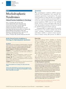
FIG. 2. SDHB mutations in pheochromocytoma/paraganglioma families. The eight exons of SDHB are represented diagrammatically. Missense mutations are shown in the upper portion of the figure, and truncating-type mutations are shown in the lower portion. The phenotype observed in each family is represented by a unit of five boxes, with the family number shown above.
index cases and 15 of 19 (79%) of the SDHD index cases were reported as familial presentations. When all SDHB and SDHD mutation carriers were analyzed (n ⫽ 112), the estimated age-related penetrance (Fig. 4B) showed that 29% of SDHB and 48% of SDHD mutation carriers are diagnosed by 30 yr of age, but by 40 yr of age, 45% of SDHB and 73% of SDHD mutation carriers are diagnosed (HR, 1.9; 95% CI, 1.2–3.1; P ⫽ 0䡠008). Clinical findings at diagnosis
Diagnosis of disease in 83 patients was made clinically (n ⫽ 62) and/or with imaging modalities (n ⫽ 71) including computed tomography, magnetic resonance imaging, and scintigraphy with meta-iodobenzylguanidine or octreotide. For SDHB patients, 42 of 50 (84%) were symptomatic at presentation, as were 23 of 27 (85%) SDHD patients (Table 3). Catecholamine excess indicating a functioning tumor was defined as one urinary catecholamine measurement raised to at least twice the normal value. Functioning tumors (mainly extraadrenal) were identified in 22 of 35 (63%) SDHB mutation carriers and six of nine (67%) tested SDHD mutation carriers. Three SDHD mutation carriers who developed only head and neck tumors were identified as having abnormal urinary catecholamines. Because head and neck tumors are generally regarded as non-catecholamine-secreting tumors, these results may indicate underlying extraadrenal tumors. Of the total group diagnosed (n ⫽ 83), urinary catecholamine measurements showed that of those with disease, 22 of 38 (58%) had an elevated norepinephrine, whereas nine of 38 (24%) had an elevated epinephrine; similarly, 14 of 24 (58%) had a raised normetanephrine compared with five of 24 (21%) with a raised metanephrine. The pattern of urinary catecholamines for SDHB mutation carriers is shown in Table 4.
Treatment and pathology at first surgery
After initial diagnosis, 57 patients were treated by surgery alone (Fig. 1) and 18 patients underwent surgery with additional therapy of chemotherapy, radiotherapy, and/or [131I]meta-iodobenzylguanidine. At first surgery, multiple tumors were observed more frequently in SDHD mutation carriers (eight of 27) than in SDHB mutation carriers (six of 49) (Fischer’s exact P ⫽ 0.02). Tumor location differed between the SDHB and SDHD mutation carriers at the first surgery. Adrenal pheochromocytomas were identified in six SDHB mutation carriers and one SDHD mutation carrier. In particular, 29 of 49 (59%) SDHB mutation carriers were diagnosed with only extraadrenal abdominal tumors, whereas 24 of 27 SDHD (89%) mutation carriers showed only head and neck tumors (P ⬍ 0.001). At this first surgery, metastases were identified in 11 of 49 (22%) SDHB mutation-positive patients, with sites of distant metastases including lung, liver, bone, and lymph nodes. No SDHD mutation carriers presented with metastases at their first surgery. Local invasion was present in seven SDHB mutation carriers who did not have distant metastases at first surgery. Overall location of tumor according to mutation type
There was a highly significant difference in the age-related penetrance for tumor type between SDHB and SDHD mutation carriers, in that by 40 yr of age, an estimated 68% of all SDHD carriers will have developed a head and neck paraganglioma, whereas only 15% of SDHB carriers will have presented with these tumors (HR, 6.2; 95% CI, 3.2–11.9; P ⬍ 0.001) (Fig. 5A). In sharp contrast, by 60 yr of age an estimated 35% of
The Endocrine Society. Downloaded from press.endocrine.org by [${individualUser.displayName}] on 18 January 2017. at 00:53 For personal use only. No other uses without permission. . All rights reserved.
832
J Clin Endocrinol Metab, March 2006, 91(3):827– 836
Benn et al. • Pheochromocytoma/Paraganglioma Syndromes
TABLE 3. Comparison of SDHB and SDHD mutation carriers at presentation and first surgery
Symptomatic at presentation Common presenting symptoms Antihypertensive treatment Location of tumors Multiple tumors Metastases identified at first surgery
SDHB mutation positive
SDHD mutation positive
Significance
42/50 (84%) Hypertension, arrhythmia, headache 29/34 (85%) Extraadrenal abdominal, 29/49 (59%) 6/49 (12%) 11/49 (22%)
23/27 (85%) Headache, dysphonia, deafness, hypertension 12/14 (86%) Head and neck, 24/27 (89%) 8/27 (30%) 0 (0%)
P ⬍ 0.001 P ⫽ 0.02 P ⫽ 0.005
ilies, in addition to head and neck tumors, extraadrenal tumors were reported. During the course of disease, the probability of a patient developing a tumor at any of the four designated sites was examined in relation to the type of mutation (SDHB or SDHD) carried (Table 5). Analysis showed that a patient with an SDHD mutation compared with an SDHB mutation has an odds ratio of 24.3 (95% CI, 7.9 –74; P ⬍ 0.001) of developing a head and neck tumor. However, an SDHD mutation carrier had an odds ratio of 0.28 (95% CI, 0.10 – 0.81; P ⫽ 0.02) of developing an extraadrenal abdominal tumor when compared with an SDHB mutation carrier. There was an almost equal chance of patients with SDHD or SDHB mutations developing a tumor in the thorax. Overall disease progression and follow-up
FIG. 3. SDHD mutations in pheochromocytoma/paraganglioma families. The four exons of SDHD are represented diagrammatically. Missense mutations are shown in the upper portion of the figure, and truncating-type mutations are shown in the lower portion. The phenotype observed in each family is represented by a unit of five boxes, with the family number shown above.
SDHD mutation carriers will have presented with an extraadrenal abdominal or thoracic tumor, as would an estimated 69% of SDHB carriers (HR, 0.46; 95% CI, 0.21–1.0; P ⫽ 0.06) (Fig. 5B). Interestingly, in seven of 19 SDHD mutation famTABLE 4. Urinary catecholamine results for 30 SDHB-positive mutation carriers Norepinephrine
Epinephrine normal Epinephrine raised Total
Normal
Raised
12 1 13
11 6 17
Total
23 7 30
Of the 11 patients with a raised norepinephrine and normal epinephrine, seven had extraadrenal tumors.
Of 75 patients who were treated surgically, 25 required subsequent surgery on one occasion, 13 required subsequent surgery on two occasions, and one patient (F-06) underwent surgery five times for related disease. Overall, SDHB mutations were associated with a higher rate of malignancy than SDHD mutations with 18 of 48 (37.5%) SDHB mutation patients reported with malignancy as opposed to two of 26 (7.7%) SDHD mutation patients (HR, 5.6; 95% CI, 1.3–24; P ⫽ 0.02). The median age of diagnosis with malignancy for SDHB mutation patients was 48 (95% CI, 32.7– 63.3) years. Importantly, two SDHD mutation-positive patients (F-03 and F-10) with head and neck tumors were diagnosed with metastases at the second surgical event, one dying from disease at the age of 17 yr having had the first surgery at 10 yr of age. The median number of years of follow-up for patients with disease was 4 yr, with an interquartile range of 1–9.75 yr. There was no statistically significant difference in the distribution of time to disease-associated death for the SDHB and the SDHD groups (HR, 2.5; 95% CI, 0.3–20; P ⫽ 0.4). Twelve of 14 deaths in the study population were from disease-related causes (tumor, metastases, or surgery), and two SDHB mutation carriers who had not developed disease died of unrelated causes, one of cardiac disease and the other of metastatic myeloma. Excluding the four presumed carriers (F-33), seven of eight died from SDHB-associated disease and one from SDHD-associated disease. Associated disease
Several additional tumor types were diagnosed in participating families. A gastrointestinal stromal tumor was present in one patient who died at 80 yr of age from ischemic heart disease. This patient (F-35) had not developed pheochromocytoma or paraganglioma, although the absence of
The Endocrine Society. Downloaded from press.endocrine.org by [${individualUser.displayName}] on 18 January 2017. at 00:53 For personal use only. No other uses without permission. . All rights reserved.
Benn et al. • Pheochromocytoma/Paraganglioma Syndromes
FIG. 4. A, Age-related penetrance for SDH mutation carriers for index cases (n ⫽ 62). The estimated median age of first diagnosis was 34 yr for SDHB mutation carriers and 28 yr for SDHD mutation carriers (P ⫽ 0.1). B, Age-related penetrance for all SDH mutation carriers. The estimated median age of first diagnosis was 47 yr for SDHB mutation carriers and 31 yr for SDHD mutation carriers (P ⫽ 0.008).
relevant disease was not confirmed by autopsy. No genomic DNA was available from this patient, but the tumor showed the SDHB I127S mutation. In this same family, two members had been diagnosed with extraadrenal abdominal paraganglioma (pheochromocytoma). In one family (F-33), angioli-
J Clin Endocrinol Metab, March 2006, 91(3):827– 836
833
FIG. 5. A, Estimated median age of first head and neck tumor for all SDH mutation carriers (n ⫽ 112). By 40 yr of age, it is estimated that 68% of all SDHD mutation carriers will have developed a head and neck paraganglioma, whereas only 15% of SDHB mutation carriers will have presented with these tumors (P ⫽ 0.001). B, Estimated median age of first extraadrenal abdominal or thoracic tumor for all SDH mutation carriers. Age-related penetrance shows that by 60 yr of age, an estimated 35% of SDHD mutation carriers will have presented with an extraadrenal abdominal or thoracic tumor, as would an estimated 69% of SDHB carriers (P ⫽ 0.06).
The Endocrine Society. Downloaded from press.endocrine.org by [${individualUser.displayName}] on 18 January 2017. at 00:53 For personal use only. No other uses without permission. . All rights reserved.
834
J Clin Endocrinol Metab, March 2006, 91(3):827– 836
Benn et al. • Pheochromocytoma/Paraganglioma Syndromes
TABLE 5. Association between mutation type (SDHB or SDHD) and location of tumor identified Classification
Tumor location
Pheochromocytoma Extraadrenal abdominal paraganglioma (pheochromocytoma)
Adrenal gland Paraaortic/pericaval Bladder Remnants of organs of Zu¨ckerkandl Perirenal Other (retroperitoneal, periadrenal) Aorticopulmonary/mediastinal Cardiac/pericardial Pulmonary parenchyma Carotid body Vagal paraganglia Jugular bulb paraganglia Jugulotympanic paraganglia
Extraadrenal thoracic paraganglioma (pheochromocytoma) Head and neck paraganglioma
SDHB (n ⫽ 51)a
SDHD (n ⫽ 28)a
9b 34b
2b 5b
9b
3b
14b
25b
Tumors were assigned one of four location classifications that were the groupings used in the statistical analysis. a Total number of patients diagnosed with disease. b Number of patients who at any time had been diagnosed with a tumor.
poma occurred in two affected presumed carriers, whereas an unaffected mutation carrier was diagnosed with both parotid adenoma and ovarian cystic teratoma. A pituitary adenoma occurred in an unaffected SDHB mutation carrier (F-49), and adenocarcinoma of the colon was present in one SDHD mutation-positive patient (F-01) who had undergone surgeries for bilateral carotid body tumors. In the cohort presented here, there was one report of thyroid paraganglioma in an SDHB mutation carrier (F-02) (14). Another SDHB mutation carrier affected by disease had undergone hemithyroidectomy for benign thyroid nodules (F53). Two affected SDHD mutation carriers reported the presence of multinodular goiter (F-58) and Graves’ disease (F-52). In contrast to another large cohort study of patients of mainly German or Polish origin, no cases of renal cell carcinoma were reported (6). Discussion
This study of an international cohort of patients with pheochromocytoma/paraganglioma syndromes allows important conclusions to be drawn regarding genotype-phenotype associations. In this referral-based study, the findings are interesting in that they confirm some of the observations of a previous population-based study (6), namely that SDHB mutations are associated with extraadrenal disease and malignancy, whereas SDHD mutations are associated with head and neck paragangliomas. However, it is important to note that in comparing the two studies, this referral-based study did not observe the greater severity of disease or higher penetrance often found in referral-based studies, although malignancy in two SDHD mutation-positive families was reported. We observed that there was no significant difference between SDHB and SDHD mutation carriers in the time to first diagnosis for the index cases alone, the estimated median age of diagnosis being 34 vs. 28 yr, respectively. However, when all mutation carriers were included, the age-related penetrance for 82 SDHB mutation carriers vs. 30 SDHD mutation carriers was different (P ⫽ 0.008). This is in contrast to another large cohort study of patients of predominantly German /Polish origin in which no differences were reported in
the overall age-related penetrance between SDHB and SDHD mutation carriers, although the authors suggested that with longer follow-up, significant differences may be observed (6). In our study, clinically unaffected mutation carriers were included in the Kaplan-Meier analysis of penetrance in an attempt to partially correct for ascertainment bias. Another crucial step will be to collect additional specific penetrance data on a sufficiently large number of families to yield an adequate number of nonindex cases for separate analysis. In relation to tumor location, SDHD mutation carriers were more likely than SDHB mutation carriers to develop head and neck tumors (P ⬍ 0.001) and have a multifocal presentation (P ⫽ 0.02). Conversely, SDHB mutation carriers were more likely than SDHD mutation carriers to present with extraadrenal abdominal tumors (P ⫽ 0.02) and with increased risk of malignancy. This observation supports recent findings that patients with germline SDHB mutations should be considered as high risk for malignant disease and recurrence (6, 13). Head and neck paragangliomas are generally regarded as benign tumors but can extend into the skull. Of importance, two SDHD mutation-positive patients with head and neck tumors developed metastases, one dying of disease. SDHD mutations have only infrequently been associated with malignant disease of head and neck paragangliomas or pheochromocytomas (15, 16). This finding should alert clinicians to the necessity of careful monitoring of these patients for the development of tumors at new locations and also for the presence of metastases. Further genotype-phenotype associations showed that in addition to head and neck paragangliomas, there was a tendency for SDHD mutation carriers with nonsense mutations to develop pheochromocytoma, consistent with a previous observation (16), but these pheochromocytoma-causing mutations did not necessarily cluster at the 5⬘ end of the SDHD gene (17). We did, however, find general agreement with the observation of a broad mutational spectrum spread across SDHB exons 2–7 (17). The hereditary nature of this disease may be masked by maternal imprinting of SDHD resulting in only paternal transmission of SDHD-associated disease (2), although recently a different mechanism has been proposed to account
The Endocrine Society. Downloaded from press.endocrine.org by [${individualUser.displayName}] on 18 January 2017. at 00:53 For personal use only. No other uses without permission. . All rights reserved.
Benn et al. • Pheochromocytoma/Paraganglioma Syndromes
for exclusive paternal transmission (18). However, in SDHB mutation-positive families, there is no parent-of-origin effect because both paternal and maternal inheritance has been observed (11). Founder effects have been reported in SDHD mutation-positive Dutch families, where three mutations (D92Y, L139P, and L95P) account for nearly all cases of hereditary paraganglioma (5). An additional SDHD founder mutation has been identified in American families (P81L), and one founder mutation (M1I) has been proposed in Chinese families (19, 20). To date, there is no evidence of founder mutations in SDHB mutation families, and therefore the observation of four presumably unrelated families with Scottish ancestry carrying the SDHB splice site mutation IVS1 ⫹ 1G3 T is of interest. It has been recommended that after the identification of an SDHB or SDHD mutation in patients with pheochromocytoma and/or paraganglioma, genetic testing should be offered to all first-degree relatives (21). Our observations confirm the value of genetic testing of relatives in families known to carry SDHB or SDHD mutations. Of the 38 asymptomatic mutation-positive relatives, five were subsequently diagnosed with underlying related disease. Of these, two SDHB mutation-positive carriers had extraadrenal paraganglioma (pheochromocytoma) and three (including one SDHD mutation-positive carrier) had head and neck paragangliomas. Therefore, as a minimum monitoring program, it would seem reasonable to suggest careful history and physical examination in association with annual measurement of blood pressure and urinary catecholamines/metanephrines in addition to biennial imaging (neck, thorax, abdomen, and pelvis) by computed tomography and/or magnetic resonance imaging. Also, 6-[18F]fluorodopamine positron emission tomographic scanning may prove to be a useful additional screening method. Penetrance data from this study would suggest that if screening commenced at 10 yr of age, disease would be detected in approximately 96% of patients (53 of 55) with an SDHB mutation and in all SDHD mutation carriers. This detection rate could be further increased by commencement of screening at an even younger age, although cumulative exposure to radiation is a consideration in such young children. This study of an ethnically diverse patient cohort has defined a clear profile of the clinical differences between SDHB and SDHD mutation carriers with respect to their age of presentation, tumor locations, occurrence of multifocal tumors, and disease progression, all of which should be considered in the clinical management and genetic counseling of families with pheochromocytoma/paraganglioma syndromes. Acknowledgments Authors A.-P.G.-R., J.Be., X.J., P.N.-S., V.R., P.H., and P.F.P. are associated with the PGL.NET network. Authors A.-P.G.-R., J.Be., X.J., and P.F.P. are associated with the COMETE network. Additional members of the International SDH Consortium and collaborators include the following from Australia: Bronwyn Crawford, Department of Endocrinology, Royal Prince Alfred Hospital, Sydney; Robyn Ward and Rachel Williams, Medical Oncology, St. Vincent’s Hospital, Sydney; Judy Kirk, Familial Cancer Service, Westmead Hospital, Sydney; Peter Pullan, Department of Endocrinology and Diabetes, Sir Charles Gairdner Hospital, Perth; Stephen Farrell, St. Vincent’s Hospital, Melbourne; Margaret Zacharin, Department of Endocrinology and
J Clin Endocrinol Metab, March 2006, 91(3):827– 836
835
Diabetes, Royal Children’s Hospital, Melbourne; Graeme Suthers, Familial Cancer Unit, SA Clinical Genetics Service, Women’s and Children’s Hospital, Adelaide; David Torpy, Endocrine and Metabolic Unit, Royal Adelaide Hospital; Guy Henry, Department of Surgery, Marcus Vowels, Hematology and Oncology Unit, and Ken Maclean, Department of Medical Genetics, Sydney Children’s Hospital, Sydney; and Grahame Smith, Department of Urology, and Paul Knight, Department of Pediatrics and Child Health, Children’s Hospital Westmead, Sydney. Members from France include Vincent Darrouzet, Department of Otorhinolaryngology, Hoˆpital de Bordeaux, Bordeaux; Be´atrice Hamon, Department of Endocrinology, Hoˆpital de Chambery, Chambery; Olivier Chabre, Department of Endocrinology, Hoˆpital de Grenoble, Grenoble; Jean-Louis Sadoul, Department of Endocrinology, Hoˆpital de l’Archet, Nice; Bernard Conte-Devolx, Department of Endocrinology, Hoˆpital de la Timone, Marseille; Jean Ribstein, Department of Nephrology, Hoˆpital Lapeyronie, Montpellier; Anne-Sophie Jehenne, Department of Genetics, and Ste´phane Hans and Daniel Brasnu, Department of Otorhinolaryngology, Hoˆpital Europe´en Georges Pompidou, Paris; Patrice Tran Ba Huy, Department of Otorhinolaryngology, Hoˆpital Lariboisie`re, Paris; Xavier Bertagna, Department of Endocrinology, Hoˆpital Cochin, Paris; Alexis Bozorg-Grayeli and Olivier Sterkers, Department of Otorhinolaryngology, Hoˆpital Beaujon, Clichy, Paris; Jean-Jacques Mourad, Department of Internal Medicine, Hoˆpital Avicenne, Bobigny, Paris; Christiane Ajzenberg, Department of Internal Medicine, Hoˆpital Henri Mondor, Cre´teil, Paris; Francis Chabolle, Department of Otorhinolaryngology, Hoˆpital Foch, Suresnes, Paris; Eric Baudin, Department of Radiation Oncology, Institut Gustave Roussy, Villejuif, Paris; Ge´rard Chabrier and Jean-Louis Schlienger, Department of Internal Medicine, Hoˆpital de Hautepierre, Strasbourg; and Bernard Chamontin, Department of Internal Medicine and Hypertension, Hoˆpital de Toulouse, Toulouse. Members and collaborators from other countries include Otto Rorstad and Mike Innes, Hereditary Endocrine Clinic, Tom Baker Cancer Centre, Calgary, Canada; Henning Dralle, Department of General, Visceral, and Vascular Surgery, Martin Luther University Halle-Wittenberg, Halle, Germany; John Conaglen, Department of Endocrinology, Waikato Hospital, Hamilton, New Zealand; John Connell, Endocrine Unit, Western Infirmary, Glasgow, Scotland, UK; Robert Dluhy, Brigham and Women’s Hospital, Harvard Medical School, Boston, Massachusetts; Sergio Toledo, University of Sao Paulo Medical School, Sao Paulo, Brazil; and Marta Barontini, Center for Endocrinologic Investigations-CEDIE, Buenos Aires, Argentina. We thank Ms. Jan Shaw for helpful statistical discussions and Ms. Anne Louise Richardson for technical assistance (both from Cancer Genetics, Kolling Institute of Medical Research, Sydney, Australia). Received August 17, 2005. Accepted November 17, 2005. Address all correspondence and requests for reprints to: Professor Bruce Robinson, Cancer Genetics Unit, Kolling Institute of Medical Research, Royal North Shore Hospital, St. Leonards, NSW 2065, Australia. E-mail:
[email protected]. This study was partly supported by Northern Sydney Health, Sydney, and the University of Sydney, Australia; by the Groupement d’Inte`reˆt Scientifique Institut National de la Sante´ et de la Recherche Me´dicale (INSERM) Institut des Maladies Rares for the PGL.NET network, in part by Projet Hospitalier de Recherche Clinique Grant AOM02068 and by grants from INSERM and Ministe`re De´le´gue´ a` la Recherche et des Nouvelles Technologies for the COMETE network. P.L.M.D. is the recipient of the Claudia Adams Barr Award. D.J.M. is a Cancer Institute NSW Fellow. J.R.R. was the recipient of the Australian Medical Association J. G. Hunter Research Fellowship.
References 1. DeLellis RA, Lloyd RV, Heitz PU, Eng C 2004 Pathology and genetics: tumours of endocrine organs. In: World Health Organization classification of tumours. Oxford, UK: Oxford University Press; 159 2. Baysal BE, Ferrell RE, Willett-Brozick JE, Lawrence EC, Myssiorek D, Bosch A, van der May A, Taschner PEM, Rubinstein WS, Myers EN, Richard III CW, Cornelisse CJ, Devilee P, Devlin B 2000 Mutations in SDHD, a mitochondrial complex II gene, in hereditary paraganglioma. Science 287:848 – 851 3. Astuti D, Latif F, Dallol A, Dahia PLM, Douglas F, George E, Sko¨ldberg F, Husebye ES, Eng C, Maher ER 2001 Gene mutations in the succinate dehydrogenase subunit SDHB cause susceptibility to familial pheochromocytoma
The Endocrine Society. Downloaded from press.endocrine.org by [${individualUser.displayName}] on 18 January 2017. at 00:53 For personal use only. No other uses without permission. . All rights reserved.
836
4. 5. 6.
7. 8. 9.
10.
11.
12.
J Clin Endocrinol Metab, March 2006, 91(3):827– 836
and to familial paraganglioma. Am J Hum Genet [Erratum (2002) 70:565] 69:49 –54 Muller U, Troidl C, Niemann S 2005 SDHC mutations in hereditary paraganglioma/pheochromocytoma. Fam Cancer 4:9 –12 Baysal BE, Willett-Brozick JE, Filho PAA, Lawrence EC, Myers EN, Ferrell RE 2004 An Alu-mediated partial SDHC deletion causes familial and sporadic paraganglioma J Med Genet 41:703–709 Neumann HP, Pawlu C, Peczkowska M, Bausch B, McWhinney SR, Muresan M, Buchta M, Franke G, Klisch J, Bley TA, Hoegerle S, Boedeker CC, Opocher G, Schipper J, Januszewicz A, Eng C; European-American Paraganglioma Study Group 2004 Distinct clinical features of paraganglioma syndromes associated with SDHB and SDHD gene mutations. JAMA [Erratum (2004) 292:1686] 292:943–951 Gimm O, Armanios M, Dziema H, Neumann HPH, Eng C 2000 Somatic and occult germ-line mutations in SDHD, a mitochondrial complex II gene, in nonfamilial pheochromocytoma. Cancer Res 60:6822– 6825 Aguiar RC, Cox G, Pomeroy SL, Dahia PLM 2001 Analysis of the SDHD gene, the susceptibility gene for familial paraganglioma syndrome (PGL1), in pheochromocytomas. J Clin Endocrinol Metab 86:2890 –2894 Gimenez-Roqueplo AP, Favier J, Rustin P, Mourad JJ, Plouin PF, Corvol P, Ro¨tig A, Jeunemaitre X 2001 The R22X mutation of the SDHD gene in hereditary paraganglioma abolishes the enzymatic activity of complex II in the mitochondrial respiratory chain and activates the hypoxia pathway. Am J Hum Genet 69:1186 –1197 Gimenez-Roqueplo AP, Favier J, Rustin P, Rieubland C, Kerlan V, Plouin PF, Ro¨tig A, Jeunemaitre X 2002 Functional consequences of a SDHB gene mutation in an apparently sporadic pheochromocytoma. J Clin Endocrinol Metab 87:4771– 4774 Benn DE, Croxson MS, Tucker K, Bambach CP, Richardson AL, Delbridge L, Pullan PT, Marsh DJ, Robinson BG 2003 Novel succinate dehydrogenase subunit B (SDHB) mutations in familial phaeochromocytomas and paragangliomas, but an absence of somatic SDHB mutations in sporadic phaeochromocytomas. Oncogene 22:1358 –1364 McDonnell CM, Benn DE, Marsh DJ, Robinson BG, Zacharin MR 2004 K40E: a novel succinate dehydrogenase (SDH)B mutation causing familial phaeochromocytoma and paraganglioma. Clin Endocrinol (Oxf) 61:510 –514
Benn et al. • Pheochromocytoma/Paraganglioma Syndromes
13. Gimenez-Roqueplo AP, Favier J, Rustin P, Rieubland C, Crespin M, Nau V, Khau Van Kien P, Corvol P, Plouin PF, Jeunemaitre X; COMETE Network 2003 Mutations in the SDHB gene are associated with extraadrenal and/or malignant phaeochromocytomas. Cancer Res 63:5615–5621 14. Zantour B, Guilhaume B, Tissier F, Louvel A, Jeunemaitre X, GimenezRoqueplo AP, Bertagna X 2004 A thyroid nodule revealing a paraganglioma in a patient with a new germline mutation in the succinate dehydrogenase gene B gene. Eur J Endocrinol 151:433– 438 15. Milunsky JM, Maher TA, Michels VV, Milunsky A 2001 Novel mutations and the emergence of a common mutation in the SDHD gene causing familial paraganglioma. Am J Med Genet 100:311–314 16. Astrom K, Cohen JE, Willett-Brozick JE, Aston CE, Baysal BE 2003 Altitude is a phenotypic modifier in hereditary paraganglioma type 1: evidence for an oxygen-sensing defect. Hum Genet 113:228 –237 17. Maher ER, Eng C 2002 The pressure rises: update on the genetics of phaeochromocytoma. Hum Mol Genet 11:2347–2354 18. Hensen EF, Jordanova ES, van Minderhout IJ, Hogendoorn PC, Taschner PE, van der Mey AG, Devilee P, Cornelisse CJ 2004 Somatic loss of maternal chromosome 11 causes parent-of-origin-dependent inheritance in SDHDlinked paraganglioma and phaeochromocytoma families. Oncogene 23:4076 – 4083 19. Taschner PE, Jansen JC, Baysal BE, Bosch A, Rosenberg EH, Brocker-Vriends AH, van Der Mey AG, van Ommen GJ, Cornelisse CJ, Devilee P 2001 Nearly all hereditary paragangliomas in the Netherlands are caused by two founder mutations in the SDHD gene. Genes Chromosomes Cancer 31:274 –281 20. Lee SC, Chionh SB, Chong SM, Taschner PEM 2003 Hereditary paraganglioma due to the SDHD M1I mutation in a second Chinese family: a founder effect? Laryngoscope 113:1055–1058 21. Neumann HP, Bausch B, McWhinney SR, Bender BU, Gimm O, Franke G, Schipper J, Klisch J, Altehoefer C, Zerres K, Januszewicz A, Eng C, Smith WM, Munk R, Manz T, Glaesker S, Apel TW, Treier M, Reineke M, Walz MK, Hoang-Vu C, Brauckhoff M, Klein-Franke A, Klose P, Schmidt H, Maier-Woelfle M, Peczkowska M, Szmigielski C, Eng C; Freiburg-WarsawColumbus Pheochromocytoma Study Group 2002 Germ-line mutations in nonsyndromic pheochromocytoma. N Engl J Med 346:1459 –1466
JCEM is published monthly by The Endocrine Society (http://www.endo-society.org), the foremost professional society serving the endocrine community.
The Endocrine Society. Downloaded from press.endocrine.org by [${individualUser.displayName}] on 18 January 2017. at 00:53 For personal use only. No other uses without permission. . All rights reserved.