Pelvic Vein Compression Syndromes S. Lakhanpal MD, FACS President & CEO Center for Vein Restoration Center for Vascular Medicine
OBJECTIVES For this CME talk • •
• •
Concept of venous hypertension: – Lower extremity and pelvis Common causes of Pelvic venous hypertension; – Pelvic Venous Compressions( Non Thrombotic compressions) – Enhanced venous inflow – Ovarian vein reflux – Thrombotic occlusions Diagnosis of Venous Compression Syndromes. Treatment of Venous Compression Syndromes
Two basic concepts before we talk about Pelvic Venous compression syndromes 1) The entire venous system is inter-connected
2) Venous pressures & venous hypertension
The entire Infra-diaphragmatic Venous system needs to be studied as one system!!
The Intra-pelvic to Extra-pelvic connections
Conditions leading to Pelvic Hypertension; Obstruction and /or Reflux •
Restriction of outflow to blood from below the diaphragm: – Non Thrombotic Obstructive compression of the veins: • May Thurner's Syndrome • Nutcracker Syndrome – Thrombotic outflow restriction • Deep Venous Thrombosis
•
‘Too much’ venous inflow into the pelvis – Ovarian vein reflux
Pathological anatomy of May Thurner’s Syndrome
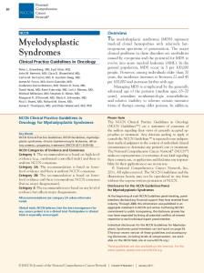
Pathological anatomy of Nutcracker syndrome •
Most typical nutcracker morphologic features imply compression of the LRV between the aorta and the Superior Mesenteric Artery (SMA)
Pathological anatomy defining PCS •
Two anatomic findings define PCS: – Ovarian vein reflux – pelvic varicosities.
•
Each may be seen without the other or both can be present in asymptomatic patients.
Clinical presentation of Venous Compressive Syndromes of the pelvis & abdomen
Venous Hypertension
I Contained within the Pelvis Pelvic Congestion syndrome
II Leaks into the legs through escape veins Chronic venous disease of the legs
Combination of I & II
When venous hypertension is contained within the pelvis: PCS – clinical presentation: •
•
•
•
Lower abdominal/pelvic pain • Chronic • Dull • May have a predisposition to the left flank with radiation to the buttock(mimicking sciatica) Relieving and aggravating factors • Relieved by laying down • Aggravated by standing, increased intra-abdominal pressure(pregnancy), premenstrual period. Associated with • Dyspareunia • Bladder irritability & urgency • Vulvar /vaginal varicosities Other non specific symptoms: • Fullness in legs, lethargy, depression, abdominal or pelvic tenderness, vaginal discharge, dysmenorrhea, swollen vulva, lumbosacral neuropathy, rectal discomfort, non specific GI symptoms. Asciutto G, Asciutto KC, Mumme A, Geier B. Pelvic venous incompetence: reflux patterns and treatment results. Eur J Vasc Endovasc Surg 2009;38:381-6.
Clinical presentation of Nutcracker syndrome • •
Asymptomatic hematuria or hematuria with severe pelvic pain. Signs are aggravated by physical activity – Hematuria – Varicocele – Orthostatic proteinuria – Orthostatic intolerance
•
Hematuria, pelvic pain, pelvic varicosities, and varicoceles are the most common clinical signs that should raise suspicion for the diagnosis
When venous hypertension is transmitted to the lower extremities -- Non Saphenous Varicosities
The ‘Escape Veins’
Incidence of PCS: The magnitude of the challenge •
Chronic pelvic pain; • Responsible for 30% of outpatient Gyn. visits in the US • Potentially affecting up to 40% of the female population during their lifetime
•
Pelvic Congestion Syndrome; – PCS accounts for 30% of the patients presenting with Chronic pelvic pain.
•
For patients with lower extremity varicose veins; – 10-15% have non saphenous varicosities
Gyn. Outpatient visits
PCS
All other
PCS – Demographics & Differential diagnosis •
Demographics – Primarily premenopausal age range 20-45 years – Most 20-40 years of age – Genetic or ethnic predilections are unclear – Family history and multiparity are both risks
•
Differential diagnosis – Pelvic Malignancy – Endometriosis (39%) – Pelvic Congestion Syndrome (31%) – Pelvic Inflammatory disease (11%) – Adhesions (10%) – Fibroids (4%) – Other (5%) Soysal et al, Hum Reprod 2001
Diagnosis of Venous Compressive Syndromes
US diagnosis for PCS
•
•
•
Helping identify etiology – Primary PCS – Secondary PCS – Lt. Common Iliac vein stenosis – Lt. Renal vein stenosis Proving sequelae of PCS • Ovarian vein (dilatation and reflux) • Peri-uterine veins(dilatation and reflux) Pelvic and lower extremity (LE) duplex ultrasound (US) examination, – performed preferably while standing. – With transabdominal 5-MHz and transvaginal probes – After 3 days of a no-residue diet and an empty stomach.
CTV/MRV for PCS •
• •
Good sensitivity, fair specificity ** Supine position can decrease size Criteria – >4 tortuous pariuterine veins – Periuterine veins > 4mm – Ovarian vein diameter > 8mm Clinical Practice Guidelines, SVS, JVS 2011
Schematic representation of nutcracker phenomenon
Hilar portion of the left renal vein and the gonadal vein are distended. Distended lumbar and azygous collaterals may be seen in some cases.
Diagnostic Venography: The Gold standard Indications: 1) Suspected PCS based on non invasive imaging, proceed with intervention in the same sitting if confirmed 2) Suspected PCS - Equivocal non invasive imaging.
Diagnostic Venography – ‘The Gold standard’
Intra Vascular Ultrasonography
Treatment of Pelvic Congestion Syndrome •
• • •
TEAM APPROACH – Gynecologist with special interest in pelvic pain – Psychologist – Vascular Specialist – Venous Disease specisalist – Administrator with special interest in treating these extremely frustrated patients Rule out Malignancy Hormonal therapy Interventional therapy
Treatment of Pelvic Congestion syndrome
Schematic – Ovarian Vein Embolization
CONCLUSIONS For this CME talk •
Concept of venous hypertension: – Lower extremity and pelvis; Pelvis -> escape veins -> lower extremities
•
Common causes of Pelvic venous hypertension; – Pelvic Venous Compressions( Non Thrombotic compressions) ; May Thurner Syndrome, Nutcracker Syndrome – Enhanced venous inflow – Ovarian vein reflux ; Ovarian vein reflux
– Thrombotic occlusions •
Diagnosis of Venous Compression Syndromes: –
•
Clinical presentation, Surface Ultrasound, CT/MRI, Venography & IVUS
Treatment of Venous Compression Syndromes: –
Rule out D/D, Medical and psychological care, Iliac vein Stenting, Embolization of the Ovarian veins
Thank You for your attention !!!