PAIN POTPOURRI: INFECTIOUS/POSTINFECTIOUS ARTHRITIS, PAIN SYNDROMES AND NONRHEUMATIC SYNDROMES Rheumatology Board Review
Tova Ronis Rheumatology Fellow
REACTIVE ARTHRITIS
REACTIVE ARTHRITIS • Follows salmonella, shigella, campylobacter, yersinia and chlamydial infection • Associated with HLA B27 • Arthritis, enthesitis characteristic features • May see acute iritis, urethritis
REACTIVE ARTHRITIS TREATMENT • Antibiotics NOT indicated with diarrheal associated reactive arthritis • Antibiotic treatment may improve rate of recovery with Chlamydial reactive arthritis
REACTIVE ARTHRITIS TREATMENT • NSAIDS, especially indomethacin if tolerated, are beneficial • May be chronic • Sulfasalazine in chronic cases
INFECTIONS MIMICKING RHEUMATIC DISEASES
INFECTIONS MIMICKING RHEUMATIC DISEASES • Septic arthritis • Osteomyelitis • Necrotizing fasciitis • Toxic shock
DISTINGUISHING INFECTION FROM ARTHRITIS • Severe pain, unable to ambulate • Point tenderness (osteomyelitis) • Remember in children most likely place for osteo is near growth plate—sympathetic sterile effusions common • Monoarticular JIA involving hip RARE in young – 3 phase Technitium bone scan: fast, helps with osteo if asymmetric. – MRI with Gad—best for osteo and imaging joint swelling
• Very toxic appearing—invasive Group A strep, fasciitis, toxic shock
POSTSTREPTOCOCCAL SYNDROMES
POSTSTREPTOCOCCAL SYNDROMES • Classical acute rheumatic fever • Poststreptococcal arthritis Issues: How to diagnose? How to treat? Prophylaxis?
REVISED JONES CRITERIA FOR DIAGNOSIS OF ACUTE RHEUMATIC FEVER MAJOR Carditis Polyarthritis Chorea Erythema marginatum Subcutaneous nodules
MINOR Fever Arthralgia Previous history of rheumatic fever or rheumatic heart disease
Plus: Supporting evidence for preceding streptococcal infection; including increased ASO or other streptococcal antibodies, positive throat culture or recent scarlet fever
ACUTE RHEUMATIC FEVER FEVER ARTHRITIS CARDITIS E. MARGINATUM CHOREA SUBCUTANEOUS NODULES
2 4 6 8 10 12 14 16 18 20 22 24 WEEKS
ARF Secondary Prophylaxis • Without carditis: 5 years or until age 21 • With carditis but no residual cardiac disease: 10 years or well into adulthood • With carditis and residual cardiac disease: over 10 years after last episode, or at least age 40, or lifelong
Regimens • Penicillin 1.2M units IM q 34 weeks, preferred (especially for carditis) • Penicillin 250 mg orally bid • Sulfasoxizole or Erythromycin for Pen allergic
POSTSTREPTOCOCCAL ARTHRITIS • Clinical picture does NOT meet the modified Jones criteria • Longer duration of arthralgias, arthritis • No skin rashes • Carditis can be seen • Prophylaxis controversial Red Book waffles – We recommend 1 yr without carditis or if carditis occurs, same as ARF
GONOCOCCAL, MENINGOCOCCAL AND OTHER INFECTIOUS ARTHRITIS
GONOCOCCAL ARTHRITIS PRESENTING FINDINGS
Migratory polyarthritis Tenosynovitis *
6670% 67%
Dermatitis
3967%
Fever
5163%
Purulent arthritis
4280%
Monarthritis
2732%
Polyarthritis
1053%
GONOCOCCAL ARTHRITIS • N. gonorrhoeae difficult to grow in culture • PCR can pick up 1 organism in joint fluid However, some joints may be immune complex mediated, especially in migratory phase
MENINGOCOCCAL ARTHRITIS 3 TYPES • Acute polyarthritis associated with meningococcemia and meningitis • mono/oligoarthritis: 7 days after onset of infection (?sterile) • Primary acute arthritis without meningococcemia Sometimes arthritis and dermatitis due to meningococcus looks similar to GC.
ARTHRITIS ASSOCIATED WITH MYCOBACTERIA AND MYCOSES Coccidioidomycoses Polyarthritis with E. nodosum Monoarthritis of knee Blastomycosis Monarthritis of large lower extremity joints associated with skin and joint involvement, spondylitis Cryptococcosis Monoarthritis secondary to osseous infection, spondylitis Histoplasmosis Polyarthritis with E. nodosum Sporotrichosis
Monoarthritis of knee, wrist or hand; Polyarthritis with disseminated skin lesions
ARTHRITIS ASSOCIATED WITH MYCOBACTERIA AND MYCOSES Tuberculosis Atypical TB
Candidiasis Actinomyces Leprosy
Spondylitis, monoarthritis of large weight bearing joints Arthritis/tendonitis in hand or wrist or bone/joint/tendon involvement in AIDS Monoarthritis of knee with serious systemic illness Spondylitis Polyarthritis with E. nodosum; Destruction of small bones of hands and feet; neuropathic joints
LYME DISEASE
LYME DISEASE • Recognized in 1975 with an unusual epidemic of JIA in Lyme, Conn. • Now recognized in North America, Europe, Russia, China, Japan and ?Australia • 3 endemic areas in US
LYME DISEASE • Organism: borrelia burgdorferi in US, b. afzalii and b. garinii in Europe and Asia • Infects ticks that infest deer and whitefooted mouse, woodrats on West Coast • TICKS: – Ixodes scapularis (Northeast US, Great Lakes) Preferred host is deer and whitefooted mice—and humans – Ixodes pacificus (Northern Calif/Oregon)preferred host is lizard (are not susceptible to infection), lesser host is woodrat (is susceptible)—and humans – Ixodes neotoma (Northern Calif/Oregon)preferred host is woodrats, but do not bite humans – Ixodes ricinis (Europe)
• Ticks need to feed for 12+ h to transmit spirochete
HOW TO REMOVE A TICK
LYME DISEASE CLINICAL STAGES STAGE 1 EARLY LOCALIZED
Erythema migrans
EARLY DISSEMINATED
Secondary skin lesions Headache Musculoskeletal pain Flulike illness
LYME DISEASE CLINICAL STAGES STAGE 2 Cardiac (5%)
Acute neurologic (15%)
Onset weeks to months 15% total cases Fluctuating AV block, first degree, Wenkebach, complete Sometime carditis Organisms can be found in tissue Meningitis +/ encephalitis Cranial neuritis, radiculoneuritis Chorea, transverse myelitis Focal demyelinating encephalitis Polyneuropathy Not clearly local infection
LYME DISEASE CLINICAL STAGES STAGE 3 CHRONIC ARTHRITIS
Months after onset, 60% of pts 12 large joints, esp. knees Popliteal cysts PCR often positive Usually resolves with treatment but 10% can become chronic
CHRONIC NERVOUS SYSTEM DISEASE
Onset months to years, 5% of pts Cognitive dysfunction Parasthesias Changes in EMG, NCV
LYME DISEASE DIAGNOSIS • Clinical syndrome and exposure • Lyme ELISAhigh false positive rate (very rare falsenegatives with arthritis) • Positive ELISA needs to be confirmed with Western Blot Need to see 5 of 10 specific bands • Serology may not be positive for a few weeks after infection • PCR may be positive in joint fluid Guideline in Ann Int Med 1997, 127: 11061108
LYME DISEASE TREATMENT Early
Amoxicillin, Doxycycline, 21 d Cefuroxime, ?Azithromycin Early neuro Bell’s palsy onlyoral antibiotics 21 d MeningitisCeftriaxone, Pen G 14 28 d IV Doxycycline Arthritis Amoxicillin + Probenecid 30 d Doxycycline 30 d Ceftriaxone, Pen G 1430 d Carditis Ceftriaxone, Pen G 14 d Doxycycline, Amoxicillin 21 d
VIRAL ARTHRITIS
VIRAL ARTHRITIS Parvovirus B19
Small single stranded DNA virus Erythema infectiosum (Fifth’s Disease) Rash fades in 10 d but may recur 7% of children have arthralgias
Arthritis occurs in up to 50% adults and 5% children Arthritis involves multiple joints, hands fingers, feet Can see chronic disease
VIRAL ARTHRITIS Hepatitis B
“Serum sickness” early Rash maculopapular or urticarial Nonmigratory polyarthritis Immune complex mediated Symptoms decrease as jaundice increases Can see arthritis following Hep B vaccination
Hepatitis C
Multiple autoimmune syndromes including arthritis
VIRAL ARTHRITIS Rubella
33% of older patients (adolescents and older), Onset 23 days following rash Polyarthritis Duration usually 1 week ? Chronic forms Virus present in blood and synovial fluid
Rubella vaccine Occurs 1028 d after immunization 511% of adult women, much lower in children
VIRAL ARTHRITIS Less common viruses associated with arthritis: Alpha viruses (Australia, S. Pacific, Africa, Asia) Mumps CMV EBV Varicella Enteroviruses HIV (Reiter’s Syndrome)
OTHER INFECTIONRELATED SYNDROMES Brucellosis
Joint involvement in 33%, sacroiliitis 47% peripheral 38%
Cat scratch fever Monoarthritis Mycoplasma and ureaplasma
Associated with hypogammaglobulinemia
PRESUMED INFECTION RELATED SYNDROMES Toxic synovitis HenochSchoenlein purpura Kawasaki syndrome Discitis
MALIGNANCIES
NEOPLASTIC CONDITIONS AFFECTING BONE • • • •
Leukemia Neuroblastoma Lymphoma Malignant tumors of bone, cartilage and synovium • Metastatic disease • Pigmented villonodular synovits • Histiocytosis
CHILDHOOD LEUKEMIA PRESENTING AS BONE PAIN • May be first and most prominent symptom in 18% of children • May have no abnormal cells in peripheral CBC • Radiographs, platelet count, Hgb, WBC may be entirely normal • ESR is usually (but not always) elevated • Presence of atypical lymphocytes on smear is suspicious
NONMALIGNANT CONDITIONS AFFECTING BONE
NONMALIGNANT CONDITIONS AFFECTING BONE • Osteoid osteoma • Osteochondroses • Slipped capital femoral epiphysis • Aneurysmal bone cyst • Chondromalacia patellae • Occult trauma
OSTEOID OSTEOMA
Osteoid Osteoma
LEGGPERTHES DISEASE
LeggCalvéPerthes disease
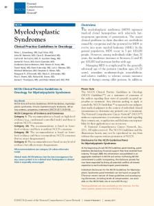
Slipped Capital Femoral Epiphysis
A Klein line is a line drawn along the superior border of the femoral neck that would normally pass through a portion of the femoral head. If not, slipped capital femoral epiphysis is diagnosed.
Neurologic disorders • Muscular dystrophies • Gait disturbances and limps secondary to neurologic disease – e..g., mild cerebral palsy
• GuillainBarre
MECHANICAL PAIN SYNDROMES
MECHANICAL PAIN SYNDROMES • Limb pains or "growing pains” • Patellofemoral syndrome • Benign hypermobility syndrome
CHARACTERISTICS OF LIMB PAINS IN CHILDHOOD • • • • • • • • • •
Age 4 to 13 years Lower extremities>>upper Nocturnal>>daytime Calves, thighs or shins>>joints No pattern of recurrence ? Association with activity Girls>boys Respond to heat, massage or mild analgesia No lab or Xray findings Fine in morning
PATELLOFEMORAL PAIN • • • •
Female>male Age at onset 1113 years Knee paingrating, catching, instability Pain increased with descending stairs, squatting, kneeling, running • Unable to sit for long periods without extending knee • Physical findingscrepitance, +patellar apprehension test, patellar compression test
NORMAL
MEDIAL
MALALIGNED
FEMORAL CONDYLES
LATERAL
Prepubertal body habitus
Postpubertal body habitus
Lateral forces
BENIGN HYPERMOBILITY SYNDROME Clinically based diagnosis: • Passive dorsiflexion at MCPs to 90 o • Apposition of the thumb to the flexor aspect of the forearm • Hyperextension of the elbow > 5 o • Hyperextension of the knee > 5 o • Forward trunk flexion placing hands flat on floor with knees extended
BENIGN HYPERMOBILITY SYNDROME • Up to 10% of normal population • Arthralgias common • Injuries common (dislocation of affected joints, back pain, meniscal and tendon injuries) • Increased incidence of mitral valve prolapse • Genetically determined • Generalized laxity of tissues? • EhlersDanlos syndrome type 3?
PAIN SYNDROMES
PAIN SYNDROMES • Myofascial pain syndrome • Fibromyalgia syndrome • Chronic fatigue syndrome • Reflex sympathetic (neurovascular) dystrophy • Psychogenic pain syndrome • Remember somatization is a symptom of depression in adolescence
CENTRALLY MEDIATED PAIN SYNDROMES • • • • • • •
Fibromyalgia Irritable bowel syndrome Migraines Tension headaches TMJ disorder Irritable bladder, interstitial cystitis Myofascial pain syndromes
“NeuroChemicalEndocrine disorders”
EVIDENCE FOR CENTRAL SENSITIZATION • Neurophysiologic testing reveals abnormal nociceptive processing • Dysautonomia • Increased (3X) substance P in CSF • Regional modulation in CNS blood flow in hemithalami, caudate nuclei, pontine tegmentum • Abnormal summation of pain signals “windup” • Abnormal somatomedin, prolactin and cortisol stress response
MYOFASCIAL PAIN • Localized, muscular pain • No generalized aching, stiffness or fatigue • Responds to local therapy
TRIGGER POINTS Subscapularis Lower trapezius Upper trapezius
FIBROMYALGIA SYNDROME • Occurs in prepubertal, pubertal and adolescents • Females>>>males • Diffuse tenderness at discrete anatomic locations termed tender points • Generalized chronic musculoskeletal pain • Fatigue • Sleep disturbances • Parasthesias • Headaches • Depression and anxiety • Relationship to chronic viral/chronic fatigue syndrome?
FIBROMYALGIA TENDER AREAS
REFLEX SYMPATHETIC DYSTROPHY • • • •
Age prepubertal through early to mid adolescence Females>males One or more extremities may be involved Diagnosis requires the presence of physical abnormalities including temperature and color changes or swelling • Diffuse exquisite pain to light touch and severe dysfunction are hallmarks • Frequently see highachieving, perfectionistic children with close (enmeshed?) family units
“Pain syndromes” • • • •
Early diagnosis—the pediatrician’s job Confidence of diagnosis Clear, concrete plan of treatment Patient/family/treating team/payer in full support of plan • Psychosocial intervention essential—stress management, coping skills • Specialized PT/OT approaches: Fibromyalgia Tender areas, aerobic exercise, “work through pain and fatigue” RSD Desensitization, normal use, “work through pain”
EDUCATIONAL GOALS • Minimize home tutoring • Maximize school attendance • Maximize least restrictive environment by: Individualized educational plans (IEP) Resource classes Coordination with therapy units Special education classes
QUESTIONS?
A 16yearold boy presents with a very swollen, painful right knee. He is a soccer player, but there is no history of recent injury. During the interview, you notice the boy has injected conjunctivae.
A 16yearold boy presents with a very swollen, painful right knee. He is a soccer player, but there is no history of recent injury. During the interview, you notice the boy has injected conjunctivae. Of the following, further evaluation MOST likely will reveal A) alopecia areata B) Gottron papules C) KayserFleischer rings D) malar rash E) urethritis
A 16yearold boy presents with a very swollen, painful right knee. He is a soccer player, but there is no history of recent injury. During the interview, you notice the boy has injected conjunctivae. Of the following, further evaluation MOST likely will reveal A) alopecia areata B) Gottron papules C) KayserFleischer rings D) malar rash E) urethritis
An 18yearold postpartum girl presents to your clinic with complaints of discomfort in her knees and hands. She denies any swelling or erythema of these areas or recent trauma. She takes no regular medications, and she has been healthy until 6 days ago, when she developed these complaints. She did receive a rubella vaccination about 1 month ago because on her first visit to an obstetrician during her pregnancy she was found to have a negative serum titer to rubella. Her physical examination findings are normal. Of the following, the MOST likely diagnosis is A) adverse effect of the rubella vaccine B) anicteric hepatitis B infection C) poststreptococcal arthritis D) reactive arthritis due to Salmonella sp E) recent infection with parvovirus
An 18yearold postpartum girl presents to your clinic with complaints of discomfort in her knees and hands. She denies any swelling or erythema of these areas or recent trauma. She takes no regular medications, and she has been healthy until 6 days ago, when she developed these complaints. She did receive a rubella vaccination about 1 month ago because on her first visit to an obstetrician during her pregnancy she was found to have a negative serum titer to rubella. Her physical examination findings are normal. Of the following, the MOST likely diagnosis is A) adverse effect of the rubella vaccine B) anicteric hepatitis B infection C) poststreptococcal arthritis D) reactive arthritis due to Salmonella sp E) recent infection with parvovirus
An 11yearold girl presents 2 weeks after an office visit for a presumed viral illness characterized by fever, malaise, and flushing of the cheeks. Today, her mother notes that she no longer has a fever, but she complains of pain in her knees and elbows. On physical examination, the left knee is slightly swollen and warm but not erythematous. The girl reports pain on movement of both elbows, but there are no physical findings on examination of the elbows or other joints. The remainder of the physical examination findings are normal, except for an oral temperature of 100.6°F (38.1°C). Results of laboratory studies include a white blood cell count of 8.9x10³/mcL (8.9x10⁹/L) with 40% polymorphonuclear leukocytes, 45% lymphocytes, and 15% monocytes; hemoglobin of 11.0 g/dL (110.0 g/L); platelet count of 472.0x10³/mcL (472.0x10⁹/L); and erythrocyte sedimentation rate of 20 mm/hr. Of the following, the MOST likely pathogen to cause this child's joint complaints is A) Borrelia burgdorferi B) Coxsackievirus C) group A betahemolytic streptococci D) influenza A virus E) parvovirus B19
An 11yearold girl presents 2 weeks after an office visit for a presumed viral illness characterized by fever, malaise, and flushing of the cheeks. Today, her mother notes that she no longer has a fever, but she complains of pain in her knees and elbows. On physical examination, the left knee is slightly swollen and warm but not erythematous. The girl reports pain on movement of both elbows, but there are no physical findings on examination of the elbows or other joints. The remainder of the physical examination findings are normal, except for an oral temperature of 100.6°F (38.1°C). Results of laboratory studies include a white blood cell count of 8.9x10³/mcL (8.9x10⁹/L) with 40% polymorphonuclear leukocytes, 45% lymphocytes, and 15% monocytes; hemoglobin of 11.0 g/dL (110.0 g/L); platelet count of 472.0x10³/mcL (472.0x10⁹/L); and erythrocyte sedimentation rate of 20 mm/hr. Of the following, the MOST likely pathogen to cause this child's joint complaints is A) Borrelia burgdorferi B) Coxsackievirus C) group A betahemolytic streptococci D) influenza A virus E) parvovirus B19
A 15yearold young woman has had joint pain for the past 3 days. She developed fever, chills, and fatigue 4 days ago, but the fever has resolved. In addition, she explains that her left elbow, right knee, and right wrist are all painful, red, and swollen, and she has a rash on her hands and feet that looks like pusfilled bumps. She is sexually active, with inconsistent condom use for contraception. Physical examination reveals an afebrile young woman who has swelling, tenderness, and mild erythema of the left elbow, right knee, and right wrist. She has a few pustules and vesicles on the right palm and bilateral soles. The abdomen is not tender and is without masses.
A 15yearold young woman has had joint pain for the past 3 days. She developed fever, chills, and fatigue 4 days ago, but the fever has resolved. In addition, she explains that her left elbow, right knee, and right wrist are all painful, red, and swollen, and she has a rash on her hands and feet that looks like pusfilled bumps. She is sexually active, with inconsistent condom use for contraception. Physical examination reveals an afebrile young woman who has swelling, tenderness, and mild erythema of the left elbow, right knee, and right wrist. She has a few pustules and vesicles on the right palm and bilateral soles. The abdomen is not tender and is without masses. Of the following, the MOST likely pathogen causing this patient's symptoms is A) Chlamydia trachomatis B) group A betahemolytic streptococci C) Neisseria gonorrhoeae D) parvovirus B19 E) Treponema pallidum
A 15yearold young woman has had joint pain for the past 3 days. She developed fever, chills, and fatigue 4 days ago, but the fever has resolved. In addition, she explains that her left elbow, right knee, and right wrist are all painful, red, and swollen, and she has a rash on her hands and feet that looks like pusfilled bumps. She is sexually active, with inconsistent condom use for contraception. Physical examination reveals an afebrile young woman who has swelling, tenderness, and mild erythema of the left elbow, right knee, and right wrist. She has a few pustules and vesicles on the right palm and bilateral soles. The abdomen is not tender and is without masses. Of the following, the MOST likely pathogen causing this patient's symptoms is A) Chlamydia trachomatis B) group A betahemolytic streptococci C) Neisseria gonorrhoeae D) parvovirus B19 E) Treponema pallidum
A 12yearold girl presents to your office for the first time with a swollen, painful, erythematous right knee joint. She tells you that her left knee felt and looked similar yesterday, but now feels normal. She also is easily fatigued and has had fever. On physical examination, she has a temperature of 101.7°F (38.7°C), a heart rate of 125 beats/min, a respiratory rate of 24 breaths/min, and a blood pressure of 120/78 mm Hg. Her lungs are clear. On auscultation, you note a 3/6 holosystolic murmur at the cardiac apex with radiation to the axilla. Of the following, the BEST plan for management of this patient's joint swelling includes A) antibiotic therapy with doxycycline B) anti/inflammatory therapy with aspirin C) aspiration of the right knee joint D) heat, elevation, and splinting of the right knee E) immunotherapy with azathioprine
A 12yearold girl presents to your office for the first time with a swollen, painful, erythematous right knee joint. She tells you that her left knee felt and looked similar yesterday, but now feels normal. She also is easily fatigued and has had fever. On physical examination, she has a temperature of 101.7°F (38.7°C), a heart rate of 125 beats/min, a respiratory rate of 24 breaths/min, and a blood pressure of 120/78 mm Hg. Her lungs are clear. On auscultation, you note a 3/6 holosystolic murmur at the cardiac apex with radiation to the axilla. Of the following, the BEST plan for management of this patient's joint swelling includes A) antibiotic therapy with doxycycline B) anti/inflammatory therapy with aspirin C) aspiration of the right knee joint D) heat, elevation, and splinting of the right knee E) immunotherapy with azathioprine
When a 14yearold girl had frequent complaints of shoulder pain made worse by pitching softball a few months ago, you diagnosed overuse injury. Nonsteroidal antiinflammatory drugs and rest have provided some relief. She presents today with complaints of recurrent upper arm pain that is unrelated to exercise and sometimes awakens her from sleep. Physical examination reveals a slightly larger circumference of the left proximal humerus compared with the right. There is minimal tenderness on palpation over the area, although the girl reports a constant ache. She has full range of motion of the arm at the shoulder and elbow. You obtain a shoulder radiograph.
When a 14yearold girl had frequent complaints of shoulder pain made worse by pitching softball a few months ago, you diagnosed overuse injury. Nonsteroidal antiinflammatory drugs and rest have provided some relief. She presents today with complaints of recurrent upper arm pain that is unrelated to exercise and sometimes awakens her from sleep. Physical examination reveals a slightly larger circumference of the left proximal humerus compared with the right. There is minimal tenderness on palpation over the area, although the girl reports a constant ache. She has full range of motion of the arm at the shoulder and elbow. You obtain a shoulder radiograph. Of the following, the MOST likely diagnosis is A) acromioclavicular separation B) acute osteomyelitis C) chronic osteomyelitis D) osteosarcoma E) supracondylar fracture of the humerus
When a 14yearold girl had frequent complaints of shoulder pain made worse by pitching softball a few months ago, you diagnosed overuse injury. Nonsteroidal antiinflammatory drugs and rest have provided some relief. She presents today with complaints of recurrent upper arm pain that is unrelated to exercise and sometimes awakens her from sleep. Physical examination reveals a slightly larger circumference of the left proximal humerus compared with the right. There is minimal tenderness on palpation over the area, although the girl reports a constant ache. She has full range of motion of the arm at the shoulder and elbow. You obtain a shoulder radiograph. Of the following, the MOST likely diagnosis is A) acromioclavicular separation B) acute osteomyelitis C) chronic osteomyelitis D) osteosarcoma E) supracondylar fracture of the humerus
THANKS!