T h e O f f i c i a l J o u r n a l o f C e n t e r f o r Ve i n R e s t o r at i o n Vol. 8, Issue 2 June 2015
inside this issue
Part 1 Lower Extremity Venous Insufficiency....................................................................... Wellness Today................................................................................................................ Q&A’s . . ............................................................................................................................ Stronger Together............................................................................................................ Community Outreach........................................................................................................ Your Career Journey......................................................................................................... Our Physicians & Locations...............................................................................................
Page Page Page Page Page Page Page
1-3, 8-9 4 5 6 7 10 11
Lower extremity venous insufficiency MUST be evaluated and treated as a part of ‘Infra-diaphragmatic venous disease’. ‘A FIVE PART SERIES’ By Sanjiv Lakhanpal, MD, FACS Summary: Our venous system from toes to the right atrium is one continuous system of fancy pipes with anatomic and physiological enhancements to facilitate venous return to the heart. Compartmentalizing the evaluation of this one single system of veins only makes sense for lower grades (CEAP 0-1) of venous insufficiency in the legs. For higher grades (CEAP 2-6) of venous insufficiency it is essential to evaluate the entire infra-diaphragmatic venous system. In this five part review I will lay out the compelling case for such clinical evaluation and for appropriate treatment tailored to the needs of the individual patient if the need is substantiated by a more detailed diagnostic workup. This review will be broken down into the following parts: Editor-in-Chief, President & CEO, Center for Vein Restoration Sanjiv Lakhanpal, MD, FACS
Associate Editor, Director of Research Director of Vascular Labs Shekeeb Sufian, MD, FACS Managing Editor • Kathleen A. Hart ISSN 2159-4767 (Print), ISSN 2159-4775 (Online)
Copyright © 2015 Center for Vein Restoration. All rights reserved.
Part I: The anatomic logic for evaluation of the entire infra-diaphragmatic venous system in patients with advanced lower extremity venous disease. Part II: The physiologic logic for evaluation of the entire infra-diaphragmatic venous system in patients with advanced lower extremity venous disease.
Part III: Pathologic conditions leading to post-ambulatory venous hypertension in the lower extremities. Part IV: Diagnosis of Infra-diaphragmatic venous insufficiency, venous diseases of the lower extremity and Pelvic Congestion syndrome.
Part V: Treatment of Infra-diaphragmatic venous insufficiency, venous diseases of the lower extremity and Pelvic Congestion syndrome.
Continued on Page 2
Lower extremity venous insufficiency MUST be evaluated and treated as a part of ‘Infra-diaphragmatic venous disease’. Part I: The Anatomic logic. Superficial venous system of the lower extremity: The skin and the subcutaneous tissues are drained by the great and the small saphenous veins. Both the GSV and the SSV start from the medial and lateral extensions of the dorsal venous arch respectively.
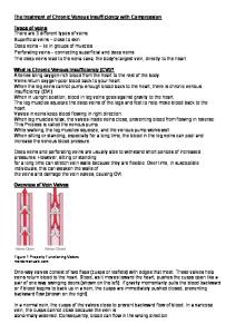
The Small Saphenous Vein (SSV) begins as the lateral continuation of the dorsal venous arch. It may drain completely into the popliteal vein, have a small extension that continues cranially as the cranial extension, or the entire SSV may continue cranially to drain into the femoral vein or the GSV. The intersaphenous vein starts below the facia and then penetrates the fascia to come superficial to it, the intersaphenous vein is seen in 2/3rds of the limbs with venous disease. SSV valves; More numerous (median 7-10) Range 4-13. Highest valve is close to the termination of the SSV. Valves in a SSV GSV connections directs flow from from SSV to GSV.
Image 001
The Great Saphenous Vein (GSV) begins just anterior to the medial ankle, ascends medial to the knee ascending along the medial side of the thigh and enters the fossa ovalis 3 cms. inferior and three cms. lateral to the pubic tubercle. It is duplicated in the calf in 25% and in the thigh in 8%. Tributaries of the GSV include: Posterior accessory GSV of the leg (>75%)– joins the GSV distal to the knee, begins posterior to the medial malleolus, anterior accessory GSV of the leg, Posterior accessory GSV of the thigh and anterior accessory GSV of the thigh. GSV usually has at least 6 valves (14-25), with a constant valve between 2-3 cms from the SFJ(85%). The frequency of valves is greater below the knee.
Image 003
Like the rest of our body, the lower extremity is drained by a system of superficial and deep veins. ‘Classic’ superficial veins lie superficial to the deep (muscular) fascia. The perforator veins, as the name implies, perforate the deep fascia to connect the superficial veins to the deep veins. Varicosities invariably lie superficial to the superficial (saphenous fascia - GSV, membranous layer - SSV) fascia, free of any fascial restraints. Reticular veins lie in the sub-dermal connective tissue and the so-called pin veins or spider veins are caused by the dilatation of the dermal plexus of veins. Saphenous fascia is an additional layer of fascia which encapsulates the great saphenous vein. The great and the small saphenous veins are interfascial veins.
Image 004
Image 002
(800) FIX-LEGS
www.centerforvein.com
2
Lower extremity venous insufficiency MUST be evaluated and treated as a part of ‘Infra-diaphragmatic venous disease’. Continued from Page 2
The deep veins in general follow the corresponding arteries. The deep plantar venous arch from around the heads of the metatarsals continues as the medial and lateral plantar veins, which both become the posterior tibial veins behind the medial ankle. In over 80% of the samples they are paired and in about 10% they are triplicate. They perforate the soleus muscle close to its bony arcade and continue as the popliteal vein. The peroneal veins originate in the distal third of the calf. The peroneal receives the large soleal veins. The peroneal and the posterior tibial have constant connections in the distal leg. The peroneal and the posterior tibial form the TP trunk which then joins the popliteal. On the dorsum of the foot the major deep veins continue as the dorsalis pedis veins. The anterior tibial veins ascend in the anterior compartment. The popliteal vein is formed by the confluence of the calf veins. The popliteal and the femoral veins run around the arteries of the same name. The femoral vein is the continuation of the popliteal vein. The deep femoral vein usually communicates with the popliteal vein either directly or through tributaries.
Image 005
The deep veins of the lower extremity: Tibial veins (deep venous system) form by the confluence of the medial and lateral planter veins while on the dorsum of the foot the major deep veins continue as the dorsalis pedis vein.
The sapheno femoral junction and communications with the superficial veins of the lower extremity and the pelvic/abdominal veins: The tributaries of the GSV, namely the superficial circumflex iliac vein, the superficial external pudendal vein and the superficial epigastric vein, communicate freely with the deep veins with similar names that drain into the External iliac vein.
Image 006
Image 009
Image 010
Image 008
(800) FIX-LEGS
www.centerforvein.com
3
Wellness Today
What is Pelvic Congestion Syndrome? By: Vinay Satwah DO, FACOI, RPVI
Many women with Pelvic Congestion Syndrome spend years trying to get an answer as to why they have this chronic pelvic pain. Pelvic Congestion Syndrome is an under-diagnosed condition which is associated with venous disease in the pelvic area, lower abdomen and thighs. Often accompanied by chronic pelvic pain and/or pressure, it is estimated that this condition affects more than one third of all women. Sometimes suffering with this condition for years, many of these women are told the problem is not due to a specific medical cause and may be “all in their head.” However, recent advancements have allowed physicians at Center for Vein Restoration to show that the pelvic pain may be due to varicose vein reflux causing pelvic venous insufficiency.
PAINFUL SYMPTOMS CAN INTERRUPT DAILY ACTIVITIES
The symptoms related to Pelvic Congestion Syndrome include pelvic pain associated with standing and sitting, which worsens throughout the day. This chronic pain is typically dull and aching in nature. Patients often experience relief from pain when lying flat and when legs are elevated. The symptoms may worsen following intercourse, during menstrual periods, and during pregnancy. Associated symptoms include heaviness/fatigue/aching of the legs with varicose veins on the vulva and/or buttocks. Similar to varicose veins in the legs, the valves in the pelvic veins that help return blood to the heart against gravity become weakened and don’t close properly. This allows blood to flow backwards and pool in the vein causing pressure and bulging veins. In the pelvis, varicose veins can cause pain, heaviness and affect the drainage of the uterus, ovaries and vulvar region.
VENOGRAM: THE MOST ACCURATE TEST FOR DIAGNOSIS
Many women with Pelvic Congestion Syndrome spend years trying to get an answer to why they have this chronic pelvic pain. Living with chronic pelvic pain is difficult and affects not only the woman directly, but also her interactions with her family, friends, and her general outlook on life. Our patients will undergo a thorough history and physical. Those with a high suspicion may undergo pelvic ultrasound and venography. Thought to be the most accurate method for diagnosis, a venogram is performed by injecting contrast dye in the veins of the pelvic organs to make them visible during an X-ray. Once a diagnosis is made by a Center for Vein Restoration physician, if the patient is symptomatic, a pelvic venogram with embolization should be done. Embolization is a minimally invasive procedure performed by the interventional team using imaging for guidance. During the outpatient procedure, the faulty, enlarged veins are sealed in order to relieve the painful pressure. After treatment, patients should expect a low level of pain and to spend a couple of days off their feet.
MULTIPLE PREGNANCIES CAN MEAN HIGHER RISK
Pelvic Congestion Syndrome typically affects women in their child bearing years. As the uterus expands during pregnancy, there is increased pressure exerted on the pelvic floor and veins. Post-partum, the uterus eventually contracts and although the pressure on the pelvic floor is relieved, there is residual damage to the pelvic veins. Therefore, the ovarian veins increase in size related to previous pregnancies. Women who’ve had two or more pregnancies and hormonal increases are at particular risk.
(800) FIX-LEGS
A venogram may be performed, involving the injection of dye in the veins of the pelvic organs.
www.centerforvein.com
4
QUESTIONS & Answers In each issue of Venous Review, our medical team answers questions we’ve received from referring physicians.
This issue’s guest Q & A Editors are
Theresa M. Soto, MD FACS, RPVI
Michelle Nguyen, MD
medication prescribed and emergency symptoms that require urgent evaluation in an emergency room. The patient is typically followed up in the office in one week to assess for compliance with and side effects of the medication. A repeat scan is completed to assess for progression/stability of the thrombus at this time as well.
Q: If I send you a STAT DVT rule-out, and it is positive, what do you do? What if it is negative?
A: If the patient’s duplex scan is positive
for DVT. A full patient history and examination is completed in order to evaluate for pulmonary embolism (PE), venous thromboembolism (VTE) risk factors and possible contraindications to the use of blood thinners. We notify the referring physician with the positive result and give the provider the option of treating/ managing the thrombus. Should the referring physician prefer, the CVR physician will start the patient on a blood thinner, typically Xarelto or Lovenox and prescription grade compression stockings. The CVR physician thoroughly counsels the patient on the diagnosis of DVT; risk and symptoms of PE; risks, benefits and side effects of the
Thereafter follow up will be completed as needed and at 4-6 months to evaluate for any residual or chronic venous changes. The patient is co-managed with the primary care physician and a hematologist in order to determine the ideal length of anticoagulant treatment. Testing for an underlying clotting disorder may be completed after completion of anticoagulant therapy pending the patient’s risk profile and history. In the rare event of an acute extensive thrombus involving a large segment of the deep vein, the patient will be referred to the emergency room for intravenous anticoagulation and possible thrombolytic therapy. If the patient’s scan is negative for DVT, the CVR physician will elicit a history from the patient and complete a limited physical examination. A full venous duplex scan may be recommended at this time in order to evaluate for venous insufficiency as a cause of the patient’s leg pain. Pending the results, the physician may recommend additional follow up with another specialist to further investigate the patient’s source of pain. The CVR physician will again notify the referring physician of the findings and recommendations; final disposition of the patient’s follow up will be coordinated with the referring physician in every case.
(800) FIX-LEGS
Q: Why don’t you need to use any anesthesia for procedures — aren’t the patients in pain?
A: At CVR, all our procedures are com-
pletely ambulatory. This means that your patients can drive themselves to their procedure and then drive themselves home. For endovenous ablations, only local anesthesia is used to numb the skin. Then, once we have accessed the vein, we will also instill tumescent anesthesia, which is a mixture of lidocaine, saline and bicarbonate around the vein. This will provide additional anesthesia. Patients are awake the entire time, conversing with us. If any discomfort is felt, we can add more tumescent anesthesia until the patient is comfortable. After the procedure, patients are able to immediately ambulate, and they experience minimal residual effects of the local anesthetic. Our goal is to improve your patient’s quality of life without causing any major interruptions in their everyday life.
www.centerforvein.com
5
Stronger Together CVR Expands in Michigan
Two great companies just joined, to make one perfect fit.
In April of this year, Center for Vein Restoration was proud to announce its expansion to Grand Rapids, Michigan through the acquisition of Grand Rapids Vein Clinic. The expansion marks an exciting milestone for CVR, which already serves western Michigan patients in Kalamazoo/Portage, along with 36 other clinical locations in Connecticut, the District of Columbia, Maryland, New Jersey, New York, Pennsylvania and Virginia. The Grand Rapids Vein Clinic was founded by Joseph Marogil, MD, a leader in the treatment of venous insufficiency in the Grand Rapids Community for over 20 years. His partner, Laura Kelsey, MD, joined the practice in 2005, and has made the treatment of this disease her sole focus. “Venous insufficiency is a condition that affects more than 30 million Americans and we believe that treating this potentially serious problem is a right, not a privilege,” said CVR President and CEO Sanjiv Lakhanpal, MD. “That’s why we’re proud to expand our practice to serve more patients than ever and bring them genuine relief. “CVR is able to bring world-class support services to this new partnership, managing billing, staffing and all the tasks that often take a physician’s focus away from treating patients. Local patients will have access to an expanded suite of advanced treatments for venous insufficiency, and access to a large Patient Services call center offering person-to-person assistance to discuss treatment options, answer insurance questions, and make appointments,” he said. “This is a perfect example how our practices can be stronger together.”
Dr. Kelsey brings extensive experience in the minimally invasive treatment of varicose veins. Board Certified in General Surgery, she was educated at the University of Michigan and Michigan State University’s College of Human Medicine, and completed her residence training in Grand Rapids. She joined the Grand Rapids Vein Clinic in 2005 to focus exclusively on the treatment of venous disease, excelling in the treatment of venous insufficiency. She is passionate about education and has presented at international meetings on the subject of blood coagulation, deep venous thrombosis, prophylaxis, and the treatment of other venous disease. She is a member of the American College of Phlebology, and serves as president of the alumni board of the M.S.U. College of Human Medicine.
The Center for Vein Restoration clinic in Grand Rapids is located at 1720 Michigan Street N.E. Contact the team there at (616) 454-8442 or (800) FIX-LEGS.
If you are a practice looking to partner with a larger organization, or if you’re a physician looking to join one of the most dynamic practices in the country, please feel free to contact us at (240) 965-3900 or e-mail us at
[email protected].
About Dr. Marogil and Dr. Kelsey Founder of Grand Rapids Vein Clinic, Dr. Marogil brings 20 years of experience in the minimally invasive treatment of varicose veins. A Board Certified general surgeon, Dr. Marogil was trained at Butterworth Hospital (currently Spectrum Health). Dr. Marogil started his practice in general surgery in Grand Rapids in 1971. In 1986, he joined the American college of Phlebology and started the Grand Rapids Vein Clinic soon after. Gradually Dr. Marogil focused his practice on the exclusive treatment of varicose and spider veins.
(800) FIX-LEGS
www.centerforvein.com
6
COMMUNITY Outreach
Center for Vein Restoration’s Team has continued its momentum of reaching local residents at events and providing free screenings.
Along with White Plains Hospital, CVR sponsored the “Indulge: Fashion + Fun for Moms” event at the Westchester Mall in White Plains, NY. The May 7 event provided local moms with a much-needed opportunity to treat themselves to a night out to celebrate motherhood through a fun, fashionable and informative shopping experience. CVR staff pictured: Nicole Pabon, Community Outreach Specialist, Nimsi Gonzalez, Community Outreach Coordinator, and Bella Rivera, Community Outreach Specialist.
Our New York CVR team was proud to provide vein health education to customers at the Hudson Square Pharmacy in Midtown Manhattan. This event was held in conjunction with WCBS 880 AM radio. Pictured is Community Outreach Coordinator Nimsi Gonzalez.
CVR’s Community Outreach Team: Helping Spread Awareness of Vein Health
Our Virginia team was out in force at the Southern Women’s Show, April 17-19 at the Richmond Raceway Complex. Along with health education, the event featured fashion, and cooking demonstrations. Celebrity appearances included GRAMMY and Stellar-nominated vocalist Cynthia Jones. Pictured greeting visitors is Meron Hagos, CVR Community Outreach Specialist.
CVR was proud to sponsor the WYCB 1340AM and WPRS 104.1FM 13th Annual Prayer Breakfast. The March event honoring 20 local pastors was held in Martins Crosswinds in Greenbelt, Maryland. Stephanie Jones, Community Outreach Manager and Marsha Withers, Clinical Coordinator represented CVR at the event attended by 1,500 guests.
(800) FIX-LEGS
www.centerforvein.com
7
Lower extremity venous insufficiency MUST be evaluated and treated as a part of ‘Infra-diaphragmatic venous disease’. Continued from Page 3
a) avalvular femoral vein, b) superior epigastric vein, c) superficial circumflex iliac vein, d) median circumflex vein, e) obturator vein, f) inferior gluteal vein, g) superficial external pudendal vein, h) branches of obturator vein, i) deep external pudendal vein, j) long saphenous vein (from phlebolymphology. org) Ext iliac vein: Begins at the inguinal ligament, courses along the pelvic brim, ends anterior to the SI joint by joining the internal iliac vein to form the common iliac vein. Tributaries anastomose freely with the corresponding superficial veins and the obturator veins: deep inferior epigastric veins, deep external pudendal veins (pubic veins), deep circumflex iliac veins. Internal iliac vein: Short trunk formed by the union of its extrapelvic (Gluteal {superior & inferior}, internal pudendal and obturator) & intrapelvic tributaries (lateral sacral visceral {rectal, vesical, uterine and vaginal}. They drain the presacral venous plexus and the pelvic visceral plexus. These plexuses and the additional superficial plexus provide free communication across the midline.
The Inferior Venae Cava: is the largest venous trunk in the body. It is formed by the union of the two common iliac veins in front of the right side of the body of the fifth lumbar vertebra about two and a half centimeters (one inch) to the right of the median plane. In its ascent, it lies upon the bodies of the lower three lumbar vertebrae, the right lumbar and renal arteries, and the right crus of the diaphragm, by which it is separated from the aorta. It passes through the caval opening in the diaphragm opposite the eighth thoracic vertebra, pierces the pericardium and immediately enters the right atrium of the heart. In its upward course it receives the lumbar, the right testicular or ovarian, the renaI, the right phrenic, the right suprarenal, and the hepatic veins. The veins tributary to the inferior vena cava generally follows the same course as the corresponding arteries. Because of the position of the inferior vena cava to the right of the median line, the veins entering it from the left are longer than those from the right side.
Image 013
Image 011
Image 014
Image 012
Common Iliac vein: From the SI joint to the right side of the fifth lumbar vertebrae. RCIV – only tributary is the right ascending lumbar vein (collects blood from the lumbar veins and drains into the Azygos vein). LCIV – left ascending lumbar vein (same as right) and the median sacral vein.
(800) FIX-LEGS
Ovarian vein: Provide drainage to the Parametrium, cervix, mesosalpinx, pampiniform plexus. It forms a rich anastomotic venous plexus with the Paraovarian, uterine, vesical, rectal and vulvar plexus. 2-3 trunks form a single ovarian vein at L4. Left ovarian vein drains into the LRV. Right Ovarian vein drains into the IVC. The ovarian veins have an average diam. of 5mm. Valves are present mainly in the distal third. Absence of valves in 15% on the left side and 6% on the right side.
www.centerforvein.com
8
Lower extremity venous insufficiency MUST be evaluated and treated as a part of ‘Infra-diaphragmatic venous disease’. Continued from Page 8
Inpatients with Pelvic venous hypertension, the Intrapelvic and extrapelvic venous communications may open up (as escape veins) to form non saphenous varicosities in the lower extremities.
Gluteal varicosities as escape veins from the pelvis Lower extremity venous insufficiency may result from a primary pathology in the veins of the lower extremity (GSV -75%, SSV -10%) or within the veins of the pelvis (15-20%). If the pathology lies in the veins of the pelvis, the venous hypertension may be transmitted from the Pelvis to the lower extremity through the escape veins (non-saphenous varicosities) and present with venous insufficiency of the lower extremity in the absence of any pathology in the lower extremity veins. In such cases it is imperative to identify the pathology in the pelvis/abdomen and the treatment has to be focused towards the pathology in the pelvis. Up to twenty percent of the patients presenting with lower extremity venous disease have the etiology of their disease in the pelvis.
Image 015
These non-saphenous veins (NSV) are located in the watershed areas as; Buttock (through the gluteal veins), perineal veins, Vulvar veins, posterolateral thigh (internal iliac system through the femoral vein), lower posterior thigh, popliteal fossa, knee (popliteal/femoral vein) and along the peripheral nerves such as the tibial and sciatic nerve (internal iliac through the gluteal veins).
Image 016
Image 001: http://www.microsurgeon.org/ An atlas of microsurgery techniques and principles. Image is from their image repository, not in an article. http://www.microsurgeon.org/images/toevenoussystem.jpg Image 002: Epidemiology of Chronic Peripheral Venous Disease. Originally in Gloviczki P, and Bergen JJ eds. Atlas of Endoscopic Perforating Vein Surgery Vhttp://intranet.tdmu.edu.ua/data/kafedra/internal/surgery2/classes_stud/en/med/lik/ptn/Surgery/6/TOPIC%2020.%20 DISEASES%20OF%20THE%20VEINS.htm Image 003: http://www.nuvelaesthetica.com/GALLERY2/NuVelaBeforeAfter/images/Saphenous-Perforator-Leg-Veins.jpg http://www.nuvelaesthetica.com/ A Medical Esthetics Laser and Vein Center. From their gallery not associated with any article or person. Image 004: The hemodynamics and diagnosis of venous disease, Journal of Vascular Surgery, Volume 46, Issue 6, Supplement, 2007, S4-S24 http://dx.dol.org/10.1016 Image 005: http://nurse-practitioners-and-physician-assistants.advanceweb.com/SharedResources/AdvanceforNP/Resources/Content/ContentImages/np060103_p28fig1.gif http://nurse-practitioners-and-physician-assistants.advanceweb.com/ In their gallery, not associated with any article or person Image 006:http://jap.physiology.org/content/109/4/1045 Venous emptying from the foot: influences of weight bearing, toe curls, electrical stimulation, passive compression, and posture Barry J. Broderick , Gavin J. Corley , Fabio Quondamatteo , Paul P. Breen , Jorge Serrador , Gearóid ÓLaighin Journal of Applied Physiology Published 1 October 2010 Vol. 109 no. 4, 1045-1052 DOI: 10.1152/japplphysiol.00231.2010 Image 008: http://www.mdanderson.es/sites/default/files/editor/varios/anatomia%20sistema%20venoso3.jpg. From MD Anderson Center website – can’t find source. Image 009: http://www.mdanderson.es/sites/default/files/editor/varios/anatomia%20sistema%20venoso3.jpg . From MD Anderson Center website – can’t find source. Image 010: Phlebolymphology.org Image 011: http://upload.wikimedia.org/wikipedia/commons/0/07/Gray586.png. Henry Gray (1821–1865). Anatomy of the Human Body. 1918 Free Use Image 012: http://lucy.stanford.edu/circulation.html. Stanford.edu Image gallery, not tied to article Image 013: Source Unknown Image 014: http://medicine.stonybrookmedicine.edu/sdmpubfiles/styles/220/public/Pelvic-cropped-shopped-220_0.jpg From Stony brook medicine’s website, cannot find original source or article.Source: Servier.com Image 015: Source: Servier.com Image 016: http://phl.sagepub.com/content/27/6/270/F20.large.jpg. Phlebology September 2012 vol. 27 no. 6 270-288. Three-dimensional modelling of the venous system by direct multislice helical computed tomography venography: technique, indications and results J F Uhl*,† ⇑ Surgical Center, 113 Avenue Victor Hugo, 75116 Paris. †URDIA Research Unit EA4465, University Paris Descartes, 45 rue des saints pères, Paris 75006, France. Correspondence: J-F Uhl MD. 113 avenue Victor Hugo – 75116 – Paris – France *Varicose Vein Email:
[email protected]
(800) FIX-LEGS
www.centerforvein.com
9
WE ARE COMMITTED • • • • • •
A Culture of Collaboration Continuous Education Opportunities for Advancement Solid Team Approach Strong Leadership Positive Employee Experience
journey Our robust growth means that We are always looking for exceptional talent:
Vascular Techs
Nurses
Medical Assistants
Center for Vein Restoration is nationally recognized as the leader in the treatment of vein disease and venous insufficiency – the cause of varicose and spider veins. Our commitment to our employees is strong throughout our culture which is built on our mission and values. We are a network that fuels individual growth, development and promotes from within. Our hiring philosophy is simple: we attract and train the most qualified individuals and provide them the opportunity to demonstrate excellence while achieving career goals!
Administrative Professionals Sales Professionals
Community Outreach
Contact us Find out more about how you can become part of the CVR team. Visit www.centerforvein.com and follow the career link to apply. Or submit your resume and cover letter to:
[email protected] Attention: Human Resources
(800) FIX-LEGS
www.centerforvein.com
10
1
2
3
4
5
6
7
8
9
10
11
12
13
14
15
16
17
18
19
20
21
22
23
24
25
26
27
28
29
30
1. Sanjiv Lakhanpal, MD, FACS 2. Shekeeb Sufian, MD, FACS 3. Thomas Militano, MD, FACS, RPVI 4. Frank Sbrocco, MD 5. Khanh Nguyen, DO, RPVI 6. Eddie Fernandez, MD 7. Stéphane Corriveau, MD 8. Rory C. Byrne, MD 9. Sean K. Stewart, MD 10. Richard Nguyen, MD 11. Arun Chowla, MD, FACS 12. Vinay Satwah, DO, FACOI 13. Michelle Nguyen, MD 14. Mark Edelman, MD 15. Henry Meilman, MD 16. Anuj Shah, MD 17.Lawrence Starin, MD 18. Shubha Varma, MD 19. Theresa Soto, MD, FACOG, FACS, RPVI 20. Duc Le, MD 21. Arvind Narasimhan, MD 22. Seema Kumar, MD, MPP 23. Lauren Best, MD 24. Michael Banker, MD, FACS, FACCP, FACP, RPVI 25. David Shevitz, MD 26. Kourosh C. Ghalili, MD 27. Alfred Jump, MD, MS 28. Laura Kelsey, MD 29. Joseph B. Marogil, MD 30. Arlen G. Fleisher, MD, FACS, RPVI, RVT
O U R
P H Y S I C I A N S
&
L O C A T I O N S
Administrative Office • Maryland Trade Center 2, 7474 Greenway Center Dr., Ste. 1000, Greenbelt, MD 20770 Ph: (240) 965-3200 Maryland Annapolis 108 Forbes St. Annapolis, MD 21401 Ph: (410) 266-3820 Baltimore/Towson 7300 York Rd., Ste. LL Towson, MD 21204 Ph: (410) 296-4876 Bel Air 620 W. MacPhaill Rd., Ste. 104 Bel Air, MD 21014 Ph: (410) 420-3604 Catonsville 1001 Pine Heights Ave., Ste., 303 Baltimore, MD 21229 Ph: (410) 525-1444 Columbia Medical Arts Building, 11085 Little Patuxent Pky., Ste. 203 Columbia, MD 21044 Ph: (410) 730-2784 Easton 401 Purdy Street, Ste. 204 Easton, MD 21601 Ph: (410) 770-9401 Frederick 178 Thomas Johnson Drive, Ste. 104 Frederick, MD 21702 Ph: (301) 662-0200 Germantown 19735 Germantown Rd., Ste. 330 Germantown, MD 20874 Ph: (301) 515-7203 Glen Burnie 1600 Crain Hwy., Ste, 408 Glen Burnie, MD 21061 Ph: (410) 424-2237
Greenbelt 7300 Hanover Dr., Ste. 303 Greenbelt, MD 20770 Ph: (301) 441-8807 North Bethesda/Rockville 11921 Rockville Pike, Ste. 401 Rockville, MD 20852 Ph: (301) 468-5781 Owings Mills 20 Crossroads Dr., Ste. 212 Owings Mills, MD 21117 Ph: (800) FIX-LEGS / (800) 349-5347 Prince Frederick 301 Steeple Chase Dr., Ste. 401 Prince Frederick, MD 20678 Ph: (410) 414-6080 Takoma Park/Silver Spring 831 University Blvd. E, Ste. 24-25 Silver Spring, MD 20903 Ph: (301) 891-6040 Waldorf 12107 Old Line Center Waldorf, MD 20602 Ph: (301) 374-2047 Connecticut Norwalk 40 Cross St., Ste. 240 Norwalk, CT 06851 Ph: (203) 229-0166 Stamford 1290 Summer St., Ste. 2100 Stamford, CT 06905 Ph: (203) 324-4220 District of Columbia DC #2 3301 New Mexico Ave. NW., Ste. 336 Washington, DC 20016 Ph: (202) 244-0783 DC #3 1160 Varnum St. NE, Ste. 212 Washington, DC 20017 Ph: (202) 526-5400
(800) FIX-LEGS
Michigan Grand Rapids 1720 Michigan St. NE Grand Rapids, MI 49503 Ph: (616) 454-8442
Virginia Alexandria 2000 N. Beauregard St., Ste. 310 Alexandria, VA 22311 Ph: (703) 379-0305
Kalamazoo/Portage 3810 West Centre Ave., Ste. A Portage, MI 49024 Ph: (269) 323-8000
Fairfax/Fair Oaks 3700 Joseph Siewick Dr., Ste. 207 Fairfax, VA 22033 Ph: (703) 453-0443
New Jersey Hackensack 211 Essex St., Ste. 403 Hackensack, NJ 07601 Ph: (201) 883-9370 Montclair 127 Pine St., Ste. 5 Montclair, NJ 07042 Ph: (973) 509-3100 North Bergen 8901 Kennedy Blvd., Ste. 3E North Bergen, NJ 07047 Ph: (201) 868-6713 Woodland Park 205 Browertown Rd., Ste. 002 Woodland Park, NJ 07424 Ph: (973) 785-4600 New York Scarsdale 700 White Plains Rd., # 241 Scarsdale, NY 10583 Ph: (914) 725-6800 White Plains 3010 Westchester Ave., Ste. 105 Purchase, NY 10577 Ph: (914) 251-0100 Pennsylvania Bristol 501 Bath Rd., Ste. 215 Bristol, PA 19007 Ph: (215) 788-7400
Fairfax/Merrifield 8316 Arlington Blvd., Ste. 514-A Fairfax, VA 22031 Ph: (703) 289-1122 Fredericksburg 211 Park Hill Drive., Ste. B Fredericksburg, VA 22401 Ph: 540-371-0700 Herndon 150 Elden St., Ste. 210 Herndon, VA 20170 Ph: (703) 437-0601 Leesburg 224 D Cornwall St., Ste. 303 Leesburg, VA 20176 Ph: (703) 443-6971 Manassas 8140 Ashton Ave., Ste. 216 Manassas, VA 20109 Ph: (703) 369-2220 Richmond 2002 Bremo Rd., Ste. 200 Richmond, VA 23226 Ph: (804) 282-4613 Tysons/Vienna 8100 Boone Blvd., Ste. 300 Vienna, VA 22182 Ph: (703) 848-2500 Woodbridge 2200 Opitz Blvd., Ste. 245 Woodbridge, VA 22191 Ph: (703) 490-8585
www.centerforvein.com
11
Maryland Trade Center 2 7474 Greenway Center Drive Suite 1000 Greenbelt, MD 20770
CVR Expands to Pennsylvania!
Visit our website: www.centerforvein.com
From The
Editor
Editor-in-Chief, President & CEO, Center for Vein Restoration Sanjiv Lakhanpal, MD, FACS
T h e O f f i c i a l J o u r n a l o f C e n t e r f o r Ve i n
R e s t o r at i o n
It’s been quite a busy time for us here at Center for Vein Restoration. Since January we’ve opened 10 new clinics, bringing our total of 38 locations across Connecticut, the District of Columbia, Maryland, Michigan, New Jersey, New York, Pennsylvania and Virginia. Such continued, vibrant growth has been a challenging and rewarding undertaking. We believe that there’s a great opportunity in the community for local, specialized medicine to complement the care people traditionally receive in larger institutions and we are committed to modernizing healthcare to be a more efficient and accessible service to patients. Thanks to our physician-led business model, which emphasizes collaboration, camaraderie and support – along with your continued referrals -- we’re proud to report we’re able to serve more patients than ever. In this edition of Venous Review we are launching the first in a 5-part original series discussing the importance of including evaluations performed below the diaphragm as part of diagnostic work-up for venous disease. Also in this edition, we examine an under-diagnosed condition in females, associated with venous disease: Pelvic Congestion Syndrome. We also check in with our Community Outreach team to see what they’ve been up to in our local communities. Finally, we showcase the acquisition of the Grand Rapids Vein Clinic in western Michigan as an example of our Stronger Together program, which highlights our unique physician-led business model not only as a way to bring superior care to local patients but also as a great place to practice medicine.
Associate Editor, Director of Research Director of Vascular Labs Shekeeb Sufian, MD, FACS
Thank you as always for reading Venous Review. We hope you find this information is useful to you and your practice. Yours in good health, Sanjiv Lakhanpal, MD, FACS Editor-in-Chief