The relation between pelvic venous dilatation and lower extremity venous insufficiency in women with chronic pelvic pain Poster No.:
C-3418
Congress:
ECR 2010
Type:
Scientific Exhibit
Topic:
Vascular
Authors:
S. Avcu, A. Bora, H. Arslan, E. Adal#, M. D. Bulut; Van/TR
Keywords:
Pelvic varicose veins, venous insufficiency, Doppler ultrasonography
DOI:
10.1594/ecr2010/C-3418
Any information contained in this pdf file is automatically generated from digital material submitted to EPOS by third parties in the form of scientific presentations. References to any names, marks, products, or services of third parties or hypertext links to thirdparty sites or information are provided solely as a convenience to you and do not in any way constitute or imply ECR's endorsement, sponsorship or recommendation of the third party, information, product or service. ECR is not responsible for the content of these pages and does not make any representations regarding the content or accuracy of material in this file. As per copyright regulations, any unauthorised use of the material or parts thereof as well as commercial reproduction or multiple distribution by any traditional or electronically based reproduction/publication method ist strictly prohibited. You agree to defend, indemnify, and hold ECR harmless from and against any and all claims, damages, costs, and expenses, including attorneys' fees, arising from or related to your use of these pages. Please note: Links to movies, ppt slideshows and any other multimedia files are not available in the pdf version of presentations. www.myESR.org
Page 1 of 20
Purpose Pelvic congestion syndrome (PCS), is defined as dilatation of veins of the broad ligament and the ovarian plexus. PCS is one of the important causes of chronic pelvic pain affecting women of reproductive age, especially multiparas [1, 2]. Pain in PCS is generally defined as tension, fullness and dullness. The duration and severity of the pain which is variable, can radiate to the hip and the thigh. Accompaning symptoms are irritable bladder, dyspareunia, and painful micturation [3]. It is diagnosed by eliminating the other pelvic pathologies by several imaging techniques in suspected cases at the clinic, since there are many clinical situations that mimic the symptoms of PCS [4]. Disorders to be eliminated in the differential diagnoses include pelvic inflammatory, endometriosis, pelvic tumors, interstitial cystitis and inflammatory bowel syndrome [5]. PCS is diagnosed by history, physical examination and use of imaging techniques. In the imaging techniques, transabdominal and transvaginal ultrasonography (US) are the first prefered procedures because they are easy and non-invasive. Meanwhile, computed tomography (CT), magnetic resonance imaging (MRI) and venography are the other diagnostic tools [6, 7]. Normally, the diameter of the parauterine and paraovarian veins which are tubular structures, is smaller than 5 mm. The specific imaging finding of pelvic venous congestion is a dilated and tortuous structure of the ovarian and parauterine veins, the diameters of which are larger than 5mm [8-10]. Pelvic varicose veins generally accompany vulvar, perineal and lower extremity varicose veins [11, 12]. There is limited data in the literature regarding the level and the rate at which pelvis varicose veins accompany lower extremity venous insufficiency [11-13]. Our aim in this study is to evaluate whether there is a relation between pelvic varicose veins and lower extremity venous insufficiency in patients with chronic pelvic pain.
Methods and Materials A total of 1029 female patients who had been referred to the Radiology Department of the Yüzüncü Y#l University School of Medicine between January 2007-October 2008 from several departments (Gynecology and Obstetrics, General Surgery, Internal
Page 2 of 20
Medicine, Urology) for abdomino-pelvic imaging (725 transabdominal US and 402 CT) were included in the study. The study protocol was performed according to the Helsinki Committee requirements and was approved by the Ethic Committee of Yuzuncu Yil University. The clinical features were questioned and it was evaluated whether or not there was venous insufficiency in the main femoral vein in both lower extremities in patients whose ovarian and parauterine venous structures had been seen to be dilated during pelvic imaging. On transabdominal ultrasonographic evaluation, a PHILIPS HD11 (Bothell, Washington, USA) with 3.5 MHz convex probe was used. The images were obtained in supine and semi-erectile position. Uterine and paraovarian pathologies were noted. In order to find the ovarian veins, the transducer was placed in the transverse plane through the left upper abdomen. The left renal and ovarian veins were viewed in this position. Then inferior vena cava was observed by placing the transducer in the middle of the abdomen. The transducer was then slid to tha lateral in order to view the right ovarian vein. The intraluminal diameter of the ovarian veins were measured and recorded. The flow direction of blood in the ovarian veins were interpreted by Coloured Duplex Doppler US. Duplex wave changes were recorded during the valsalva maneuver on the patients. The images of US were analysed by an experienced investigator. The endometrial thickness was also measured. Circular or linear, non-pulsative, anechoic parauterine and paraovarian structures greater than 5 mm were accepted as pelvic varicosis. CT imaging and scaning were performed by 4 channel MDBT scanner (SOMATOM Sensation 4; Siemens, Erlangen, Germany). The portal phase was used for evaluation of venous structures. Portal phase images were recorded 70 sec following the infusion of 100 ml ionic contrast agent at a rate of 2.5 ml/s. The abdomen was scanned from the superior part of the diaphragm to the pelvic floor with the patient holding his/her breath. The scanning paramteres were: collimation: 2.5 mm, pitch value: 1.25, kVp: 120, mA: 100. Data collecting and imaging recordings were performed at a study station where there was suitable software (Leonardo, Siemens Medical Systems). Images of each patient were evaluated by an investigator experienced in abdominal radiology. The diameter of ovarian and parauterine veins were measured in the axial plane. Nutcracker syndrome and the relation between ovarian vein and left renal vein were evaluated by coronal and sagittal mutiplanar reformate images. The widest vein diameter which was greater than 5 mm at any plane was defined as dilated and accepted as pelvic varicosis (Figures 1-3). Page 3 of 20
Lower extremity venous system was evaluated by high resolution 7.5 MHz linear probe of PHILIPS HD11 (Bothell, Washigton, USA) in the second step examnation of patients who had dilated ovarian and parauterine veins on the transabdominal US and CT imagings. Vein wall texture, intraluminal echogeneity and compressibility of bilateral main femoral veins were investigated in the supine position. The related venous structures were then evaluated according to their color and spectrum. As the reflux time changes according to the position of the patient, to the diameter of the lumen, and according to the numbers and locations of the valves, a definite limit has not been set for the venous reflux time in the literature, and a reflux time greater than 0.5-1sec was accepted as pathological [14]. We also accepted a reciprocal flow of over 0,7 sec during the valsalva and mechanical compression as pathological in our study (Figure 4). Statistical analysis All data were analyzed using the Statistical Package for the Social Sciences for Windows 10.0 software (SPSS, Chicago, IL, USA). Data were presented as mean±SD or percentage. Comparisons between groups were analyzed by Student's t-test and chisquare test. Statistical significance was defined as p < 0.05.
Images for this section:
Page 4 of 20
Fig. 1: On axial Ct image, left ovarian vein dilatation is seen at the point of drainage into the renal vein.
Page 5 of 20
Fig. 2: On this axial CT image of the pelvis, bilateral dilated venous structures, the diameters of which were greater than 5 mm, and a 4 cm cyst on the right ovary are seen.
Page 6 of 20
Fig. 3: Coronal Multiplanar Reformat (MPR) CT image shows bilateral dilated venous structures, the diameters of which are greater than 5 mm.
Page 7 of 20
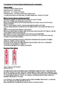
Fig. 4: It was seen that the main femoral vein flow was not ceased during the valsalva and an opposite flow consistent with reflux was seen in the lower extremity Spectral Doppler sonographic evaluation. The actual appearance was in favor of venous insufficiency.
Page 8 of 20
Results Lower extremity venous system insufficiency was found in 44 of 56 patients who had pelvic venous dilatation. Lower extremity venous system insufficiency was found in 12 cases even though they had pelvic vein diameter of lower than 5 mm. Pelvic veins whose diameters were greater than 5 mm, were seen bilaterally in 21 cases, on the right side in 9 cases, and on the left side in 14 cases (Figures 1, 2). The mean number of births was 4,8±2,5 (0-10) in the study group which was composed of 4 nulliparous and 52 multiparous cases. In the statistical analysis, there was a positive relationship between the number of births and pelvic venous dilatation (p