Treatment of Varicose Veins/Venous Insufficiency Policy Number: 7.01.124 Origination: 7/2010
Last Review: 7/2014 Next Review: 7/2015
Policy Blue Cross and Blue Shield of Kansas City (Blue KC) will provide coverage for the treatment of varicose veins and venous insufficiency when it is determined to be medically necessary because the criteria shown below are met.
Policy Statements Greater or Lesser Saphenous Veins Treatment of the greater or lesser saphenous veins by surgery (ligation and stripping) or endovenous radiofrequency or laser ablation may be considered medically necessary for symptomatic varicose veins/venous insufficiency when the following criteria have been met: There is demonstrated saphenous reflux; AND There is documentation of one or more of the following indications: o Ulceration secondary to venous stasis that fails to respond to compressive therapy; OR o Recurrent superficial thrombophlebitis that fails to respond to compressive therapy; OR o Hemorrhage or recurrent bleeding episodes from a ruptured superficial varicosity; OR o Persistent pain, swelling, itching, burning, or other symptoms are associated with saphenous reflux, AND the symptoms significantly interfere with activities of daily living, AND conservative management including compression therapy for at least 3 months has not improved the symptoms*. Treatment of greater or lesser saphenous veins by surgery or endovenous radiofrequency or laser ablation that do not meet the criteria described above is considered not medically necessary. Accessory Saphenous Veins Treatment of accessory saphenous veins by surgery (ligation and stripping) or endovenous radiofrequency or laser ablation may be considered medically necessary for symptomatic varicose veins/venous insufficiency when the following criteria have been met: Incompetence of the accessory saphenous vein is isolated, OR the greater or lesser saphenous veins had been previously eliminated (at least 3 months); AND There is demonstrated accessory saphenous reflux; AND There is documentation of one or more of the following indications: o Ulceration secondary to venous stasis that fails to respond to compressive therapy; OR o Recurrent superficial thrombophlebitis that fails to respond to compressive therapy; OR o Hemorrhage or recurrent bleeding episodes from a ruptured superficial varicosity; OR o Persistent pain, swelling, itching, burning, or other symptoms are associated with saphenous reflux, AND the symptoms significantly interfere with activities of daily living, AND conservative management including compression therapy for at least 3 months has not improved the symptoms. * Treatment of accessory saphenous veins by surgery or endovenous radiofrequency or laser ablation that do not meet the criteria described above is considered not medically necessary.
Symptomatic Varicose Tributaries The following treatments are considered medically necessary as a component of the treatment of symptomatic varicose tributaries when performed either at the same time or following prior treatment (surgical, radiofrequency or laser) of the saphenous veins (none of these techniques has been shown to be superior to another): Stab avulsion Hook phlebectomy Sclerotherapy Transilluminated powered phlebectomy Treatment of symptomatic varicose tributaries when performed either at the same time or following prior treatment of saphenous veins using any other techniques than noted above is considered investigational. Perforator Veins Surgical ligation (including subfascial endoscopic perforator surgery) or endovenous radiofrequency or laser ablation of incompetent perforator veins may be considered medically necessary as a treatment of leg ulcers associated with chronic venous insufficiency when the following conditions have been met: There is demonstrated perforator reflux; AND The superficial saphenous veins (greater, lesser, or accessory saphenous and symptomatic varicose tributaries) have been previously eliminated; AND Ulcers have not resolved following combined superficial vein treatment and compression therapy for at least 3 months*; AND The venous insufficiency is not secondary to deep venous thromboembolism. Ligation or ablation of incompetent perforator veins performed concurrently with superficial venous surgery is not medically necessary. Telangiectasia Treatment of telangiectasia such as spider veins, angiomata, and hemangiomata is considered cosmetic. Other Techniques for conditions not specifically listed above are investigational, including, but not limited to: Sclerotherapy of perforator, greater or lesser saphenous, or accessory saphenous veins Sclerotherapy of isolated tributary veins without prior or concurrent treatment of saphenous veins Stab avulsion, hook phlebectomy, or transilluminated powered phlebectomy of perforator, greater or lesser saphenous, or accessory saphenous veins Endovenous radiofrequency or laser ablation of tributary veins Endovenous cryoablation of any vein Mechanochemical ablation of any vein
Considerations There is no specific CPT code for transilluminated powered phlebectomy. Providers might elect to use CPT codes describing stab phlebectomy (37765 or 37766) or unlisted vascular surgery procedure (37799). Note: If ultrasound guidance (CPT code 76942) is used to guide sclerotherapy of the varicose tributaries, it would be considered incidental to the injection procedure. Treatment of some varicose veins may be considered cosmetic in nature if not associated with significant clinical symptoms and documented reflux at the saphenofemoral or saphenopopliteal junction, and thus contract exclusions for cosmetic therapies may apply to coverage eligibility. Photographs or chart notes in conjunction with the results of duplex ultrasound scanning demonstrating incompetent veins may be required to establish medical necessity. Note that the term "varicose veins"
does not apply to the telangiectatic dermal veins, which may be described as "spider veins" or "broken blood vessels" or veins measuring 4 mm or less in diameter. While abnormal in appearance, these veins typically are not associated with any other symptoms (such as pain or heaviness), and their treatment is considered cosmetic. *The requirement for compression stockings may be waived if the requested veins measure greater than 8mm in diameter. If the member has had prior medically necessary vein treatment (i.e. failed compression therapy) the requirement for compression stockings attempt may be waived for subsequent vein treatments (ipsilateral or contralateral).
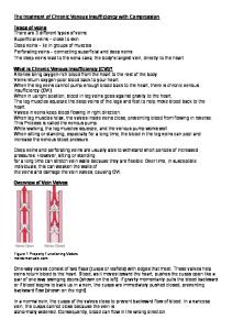
Description of Procedure or Service A variety of treatment modalities are available to treat varicose veins/venous insufficiency, including surgical approaches, thermal ablation, and sclerotherapy. The application of each of these treatment options is influenced by the severity of the symptoms, the type of vein, the source of venous reflux, and the use of other (prior or concurrent) treatments. Background The venous system of the lower extremities consists of the superficial veins (this includes the greater and lesser saphenous, and accessory or duplicate veins that travel in parallel with the greater and lesser saphenous veins), the deep system (popliteal and femoral veins), and perforator veins that cross through the fascia and connect the deep and superficial systems. One-way valves are present within all veins to direct the return of blood up the lower limb. Since venous pressure in the deep system is generally greater than that of the superficial system, valve incompetence at any level may lead to backflow (venous reflux) with pooling of blood in superficial veins. Varicose veins with visible varicosities may be the only sign of venous reflux, although itching, heaviness, tension, and pain may also occur. Chronic venous insufficiency secondary to venous reflux can lead to thrombophlebitis, leg ulcerations and hemorrhage. The CEAP classification considers the clinical, etiologic, anatomic, and pathologic characteristics of venous insufficiency, ranging from class 0 (no visible sign of disease) to class 6 (active ulceration). Treatment of venous reflux/venous insufficiency is aimed at reducing abnormal pressure transmission from the deep to the superficial veins. Conservative medical treatment consists of elevation of the extremities, graded compression, and wound care when indicated. Conventional surgical treatment consists of identifying and correcting the site of reflux by ligation of the incompetent junction followed by stripping of the vein to redirect venous flow through veins with intact valves. While most venous reflux is secondary to incompetent valves at the saphenofemoral or saphenopopliteal junctions, reflux may also occur at incompetent valves in the perforator veins or in the deep venous system. The competence of any single valve is not static and may be pressure dependent. For example, accessory saphenous veins may have independent saphenofemoral or saphenopopliteal junctions that become incompetent when the greater or lesser saphenous veins are eliminated and blood flow is diverted through the accessory veins. Saphenous Veins and Tributaries Saphenous veins include the greater and lesser saphenous, and accessory saphenous veins that travel in parallel with the greater or lesser saphenous veins. Tributaries are veins that empty into a larger vein. Treatment of venous reflux typically includes the following: 1. Identification by preoperative Doppler ultrasonography of the valvular incompetence 2. Control of the most proximal point of reflux, traditionally by suture ligation of the incompetent saphenofemoral or saphenopopliteal junction 3. Removal of the superficial vein from circulation, for example by stripping of the greater and/or lesser saphenous veins 4. Removal of varicose tributaries (at the time of the initial treatment or subsequently) by stab avulsion (phlebectomy) or injection sclerotherapy.
Minimally invasive alternatives to ligation and stripping have been investigated. These include sclerotherapy, transilluminated powered phlebotomy, and thermal ablation using cryotherapy, high frequency radiowaves (200–300 kHz), or laser energy. Sclerotherapy The objective of sclerotherapy is to destroy the endothelium of the target vessel by injecting an irritant solution (either a detergent, osmotic solution, or chemical irritant), ultimately resulting in the occlusion of the vessel. The success of the treatment depends on accurate injection of the vessel, an adequate injectate volume and concentration of sclerosant, and compression. Historically, larger veins and very tortuous veins were not considered to be good candidates for sclerotherapy due to technical limitations. Technical improvements in sclerotherapy have included the routine use of Duplex ultrasound to target refluxing vessels, luminal compression of the vein with anesthetics, and a foam/sclerosant injectate in place of liquid sclerosant. Foam sclerosants are produced by forcibly mixing a gas (e.g., air or carbon dioxide) with a liquid sclerosant (e.g., polidocanol or sodium tetradecyl sulfate). The foam is produced at the time of treatment and is considered an off-label use. A proprietary microfoam sclerosant (Varisolve, BTG PLC, London) with a controlled density and more consistent bubble sizes is being developed in Europe. Endovenous Mechanochemical Ablation Endovenous mechanochemical ablation utilizes both sclerotherapy and mechanical damage to the lumen. Following ultrasound imaging, a disposable catheter with a motor drive is inserted into the distal end of the target vein and advanced to the saphenofemoral junction. As the catheter is pulled back, a wire rotates at 3,500 rpm within the lumen of the vein, abrading the lumen. At the same time, a liquid sclerosant (sodium tetradecyl sulfate) is infused near the rotating wire.It is proposed that mechanical ablation allows for better efficacy of the sclerosant, without the need for the tumescent anesthesia used in radiofrequency (RF) ablation or endovenous laser ablation (EVLT). Thermal Ablation Radiofrequency ablation is performed by means of a specially designed catheter inserted through a small incision in the distal medial thigh to within 1–2 cm of the saphenofemoral junction. The catheter is slowly withdrawn, closing the vein. Laser ablation is performed similarly; a laser fiber is introduced into the greater saphenous vein under ultrasound guidance; the laser is activated and slowly removed along the course of the saphenous vein. Cryoablation uses extreme cold to cause injury to the vessel. The objective of endovenous techniques is to cause injury to the vessel, causing retraction and subsequent fibrotic occlusion of the vein. Technical developments since thermal ablation procedures were initially introduced include the use of perivenous tumescent anesthesia, which allows successful treatment of veins larger than 12 mm in diameter and helps to protect adjacent tissue from thermal damage during treatment of the lesser saphenous vein. Transilluminated Powered Phlebectomy Transilluminated powered phlebectomy (TIPP) is an alternative to stab avulsion or hook phlebectomy. This procedure uses 2 instruments: an illuminator which also provides irrigation, and a resector, which has an oscillating tip and can perform suction. Following removal of the saphenous vein, the illuminator is introduced via a small incision in the skin and tumescence solution (anesthetic and epinephrine) is infiltrated along the course of the varicosity. The resector is then inserted under the skin from the opposite direction, and the oscillating tip is placed directly beneath the illuminated veins to fragment and loosen the veins from the supporting tissue. Irrigation from the illuminator is used to clear the vein fragments and blood through aspiration and additional drainage holes. The illuminator and resector tips may then be repositioned, thereby reducing the number of incisions needed when compared with stab avulsion or hook phlebectomy. It has been proposed that TIPP might result in decreased operative time, decreased complications such as bruising, and faster recovery compared to the established procedures. Treatment of Perforator Veins
Perforator veins cross through the fascia and connect the deep and superficial venous systems. Incompetent perforating veins were originally addressed with an open surgical procedure, called the Linton procedure, which involved a long medial calf incision to expose all posterior, medial, and paramedial perforators. While this procedure was associated with healing of ulcers, it was largely abandoned due to a high incidence of wound complications. The Linton procedure was subsequently modified by using a series of perpendicular skin flaps instead of a longitudinal skin flap to provide access to incompetent perforator veins in the lower part of the leg. The modified Linton procedure may be occasionally utilized for the closure of incompetent perforator veins that can not be reached by less invasive procedures. Subfascial endoscopic perforator surgery (SEPS) is a less-invasive surgical procedure for treatment of incompetent perforators and has been reported since the mid-1980s. Guided by Duplex ultrasound scanning, small incisions are made in the skin and the perforating veins are clipped or divided by endoscopic scissors. The operation can be performed as an outpatient procedure. Endovenous ablation of incompetent perforator veins with sclerotherapy and radiofrequency has also been reported. Other Deep vein valve replacement is being investigated. Outcomes of interest for venous interventions include healing and recurrence, recannulation of the vein, and neovascularization. Recannulation (recanalization) is the restoration of the lumen of a vein after it has been occluded; this occurs more frequently following treatment with endovenous techniques. Neovascularization is the proliferation of new blood vessels in tissue, and occurs more frequently following vein stripping. Direct comparisons of durability for endovenous and surgical procedures are complicated by these different mechanisms of recurrence. Relevant safety outcomes include the incidence of paresthesia, thermal skin injury, thrombus formation, thrombophlebitis, wound infection, and transient neurologic effects. Regulatory Status The following devices have received specific U.S. Food and Drug Administration (FDA) marketing clearance for the endovenous treatment of superficial vein reflux: In 1999, the VNUS® Closure™ system (a radiofrequency device) received FDA clearance through the 510(k) process for "endovascular coagulation of blood vessels in patients with superficial vein reflux." The VNUS RFS and RFSFlex devices received FDA clearance in 2005 for “use in vessel and tissue coagulation including: treatment of incompetent (i.e., refluxing) perforator and tributary veins. The modified VNUS® ClosureFAST™ Intravascular Catheter received FDA clearance through the 510(k) process in 2008. In 2002, the Diomed 810 nm surgical laser and EVLT™ (endovenous laser therapy) procedure kit received FDA clearance through the 510(k) process, "… for use in the endovascular coagulation of the greater saphenous vein of the thigh in patients with superficial vein reflux." A modified Erbe Erbokryo® cryosurgical unit (Erbe USA) received FDA clearance for marketing in 2005. A variety of clinical indications are listed, including cryostripping of varicose veins of the lower limbs. The Trivex system is a device for transilluminated powered phlebectomy that received FDA clearance through the 510(k) process in October 2003. According to the label, the intended use is for “ambulatory phlebectomy procedures for the resection and ablation of varicose veins.” Varisolve® (BTG PLC, London) is a sclerosant microfoam made with a proprietary gas mix. A phase II safety study for the FDA has been completed. In late October 2009, the sponsor submitted a request to the FDA for a protocol assessment to agree on the design, endpoints and statistical analyses for the phase III trial. The ClariVein® Infusion Catheter received marketing clearance through the 510(k) process in 2008 (K071468). It is used for mechanochemical ablation. Predicate devices were listed as the Trellis® Infusion System (K013635) and the Slip-Cath® Infusion Catheter (K882796). The system includes an infusion catheter, motor drive, stopcock and syringe and is intended for the infusion of physicianspecified agents in the peripheral vasculature.
Rationale This policy was created in 2010 and updated periodically using the MEDLINE database. The most recent update was performed through December 3, 2013. Following is a summary of key studies to date. Treatment of Saphenous Reflux Compression Therapy A 2009 Cochrane review on compression for venous leg ulcers included a total of 39 randomized controlled trials (RCTs), with 47 different comparisons.(1) The review was updated in 2012, and included 48 RCTs with 59 different comparisons.(2) Most of the RCTs were small. Objective measures of healing were the time to complete healing, the proportion of ulcers healed within the trial period (typically 12 weeks), the change in ulcer size, and the rate of change in ulcer size. Evidence from 8 trials indicated that venous ulcers healed more rapidly with compression than without. Findings suggested that multicomponent systems (bandages or stockings) were more effective than singlecomponent compression. In addition, multicomponent systems containing an elastic bandage appeared more effective than those composed mainly of inelastic constituents. Although these meta-analyses did not include time to healing, studies included in the review reported that the mean time to ulcer healing was approximately 2 months, while the median time to healing in other reports was 3 to 5 months. A Cochrane review on compression stockings for the initial treatment of varicose veins in patients without venous ulceration was published in 2011.(3) Included in the review were 7 studies involving 356 participants with varicose veins without healed or active venous ulceration (CEAP [clinical, etiology, anatomy, pathophysiology] classification C2 to C4). Six of the studies compared different types or pressures of stockings. Subjectively, participants’ symptoms improved, but results were not compared with a control arm. Due primarily to inadequate reporting, the methodologic quality of the included trials was unclear. Meta-analyses were not performed due to inadequate reporting and suspected heterogeneity. The authors concluded that there is insufficient high-quality evidence to determine whether or not compression stockings are effective as the sole and initial treatment of varicose veins in patients without venous ulceration, or whether any type of stocking is superior to any other type. Ligation and Stripping Systematic literature reviews published in 2008 indicate a similar healing rate of venous ulcers with superficial vein surgery and conservative compression treatments but a reduction in ulcer recurrence rate with surgery.(4,5) In general, recurrence rates after ligation and stripping are estimated at around 20%. Jones et al reported on the results of a study that randomized 100 patients with varicose veins to undergo either ligation alone or ligation in conjunction with stripping.(6) At 1 year, reflux was detected in 9% of patients, rising to 26% at 2 years. Rutgers and Kitslaar reported on the results of a trial that randomized 181 limbs to undergo either ligation and stripping or ligation combined with sclerotherapy.(7) At 2 years, Doppler ultrasound demonstrated reflux in approximately 10% of patients after ligation and stripping, increasing to 15% at 3 years. A 2011 Cochrane review compared endovenous ablation (radiofrequency and laser) and foam sclerotherapy versus ligation/stripping for saphenous vein varices.(8) Included in the review were 13 reports from 5 studies with a combined total of 450 patients. Many of the comparisons between endovenous ablation and ligation/stripping failed to reach statistical significance. The authors concluded that current evidence suggests that endovenous radiofrequency ablation (RFA) and endovenous laser ablation (EVLA) are at least as effective as surgery in the treatment of great saphenous varicose veins. No randomized trials comparing sclerotherapy with ligation/stripping met the study inclusion criteria, and there were thus insufficient data to comment on ultrasound-guided sclerotherapy. Endovenous RFA
In 2008, Luebke et al reported a meta-analysis of 8 studies that included a total of 224 patients who underwent RFA and 204 patients who underwent stripping.(9) There was no significant difference between RFA and surgery in immediate or complete greater saphenous vein occlusion, incomplete greater saphenous vein closure, freedom from reflux, recurrent varicose veins, recanalization, or neovascularization between the 2 treatments. There were significant reductions in tenderness and ecchymosis at 1 week and fewer hematomas at 72 hours, 1 week, and 3 weeks with RFA. Quality-oflife results, including return to normal activity and return to work, favored RFA over surgery. The authors noted that rates of recanalization, retreatment, occlusion, and reflux may alter with longer follow-up and that further RCTs with longer follow-up are needed. Long-term outcomes of endovenous RFA were reported from the Closure Study Group clinical registry in 2005.(10) Thirty-four centers (1006 patients, 1222 limbs) participated in the registry, with 12 centers contributing 5-year data (406 limbs). The registry included data on the treatment of 52 lesser saphenous veins and 16 accessory saphenous veins. Follow-up at 1 week showed a 97% anatomical success rate and a decrease in pain in 50% (from 85% to 30%) of patients. An additional 162 failures were identified over the 5 years of follow-up; 129 veins were found to have recanalization, and 33 limbs had reflux in the groin. Logistic regression analysis (risk factors of gender, age, body mass index [BMI]), vein diameter, and catheter pullback speed) showed that BMI was associated with long-term failure. The rate of pull-back speed of the catheter during treatment was associated with failure to occlude or recanalization. Endovenous Laser Ablation A systematic review of EVLA versus surgery was published in 2009.(11) Fifty-nine studies were included, with 7 studies that directly compared EVLA and surgery. Randomized and nonrandomized studies directly comparing outcomes for EVLA or surgery were included for the assessment of safety or effectiveness, while case series with a minimum patient population of 100 were included for the assessment of safety alone. For all studies, it was calculated that 5759 patients (6702 limbs) were treated with EVLA and 6395 patients (7727 limbs) underwent surgery. Few differences were apparent between treatments with respect to clinical effectiveness outcomes, although long-term follow-up was lacking. Nonclinical effectiveness outcomes generally favored EVLA over surgery in the first 2 months after treatment. The authors concluded that while EVLA offers short-term benefits and appears to be as clinically effective as surgery up to 12 months after treatment, clinical trials with a minimum of 3 years of follow-up are required to establish the enduring effectiveness of EVLA. Large clinical trials published subsequent to the systematic review are described below. The 2012 RELACS study was a practical design that randomized 400 patients to EVLA performed by a surgeon at 1 site or to ligation and stripping performed by a different surgeon at a second location.(12) Fifty-four patients withdrew from the study after receiving the randomization result (from an independent site), due primarily to preference for the other treatment. At 2-year follow-up, there was no significant difference between the groups for clinically recurrent varicose veins, medical condition on the Homburg Varicose Vein Severity Score, or disease-related quality of life. Saphenofemoral reflux was detected by ultrasonography more frequently after endovenous laser treatment (EVLT) (17.8% vs 1.3%). This study will follow patients for 5 years. Another trial compared EVLA with ligation and stripping in 200 limbs (100 in each group).(13) At 1-year follow-up, 98% of the limbs were reported to be free of symptoms. At 2-year follow-up, the EVLA group had 2 veins completely reopened and 5 partially reopened, which was significantly greater than in the ligation and stripping group. In the 2013 MAGNA trial, 223 consecutive patients (240 legs) with great saphenous vein reflux were randomized to EVLA, ligation and stripping, or foam sclerotherapy.(14) At 1-year follow-up, the anatomic success rates were similar between EVLA and stripping (88.5% and 88.2%, respectively), which were superior to foam sclerotherapy (72.2%). Ten percent of the stripping group showed neovascularization. Healthrelated quality of life improved in all groups. The CEAP classification improved in all groups with no significant difference between the groups. Transient adverse events were reported in 11 patients after stripping, 7 after EVLA, and 5 after sclerotherapy.
In 2009 Theivacumar et al reported 2-year follow-up from 118 consecutive patients treated with either EVLA (69 limbs) or ligation and stripping (n=60 limbs).(15) Sixty-eight of the patients agreed to be randomized to treatment; the remainder declined randomization but received one of the 2 treatments and agreed to follow-up. The rationale for the selection of treatment in the nonrandomized population was not described. Rates of clinical recurrence (7%) were similar in the 2 treatment groups at 2 years. Recanalization of the residual greater saphenous vein, reflux in the accessory greater saphenous vein, and reflux in incompetent perforator veins accounted for the majority of cases of clinical recurrence (6%) in both groups. Neovascularization was observed in only 1% of limbs treated with endoluminal ablation and 18% of limbs treated with ligation and stripping (2% were clinically significant at 2 years). Early neovascularization has been associated with clinical recurrence at 5 years. Rasmussen et al randomized 121 patients (137 legs) to EVLA (n=69) or to ligation and stripping (n=68).(16) The incidence of clinical recurrence was found to increase gradually from 6 months onward. At 2-year follow-up, outcomes were similar between the 2 groups. In the EVLA group, there were 3 (4%) technical failures, 18 (26%) clinical recurrences, 6 (8%) cases of reflux into the anterior accessory saphenous vein, 2 (3%) cases of reflux in the groin, and 7 (10%) cases of reflux in perforator veins. In the ligation and stripping group, there were 2 (3%) technical failures, 25 (37%) recurrences, 3 (4%) cases of reflux in the accessory vein, 3 (4%) cases of reflux in the groin, and 15 (22%) cases of reflux in perforator veins. Ninety-eight percent of patients were available for 5-year follow-up.(17) There was a trend toward greater recanalization following EVLA, but this was not associated with increased clinical recurrence (EVLA, 36%; stripping, 35%) or in the percentage of reoperations (EVLA, 38.6%; stripping, 37.7%). Literature on isolated treatment of the anterior accessory saphenous vein is limited. In a 2009 study, outcomes from a cohort of 33 patients who underwent EVLA of the anterior accessory saphenous vein were compared with 33 matched controls undergoing EVLA of the greater saphenous vein.(18) In 21 of the patients (64%) in the accessory saphenous vein group, there had been no previous treatment of the greater saphenous vein. At 12-month follow-up, there was no evidence of reflux in these patients, and the treated accessory saphenous vein was not visible with ultrasound. The Aberdeen Varicose Vein Symptom Severity Score had improved in both groups, with no significant difference between the 2 groups. Patient satisfaction scores were also similar. Endovenous Cryoablation Klem et al reported a randomized trial in 2009 that found endovenous cryoablation (n=249) to be inferior to conventional stripping (n=245) for treating patients with symptomatic varicose veins. (19) The percentage of patients with greater saphenous vein remaining was 44% in the endovenous cryoablation group and 15% in the conventional stripping group. The Aberdeen Varicose Vein Questionnaire also showed better results for conventional stripping (score, 11.7) in comparison with cryoablation (score, 8.0). There were no differences between the groups in SF-36 subscores, and neural damage was the same (12%) in both groups. Disselhoff et al reported 2 and 5 year outcomes from a randomized trial that compared cryostripping with EVLA.(20,21) Included were 120 patients with symptomatic uncomplicated varicose veins (CEAP C2) with saphenofemoral incompetence and greater saphenous vein reflux. At 10 days after treatment, EVLA had better results than cryostripping with respect to pain score over the first 10 days (2.9 vs 4.4), resumption of normal activity (75% vs 45%) and induration (15% vs 52%). At 2-year follow-up, freedom from recurrent incompetence was observed in 77% of patients after EVLA and 66% of patients after cryostripping (not significantly different). At 5 years, 36.7% of patients were lost to follow-up; freedom from incompetence and neovascularization was found in 62% of patients treated with EVLA and 51% of patients treated with cryostripping (not significantly different). Neovascularization was more common after cryostripping, but incompetent tributaries were more common after EVLA. There was no significant difference between groups in the Venous Clinical Severity Score or Aberdeen Varicose Vein Severity Score at either 2 or 5 years.
Sclerotherapy A comprehensive systematic review of sclerotherapy commissioned and funded by the U.K.’s National Institute for Health and Clinical Excellence in 2006 reviewed 67 studies, including 9 RCTs, 1 registry report, 8 nonrandomized comparative studies, 43 case series, and 6 case reports.(22,23) The report concluded that sclerotherapy “appears to be efficacious in occluding incompetent veins, including both main trunk and minor vein disease, however its longer-term efficacy in terms of recurrence or new varicosities is less certain,” and that “Estimates were based mainly on data from nonrandomized studies with a high dropout rate and no details of methods of follow-up, and as such may be prone to attrition bias.” More recent randomized trials using ultrasound-guided foam sclerotherapy of the greater saphenous vein (with or without ligation) showed high variability in success rates between centers (ranging from 25% to 100%) and a decline in success rates from 85% at 3-week follow-up to 53% at 2 years.(24,25) Other studies indicate efficacy rates ranging from 12% to 76% for liquid sclerosant and from 57% to 84% for foam sclerosant.(26) A systematic review from 2008 found that foam sclerotherapy of varicose veins is associated with a higher recurrence rate in patients with saphenofemoral incompetence compared to the rates of endovenous laser therapy or RFA obliteration, while a 2009 systematic review suggested that outcomes from sclerotherapy are worse than those of surgery (ligation and stripping) for saphenous vein reflux.(27,28) Although long-term sequelae have not been reported, transient adverse effects have been found in up to 8% of patients, including visual disturbance, migraine, shortness of breath, dizziness, and numbness.(29) Bubbles appear in the right heart between 9 and 59 seconds after injection, and emboli have been detected in the middle cerebral artery following sclerotherapy of saphenous trunks and varices.(29) Deep venous occlusion after ultrasound-guided sclerotherapy has also been reported; risk was found to be greater when treating veins 5 mm in diameter or greater (odds ratio [OR], 3.7) and injecting 10 mL or more of foamed sclerosant (OR=3.6).(30) In the 2013 MAGNA trial (described above), 223 consecutive patients (240 legs) with great saphenous vein reflux were randomized to EVLA, ligation and stripping, or foam sclerotherapy.(14) At 1-year follow-up, the anatomic success rate of foam sclerotherapy (72.2%) was inferior to both EVLA and stripping (88.5% and 88.2%, respectively). Twenty-one patients in the sclerotherapy group had partial occlusion with reflux, though the clinical complaint was completely relieved. A 2012 study was a noninferiority trial of foam sclerotherapy versus ligation and stripping in 430 patients. (31) Analysis was per protocol. Forty patients (17%) had repeat sclerotherapy. At 2 years, the probability of clinical recurrence was similar in the 2 groups (11.3% sclerotherapy vs 9.0% ligation and stripping), although reflux was significantly more frequent in the sclerotherapy group (35% vs 21%). Thrombophlebitis occurred in 7.4% of patients after sclerotherapy. There were 2 serious adverse events in the sclerotherapy group (deep venous thrombosis and pulmonary emboli) that occurred within 1 week of treatment. Blaise et al reported 3-year follow-up from a multicenter double-blind randomized trial (143 patients) that compared treatment of the greater saphenous vein with either 1% or 3% polidocanol foam.(32) Additional treatment with foam sclerotherapy was carried out at 6 weeks, 3 and 6 months if required to abolish persistent venous reflux. There were 49 additional injections in the 1% polidocanol group and 29 additional injections in the 3% group. At 3-year follow-up, venous reflux was observed in 21% of patients in the 1% group and 22% of patients in the 3% polidocanol group. The VANISH-2 study is an FDA-regulated 5-year randomized, double-blind multicenter trial that compares 0.5% or 1.0% polidocanol microfoam versus either endovenous placebo or a subtherapeutic dose of polidocanol foam.(33) Patients with unsuccessful or incomplete treatment at the primary outcome assessment (week 8) could receive open-label foam sclerotherapy. At the 8-week assessment, there was elimination of reflux and/or occlusion of the previously incompetent vein in 85.6% of the combined 0.5% and 1.0% groups, 59.6% of patients in the 0.125% “subtherapeutic” group, and 1.8% of the placebo group. The improvement in the venous clinical severity score was significantly better in the 0.5% and 1.0% groups (-5.10) compared with placebo (-1.52), but was not
reported for the 0.125% group. This is 1 of 2 pivotal Phase 3 studies undertaken to obtain marketing approval from FDA. Longer-term evaluation is in progress. Mechanochemical Ablation No RCT results were identified with mechanochemical ablation (MCA). Several prospective series and cohort studies have been reported. One study compared postoperative pain and early quality of life in 68 patients treated with either RFA or MCA of great saphenous veins.(34) Patients who did not want to be treated with MCA were offered treatment with RFA; this study design could potentially lead to selection bias. There was no significant difference between the groups in procedural pain. Compared with RFA, patients treated with MCA had a 14.3-mm reduction in pain measured on a 100-mm visual analog scale (VAS) measured over the first 3 postoperative days (6.2 vs 20.5) and a 13.8-mm reduction in pain (4.8 vs 18.6 mm) over the first 2 weeks. Patients treated with ClariVein also had an earlier return to normal activities (1.2 vs 2.4 days) and return to work (3.3 vs 5.6 days). There was a similar improvement in quality of life for the 2 groups when measured at 6 weeks. A prospective multicenter series evaluated the efficacy of MCA of the great saphenous vein in 126 patients in a community setting.(35) Veins were selected that were greater than 4 mm and less than 12 mm in diameter, with an average diameter of 7.3 mm. Closure rates were 100% at 1 week, 98% at 3 months, and 94% at 6 months. The venous clinical severity score decreased from a score of approximately 9 pretreatment to about 3 at 6 months. In 2012, Elias and Raines reported an industrysponsored safety and efficacy study of MCA with the ClariVein® system.(36) Thirty greater saphenous veins in 29 patients were treated with this device. Greater saphenous veins with diameters greater than 12 mm were excluded. At 6-month follow-up, 1 vein had recanalized, for a primary closure rate of 96.7%. No pain during the procedure or adverse events were reported. Another prospective series evaluated MCA of the small saphenous vein in 50 consecutive patients.(37) Only patients with a vein diameter of 2.5 to 11 mm were included. The dose of sclerosant was increased after the first 15 patients. At the 6-week assessment, all treated veins were occluded and at 1-year follow-up, 94% remained occluded. The median VAS score for pain during the procedure was 2 of 10. There were no major complications. Controlled studies with a greater number of subjects and longer follow-up are needed. A 2013 review of MCA notes that a 5-year 840 patient randomized trial comparing ClariVein with RFA began in 2012 in Europe.(38) This trial will provide needed data on the comparative effectiveness of MCA, measured at a longer duration and in a larger population. Other Treatments Both steam injection and microwave ablation for endovenous treatment of varicose veins were reported outside of the U.S.(39,40) These procedures have not been approved or cleared for marketing by FDA. Section Summary There are a number of large randomized trials on endovenous ablation of the saphenous veins. Comparison with ligation and stripping at 2-year follow-up supports use of both RFA and EVLT. Evidence suggests that ligation and stripping may lead to neovascularization, while thermal ablation may lead to recanalization. Controlled studies with longer follow-up are needed to determine the longterm efficacy of these treatments with greater certainty. Two RCTs suggest that cryotherapy is not as effective as available alternatives. For sclerotherapy, there is high variability in success rates of the procedure and reports of serious adverse events. MCA is a combination of liquid sclerotherapy with mechanical abrasion. A potential advantage of this procedure compared to thermal ablation techniques is that it does not require tumescent anesthesia and may result in less postoperative pain. To date, the ClariVein procedure has been assessed in relatively few patients and for short durations. Thus, there is insufficient evidence to permit conclusions regarding the safety and efficacy of mechanochemical ablation.
Treatment of Tributary Varicosities Sclerotherapy and Phlebectomy Early studies established ligation and stripping as the gold standard for the treatment of saphenofemoral incompetence based on improved long-term recurrence rates, with sclerotherapy used primarily as an adjunct to treat varicose tributaries. A 2006 Cochrane Review, based primarily on RCTs from the 1980s, concluded that, “The evidence supports the current place of sclerotherapy in modern clinical practice, which is usually limited to treatment of recurrent varicose veins following surgery and thread veins.”(41) Sclerotherapy and phlebectomy are considered appropriate in the absence of reflux of the saphenous system, eg, post- or adjunctive treatment to other procedures such as surgery.(28) A small proportion of patients may present with tributary varicosities in the absence of saphenous reflux. For example, of 1009 patients recruited for an RCT, 64 patients were found to have minor varicose veins without reflux, 34 of whom agreed to be randomized to sclerotherapy or conservative treatment.(42) At baseline, 92% had symptoms of heaviness, 69% had cosmetic concerns, 53% reported itching, and 30% reported relief of symptoms through the use of compression hosiery. At 1year follow-up, there was an improvement in clinicians’ assessment of the anatomical extent of varicose veins, with 85% of patients in the sclerotherapy group improved compared to 29% of patients in the conservative-therapy group. Symptoms of aching were better or eliminated in 69% of the sclerotherapy group and 28% of the group treated with conservative therapy. Cosmetic concerns were improved in 85% of the sclerotherapy patients and 14% of controls. The bulk of the literature discussing the role of ultrasound guidance refers to sclerotherapy of the saphenous vein, as opposed to the varicose tributaries. In 2012, Yamaki et al reported a prospective RCT that compared visual foam sclerotherapy versus ultrasound-guided foam sclerotherapy of the greater saphenous vein together with visual foam sclerotherapy for varicose tributary veins.(43) A total of 51 limbs in 48 patients were treated with ultrasound-guided foam sclerotherapy plus visual foam sclerotherapy of the varicose tributaries, and 52 limbs in 49 patients were treated with foam sclerotherapy alone. At 6-month follow-up, complete occlusion was found in 23 limbs (45.1%) treated with ultrasound-guided and visual-guided foam sclerotherapy and in 22 limbs (42.3%) treated with visual sclerotherapy alone. Reflux was absent in 30 limbs (58.8%) treated with ultrasound and visual guidance and in 37 (71.2%) treated with visual guidance alone (not significantly different). The authors note that for the treatment of tributary veins in clinical practice, most patients receive direct injection of foam without ultrasound guidance. Transilluminated Powered Phlebectomy A 2008 meta-analysis included 5 studies that compared transilluminated powered phlebectomy (TIPP) with conventional surgery.(44) Results showed a significant advantage of TIPP over the conventional treatment for number of incisions, mean cosmetic score, and duration of the procedure. However, TIPP also increased the incidence of hematoma and resulted in worse mean pain scores. Included in the meta-analysis was an RCT by Chetter et al that compared TIPP (n=29) with a multiple stab incision procedure (n=33).(45) A single surgeon performed all but 2 of the procedures, and there was no difference in operating time. Patients treated with TIPP had an average of 5 incisions, compared with 20 for the multiple stab procedure. However, blinded evaluation revealed that bruising or discoloration was higher for the TIPP group at both 1 and 6 weeks after surgery. At 6 weeks after surgery, patients in the TIPP group showed no improvement in pain (-2 points on the Burford pain scale), while patients in the multiple stab incision group had a significant improvement in pain score compared with presurgical baseline (-20 points). At 6 weeks after surgery, quality-of-life measures had improved in the multiplestab incision group but not in the TIPP group. Thus, although TIPP had the advantage of fewer surgical incisions, in this single-center study, it was associated with a more prolonged recovery due to more extensive bruising, prolonged pain, and reduced early postoperative quality of life. The current literature does not show an advantage of TIPP over conventional treatment.
Section Summary Evidence indicates that both sclerotherapy and TIPP are as effective as stab phlebectomy in eliminating varicose tributaries. However, there is limited evidence that TIPP is associated with more pain, bruising, discoloration, and a longer recovery. Treatment of Perforator Reflux A systematic literature review published in 2008 indicates insufficient evidence for the role of incompetent perforator vein surgery.(5) These conclusions were based on 4 RCTs published since 2000 that compared superficial vein surgery with conservative therapy in advanced chronic venous insufficiency (CEAP category C5/6). The 4 trials included 2 level I (large subject population) and 2 level II (small subject population) studies. Two of the trials combined surgical treatment of the incompetent perforator veins with concurrent or prior treatment of the superficial saphenous veins; the other 2 treated the greater saphenous vein alone. The 2 randomized studies in which the greater saphenous vein alone was treated (including the ESCHAR trial) showed a significant reduction in ulcer recurrence in comparison with conservative therapy.(46,47) A 2011 community hospital-based multicenter, doubleblind, randomized trial found no clinical benefit (self-reported symptoms) from adding subfascial endoscopic perforator surgery (SEPS) to saphenous surgery in 75 patients with varicose ulcers (CEAP C5 or C6) and incompetent perforators.(48) Treatment of the great saphenous vein alone has been reported to improve perforator function. For example, 1 study showed that reversal of perforator vein incompetence (41% of 68 previously incompetent perforators) was more common than new perforator vein incompetence (22% of 183 previously competent perforators) following superficial vein surgery.(49) O’Donnell discusses additional (lower quality) evidence to suggest deep venous valvular involvement rather than incompetent perforators in venous insufficiency.(5) Thus, although incompetence of perforator veins is frequently cited as an important etiologic factor in the pathogenesis of venous ulcer, current evidence does not support the routine ligation or ablation of perforator veins. Subfascial Endoscopic Perforator Surgery In 2004, Tenbrook et al published a review of the literature of subfascial endoscopic perforator surgery (SEPS), which included 19 case series and 1 randomized trial.(50) In total, the reviewed studies included 1031 patients with 1140 treated limbs. The authors concluded that SEPS was associated with excellent results in terms of ulcer healing and prevention of recurrence. However, the authors also noted that randomized trials are required to define the relative contributions of compression therapy, superficial venous surgery, and SEPS in the management of severe venous disease. A 2009 metaanalysis of SEPS for chronic venous insufficiency concludes that “Its [SEPS] use should not be employed routinely and could only be justified in patients with persistent ulceration thought to be of venous origin, and in whom any superficial reflux has already been ablated and postthrombotic changes excluded.”(51) The authors also state that “introduction of less invasive techniques for perforator vein ablation, such as ultrasound-guided sclerotherapy or radiofrequency ablation, may diminish the role of SEPS in the future." Other Treatments A 2008 review of procedures for management of varicose veins recommends duplex-guided foam sclerotherapy, microincision phlebectomy, or thermal ablation using a new short RFA catheter for the treatment of symptomatic residual perforator vein incompetence.(52) Ablation of incompetent perforator veins with laser or RFA had been shown to be technically feasible, although no studies had been identified that showed an improvement in clinical outcomes (eg, ulcer healing or recurrence).(18-20,26) The 2011 literature update identified 1 study of EVLA for perforating veins in 33 patients with a CEAP classification of 4 (skin changes), 5 (healed ulcer), or 6 (active ulcer).(53) All incompetent saphenous trunks were treated simultaneously (63% of limbs). At 3-month follow-up, occlusion was achieved in
78% of the perforating veins. Five patients (15%) had active ulcers at baseline; 4 of the 5 ulcers had healed by 6 weeks after EVLA. Evidence regarding the treatment of perforator veins with ultrasoundguided sclerotherapy is limited, and there is a risk of deep venous occlusion.(30) Summary Although randomized controlled trials (RCTs) with longer follow-up are needed to evaluate long-term durability, and repeat treatments may be required, evidence indicates that endovenous treatment of saphenous veins with radiofrequency or laser ablation improves short-term clinical outcomes (eg, pain and return to work) in comparison with surgery. In contrast, results from a recent RCT of cryoablation indicate that this therapy is inferior to conventional stripping. Sclerotherapy as the sole treatment of saphenofemoral or saphenopopliteal reflux has not been demonstrated to be as effective as available alternatives, and there is insufficient evidence on mechanochemical ablation. The literature indicates that sclerotherapy of tributaries following occlusion of the saphenofemoral or saphenopopliteal junction and saphenous veins may be considered medically necessary. Evidence is insufficient to evaluate the health benefit of sclerotherapy as a sole treatment of varicose tributaries without prior or concurrent treatment of the saphenous veins. No studies have been identified that compare radiofrequency or laser ablation of tributary veins with standard procedures (microphlebectomy and/or sclerotherapy). Transilluminated powered phlebectomy is effective at removing varicosities; outcomes are comparable to available alternatives such as stab avulsion and hook phlebectomy. The literature indicates that the routine ligation/ablation of incompetent perforator veins is not medically necessary for the treatment of varicose veins/venous insufficiency at the time of superficial vein procedures. However, when combined superficial vein procedures and compression therapy have failed to improve symptoms (ie, ulcers), treatment of perforator vein reflux may be as beneficial as any alternative (eg, deep vein valve replacement). Therefore, treatment of incompetent perforator veins may be considered medically necessary in this specific situation. Comparative studies are needed to determine the most effective method of ligating/ablating incompetent perforator veins. SEPS has been shown to be as effective as the Linton procedure with a reduction in adverse events. Although only 1 case series has been identified showing an improvement in health outcomes, endovenous ablation with specialized laser or radiofrequency probes has been shown to effectively ablate incompetent perforator veins with a potential decrease in morbidity in comparison with surgical interventions. For sclerotherapy, concerns have been raised about the risk of deep vein occlusion, and evidence is currently insufficient to evaluate the safety or efficacy of this treatment for incompetent perforator veins. Practice Guidelines and Position Statements The Society for Vascular Surgery and the American Venous Forum published clinical practice guidelines in 2011.(54) The recommendations are rated as strong=1 or weak=2, based on a level of evidence that is either high quality=A, moderate quality=B, or low quality=C, and include the following: Compression therapy for venous ulcerations and varicose veins: Compression therapy is recommended as the primary treatment to aid healing of venous ulceration (Grade 1B, strong recommendation, moderate quality evidence). To decrease the recurrence of venous ulcers, they recommend ablation of the incompetent superficial veins in addition to compression therapy (Grade 1A, strong recommendation, high-quality evidence). They recommend use of compression therapy for patients with symptomatic varicose veins (Grade 2C, weak recommendation, low-quality evidence) but recommend against compression therapy as the primary treatment if the patient is a candidate for saphenous vein ablation (Grade 1B, strong recommendation, moderate quality evidence).
Treatment of the incompetent great saphenous vein: Endovenous thermal ablation (radiofrequency or laser) is recommended over chemical ablation with foam (Grade 1B, strong recommendation, moderate quality evidence) or high ligation and stripping (Grade 1B, strong recommendation, moderate quality evidence) due to reduced convalescence and less pain and morbidity. Cryostripping is a technique that is new in the United States, and it has not been fully evaluated. Varicose tributaries: Phlebectomy or sclerotherapy are recommended to treat varicose tributaries (Grade 1B, strong recommendation, moderate quality evidence). Transilluminated powered phlebectomy using lower oscillation speeds and extended tumescence is an alternative to traditional phlebectomy (Grade 2C, weak recommendation, low-quality evidence). Perforating vein incompetence: Selective treatment of perforating vein incompetence in patients with simple varicose veins is not recommended (CEAP class C2; Grade 1B, strong recommendation, moderate quality evidence), but there is a Grade 2B recommendation (weak recommendation, moderate quality evidence) for treatment of pathologic perforating veins (outward flow of ≥500 ms duration, with a diameter of ≥3.5 mm) located underneath healed or active ulcers (CEAP class C5-C6) by subfascial endoscopic perforating vein surgery, sclerotherapy, or thermal ablations (Grade 1C, weak recommendation, low-quality evidence). In 2009, the American College of Radiology published appropriateness criteria for the treatment of lower-extremity venous insufficiency.(55) The following is a summary of treatment options: Compression Stockings: Graduated compression stockings are routinely used to control venous insufficiency symptoms. They provide external support that can constrict dilated veins and restore competence to incompetent valves. Compression stockings are particularly helpful during pregnancy, and they are frequently used following venous ablation treatment. Surgery: Great saphenous vein (GSV) stripping with branch ligation had historically been the primary treatment option for venous insufficiency. The GSV is ligated near the groin. Ligation alone can preserve the vein for subsequent harvesting in case of arterial bypass; however, ligation alone has proven unsatisfactory for preventing the occurrence of reflux, so it is often supplemented by vein stripping. Ambulatory phlebectomy is primarily used to treat surface varicose veins. It can be performed as an adjunct to endovenous ablation or stripping. This procedure involves making tiny punctures or incisions through which the varicose veins are removed. Other surgical methods to treat venous insufficiency have been described, including SEPS for treating venous ulcers and valvular surgery for treating reflux caused by incompetent valves of the deep veins. Injection Sclerotherapy: Injection sclerotherapy is a common treatment for telangiectasias and can be used to treat smaller varicose veins. The sclerotherapy solution can be in liquid form or can be injected as "foam" (mixed with a gas such as air). Sclerotherapy has not been shown to have long-term effectiveness for large veins, such as the GSV. Endovenous Ablation: Endovenous ablation is a minimally invasive alternative to surgery. It is a percutaneous procedure that can be used to treat the GSV, small saphenous vein, and other superficial veins. Endovenous ablation uses RFA or laser energy (EVLA) applied inside the vein to cause occlusion. Small prospective trials comparing EVLA and RFA with conventional surgery in patients with GSV reflux have shown favorable results. One study demonstrated that EVLA is comparable to surgery in abolishing reflux and improving disease-specific quality of life and that it allows earlier return to normal activity. A recent systematic literature review comparing the safety and efficacy of EVLA and surgery involving saphenous ligation and stripping as treatments for varicose veins showed few differences in clinical effectiveness outcomes, although long-term follow-up was lacking. A metaanalysis suggested that EVLA and RFA are at least as effective as surgery in treating lower-extremity varicose veins. After 3 years, the estimated pooled success rates for treatment were 78% for surgical stripping, 77% for foam sclerotherapy, 84% for RFA, and 94% for laser therapy.
Adjunctive Treatments: Adjunctive treatments may be required to help eliminate venous insufficiency. Patients with venous insufficiency and associated venous occlusion or stenosis of the common iliac vein (eg, May-Thurner syndrome) may require venous recanalization with angioplasty and stenting to achieve a patent conduit for venous return. Patients with pelvic venous insufficiency may require percutaneous embolization of the ovarian veins. Patients with deep venous thrombosis are typically treated with anticoagulation to reduce the risk of thrombus propagation, embolization, and postthrombotic syndrome. One study suggested that endovenous ablation of the saphenous vein can be considered as a viable treatment alternative in patients with venous insufficiency and previous deep venous thrombosis. Complications: All forms of lower-extremity venous insufficiency treatment are subject to recurrence. Additional risks of vein ligation and stripping surgery include: anesthetic risk, scarring, pain, bleeding, deep venous injury or thrombosis, nerve injury, and infection. Complications of the endovenous ablation procedure include bruising, swelling, transient numbness, and rarely deep venous thrombosis. In 2003, the Society of Interventional Radiography (SIR) published a position statement(56) that considered endovenous ablation therapy, using either laser or radiofrequency devices under imaging guidance and monitoring, an effective treatment of extremity venous reflux and varicose veins under the following conditions: 1. The endovenous treatment of varicose veins may be medically necessary when one of the following indications (A–E) is present: A. Persistent symptoms interfering with activities of daily living in spite of conservative/nonsurgical management. Symptoms include aching, cramping, burning, itching, and/or swelling during activity or after prolonged standing. B. Significant recurrent attacks of superficial phlebitis C. Hemorrhage from a ruptured varix D. Ulceration from venous stasis where incompetent varices are a contributing factor E. Symptomatic incompetence of the great or small saphenous veins (symptoms as in A above) and; 2. A trial of conservative, nonoperative treatment has failed. This would include mild exercise, avoidance of prolonged immobility, periodic elevation of legs, and compressive stockings. and; 3. The patient's anatomy is amenable to endovenous ablation. In a joint statement published in 2007, the American Venous Forum and SIR recommended reporting standards for endovenous ablation for the treatment of venous insufficiency.(57) The document recommended that reporting in clinical studies should include the symptoms of venous disease, history of disease and prior treatment, the presence of major comorbidities, and any exclusion criteria. It was noted that potential candidates for endovenous ablation may include patients with reflux in an incompetent greater saphenous vein or smaller saphenous vein or in a major tributary branch of the greater or smaller saphenous veins such as the anterior thigh circumflex vein, posterior thigh circumflex vein, or anterior accessory greater saphenous vein. The presence of reflux in these veins is important to document using duplex ultrasound imaging, and the ultrasound criteria used to define reflux should be indicated. It was also stated that in current practice, most vascular laboratories consider the presence of venous flow reversal for greater than 0.5 to 1.0 second with proximal compression, Valsalva maneuver, or distal compression and release to represent pathologic reflux. In 2003 and 2004, the U.K.’s National Institute for Health and Care Excellence (NICE) published guidance on radiofrequency ablation of varicose veins and on endovenous laser treatment of the long saphenous vein.(58,59) NICE concluded that the evidence on the safety and efficacy appeared adequate to support the use of these procedures provided that the normal arrangements were in place
for consent, audit, and clinical governance. The evidence on efficacy at this time was limited to case series with limited follow-up. Clinicians were encouraged to collect longer-term follow up data. NICE issued updated guidance on ultrasound-guided foam sclerotherapy for varicose veins in 2013.(60) The guidance states that: “1.1 Current evidence on the efficacy of ultrasound-guided foam sclerotherapy for varicose veins is adequate. The evidence on safety is adequate, and provided that patients are warned of the small but significant risks of foam embolization (see section 1.2), this procedure may be used with normal arrangements for clinical governance, consent and audit. 1.2 During the consent process, clinicians should inform patients that there are reports of temporary chest tightness, dry cough, headaches and visual disturbance, and rare but significant complications including myocardial infarction, seizures, transient ischaemic attacks and stroke.” NICE issued guidance on endovenous mechanochemical ablation in 2013, concluding that current evidence on the safety and efficacy of endovenous mechanochemical ablation for varicose veins is inadequate in quantity and quality.(61) Therefore this procedure should only be used with special arrangements for clinical governance, consent, and audit or research. In 2013, NICE published practice guideline on the diagnosis and management of varicose veins in the leg.(62) NICE recommends a study of the clinical and cost effectiveness of
Concurrent phlebectomies or foam sclerotherapy for varicose tributaries during truncal endothermal ablation for varicose veins Truncal endothermal ablation without concurrent phlebectomies or foam sclerotherapy Truncal endothermal ablation with phlebectomies or foam sclerotherapy, if needed, 6-12 weeks later
Medicare National Coverage There is no national coverage determination (NCD). In the absence of an NCD, coverage decisions are left to the discretion of local Medicare carriers. References 1. O'Meara S, Cullum NA, Nelson EA. Compression for venous leg ulcers. Cochrane Database Syst Rev 2009; (1):CD000265. 2. O'Meara S, Cullum N, Nelson EA et al. Compression for venous leg ulcers. Cochrane Database Syst Rev 2012; 11:CD000265. 3. Shingler S, Robertson L, Boghossian S et al. Compression stockings for the initial treatment of varicose veins in patients without venous ulceration. Cochrane Database Syst Rev 2011; 11:CD008819. 4. Howard DP, Howard A, Kothari A et al. The role of superficial venous surgery in the management of venous ulcers: a systematic review. Eur J Vasc Endovasc Surg 2008; 36(4):458-65. 5. O'Donnell TF, Jr. The present status of surgery of the superficial venous system in the management of venous ulcer and the evidence for the role of perforator interruption. J Vasc Surg 2008; 48(4):1044-52. 6. Jones L, Braithwaite BD, Selwyn D et al. Neovascularisation is the principal cause of varicose vein recurrence: results of a randomised trial of stripping the long saphenous vein. Eur J Vasc Endovasc Surg 1996; 12(4):442-5. 7. Rutgers PH, Kitslaar PJ. Randomized trial of stripping versus high ligation combined with sclerotherapy in the treatment of the incompetent greater saphenous vein. Am J Surg 1994; 168(4):311-5.
8. Nesbitt C, Eifell RK, Coyne P et al. Endovenous ablation (radiofrequency and laser) and foam sclerotherapy versus conventional surgery for great saphenous vein varices. Cochrane Database Syst Rev 2011; (10):CD005624. 9. Luebke T, Gawenda M, Heckenkamp J et al. Meta-analysis of endovenous radiofrequency obliteration of the great saphenous vein in primary varicosis. J Endovasc Ther 2008; 15(2):213-23. 10. Merchant RF, Pichot O. Long-term outcomes of endovenous radiofrequency obliteration of saphenous reflux as a treatment for superficial venous insufficiency. J Vasc Surg 2005; 42(3):5029; discussion 09. 11. Hoggan BL, Cameron AL, Maddern GJ. Systematic review of endovenous laser therapy versus surgery for the treatment of saphenous varicose veins. Ann Vasc Surg 2009; 23(2):277-87. 12. Rass K, Frings N, Glowacki P et al. Comparable effectiveness of endovenous laser ablation and high ligation with stripping of the great saphenous vein: two-year results of a randomized clinical trial (RELACS study). Arch Dermatol 2012; 148(1):49-58. 13. Christenson JT, Gueddi S, Gemayel G et al. Prospective randomized trial comparing endovenous laser ablation and surgery for treatment of primary great saphenous varicose veins with a 2-year follow-up. J Vasc Surg 2010; 52(5):1234-41. 14. Biemans AA, Kockaert M, Akkersdijk GP et al. Comparing endovenous laser ablation, foam sclerotherapy, and conventional surgery for great saphenous varicose veins. J Vasc Surg 2013; 58(3):727-34 e1. 15. Theivacumar NS, Darwood R, Gough MJ. Neovascularisation and recurrence 2 years after varicose vein treatment for sapheno-femoral and great saphenous vein reflux: a comparison of surgery and endovenous laser ablation. Eur J Vasc Endovasc Surg 2009; 38(2):203-7. 16. Rasmussen LH, Bjoern L, Lawaetz M et al. Randomised clinical trial comparing endovenous laser ablation with stripping of the great saphenous vein: clinical outcome and recurrence after 2 years. Eur J Vasc Endovasc Surg 2010; 39(5):630-5. 17. Rasmussen L, Lawaetz M, Bjoern L et al. Randomized clinical trial comparing endovenous laser ablation and stripping of the great saphenous vein with clinical and duplex outcome after 5 years. J Vasc Surg 2013; 58(2):421-6. 18. Theivacumar NS, Darwood RJ, Gough MJ. Endovenous laser ablation (EVLA) of the anterior accessory great saphenous vein (AAGSV): abolition of sapheno-femoral reflux with preservation of the great saphenous vein. Eur J Vasc Endovasc Surg 2009; 37(4):477-81. 19. Klem TM, Schnater JM, Schutte PR et al. A randomized trial of cryo stripping versus conventional stripping of the great saphenous vein. J Vasc Surg 2009; 49(2):403-9. 20. Disselhoff BC, der Kinderen DJ, Kelder JC et al. Randomized clinical trial comparing endovenous laser with cryostripping for great saphenous varicose veins. Br J Surg 2008; 95(10):1232-8. 21. Disselhoff BC, der Kinderen DJ, Kelder JC et al. Five-year results of a randomized clinical trial comparing endovenous laser ablation with cryostripping for great saphenous varicose veins. Br J Surg 2011; 98(8):1107-11. 22. National Institute for Health and Care Excellence (NICE). IPG440 Ultrasound-guided foam sclerotherapy for varicose veins. 2013. Available online at: http://guidance.nice.org.uk/IPG440/Guidance/pdf/English 23. Jia X, Mowatt G, Burr J. Systematic review of foam sclerotherapy for varicose veins. . Br J Surg 2007; 94(8):925-36. 24. Ouvry P, Allaert FA, Desnos P et al. Efficacy of polidocanol foam versus liquid in sclerotherapy of the great saphenous vein: a multicentre randomised controlled trial with a 2-year follow-up. Eur J Vasc Endovasc Surg 2008; 36(3):366-70. 25. Rabe E, Otto J, Schliephake D et al. Efficacy and safety of great saphenous vein sclerotherapy using standardised polidocanol foam (ESAF): a randomised controlled multicentre clinical trial. Eur J Vasc Endovasc Surg 2008; 35(2):238-45. 26. Hamel-Desnos C, Allaert FA. Liquid versus foam sclerotherapy. Phlebology 2009; 24(6):240-6. 27. Luebke T, Brunkwall J. Systematic review and meta-analysis of endovenous radiofrequency obliteration, endovenous laser therapy, and foam sclerotherapy for primary varicosis. J Cardiovasc Surg (Torino) 2008; 49(2):213-33. 28. Leopardi D, Hoggan BL, Fitridge RA et al. Systematic review of treatments for varicose veins. Ann Vasc Surg 2009; 23(2):264-76.
29. Coleridge Smith P. Sclerotherapy and foam sclerotherapy for varicose veins. Phlebology 2009; 24(6):260-9. 30. Myers KA, Jolley D. Factors affecting the risk of deep venous occlusion after ultrasound-guided sclerotherapy for varicose veins. Eur J Vasc Endovasc Surg 2008; 36(5):602-5. 31. Shadid N, Ceulen R, Nelemans P et al. Randomized clinical trial of ultrasound-guided foam sclerotherapy versus surgery for the incompetent great saphenous vein. Br J Surg 2012; 99(8):1062-70. 32. Blaise S, Bosson JL, Diamand JM. Ultrasound-guided sclerotherapy of the great saphenous vein with 1% vs. 3% polidocanol foam: a multicentre double-blind randomised trial with 3-year follow-up. Eur J Vasc Endovasc Surg 2010; 39(6):779-86. 33. Todd KL, 3rd, Wright D, for the V-IG. The VANISH-2 study: a randomized, blinded, multicenter study to evaluate the efficacy and safety of polidocanol endovenous microfoam 0.5% and 1.0% compared with placebo for the treatment of saphenofemoral junction incompetence. Phlebology 2013. 34. van Eekeren RR, Boersma D, Konijn V et al. Postoperative pain and early quality of life after radiofrequency ablation and mechanochemical endovenous ablation of incompetent great saphenous veins. J Vasc Surg 2013; 57(2):445-50. 35. Bishawi M, Bernstein R, Boter M et al. Mechanochemical ablation in patients with chronic venous disease: A prospective multicenter report. Phlebology 2013. 36. Elias S, Raines JK. Mechanochemical tumescentless endovenous ablation: final results of the initial clinical trial. Phlebology 2012; 27(2):67-72. 37. Boersma D, van Eekeren RR, Werson DA et al. Mechanochemical endovenous ablation of small saphenous vein insufficiency using the ClariVein((R)) device: one-year results of a prospective series. Eur J Vasc Endovasc Surg 2013; 45(3):299-303. 38. Mueller RL, Raines JK. ClariVein mechanochemical ablation: background and procedural details. Vasc Endovascular Surg 2013; 47(3):195-206. 39. Milleret R, Huot L, Nicolini P et al. Great saphenous vein ablation with steam injection: results of a multicentre study. Eur J Vasc Endovasc Surg 2013; 45(4):391-6. 40. Yang L, Wang XP, Su WJ et al. Randomized clinical trial of endovenous microwave ablation combined with high ligation versus conventional surgery for varicose veins. Eur J Vasc Endovasc Surg 2013; 46(4):473-9. 41. Tisi PV, Beverley C, Rees A. Injection sclerotherapy for varicose veins. Cochrane Database Syst Rev 2006; (4):CD001732. 42. Michaels JA, Campbell WB, Brazier JE et al. Randomised clinical trial, observational study and assessment of cost-effectiveness of the treatment of varicose veins (REACTIV trial). Health Technol Assess 2006; 10(13):1-196, iii-iv. 43. Yamaki T, Hamahata A, Soejima K et al. Prospective Randomised Comparative Study of Visual Foam Sclerotherapy Alone or in Combination with Ultrasound-guided Foam Sclerotherapy for Treatment of Superficial Venous Insufficiency: Preliminary Report. Eur J Vasc Endovasc Surg 2012. 44. Luebke T, Brunkwall J. Meta-analysis of transilluminated powered phlebectomy for superficial varicosities. J Cardiovasc Surg (Torino) 2008; 49(6):757-64. 45. Chetter IC, Mylankal KJ, Hughes H et al. Randomized clinical trial comparing multiple stab incision phlebectomy and transilluminated powered phlebectomy for varicose veins. Br J Surg 2006; 93(2):169-74. 46. Barwell JR, Davies CE, Deacon J et al. Comparison of surgery and compression with compression alone in chronic venous ulceration (ESCHAR study): randomised controlled trial. Lancet 2004; 363(9424):1854-9. 47. Gohel MS, Barwell JR, Taylor M et al. Long term results of compression therapy alone versus compression plus surgery in chronic venous ulceration (ESCHAR): randomised controlled trial. BMJ 2007; 335(7610):83. 48. Nelzen O, Fransson I. Early results from a randomized trial of saphenous surgery with or without subfascial endoscopic perforator surgery in patients with a venous ulcer. Br J Surg 2011; 98(4):495500. 49. Blomgren L, Johansson G, Dahlberg-Akerman A et al. Changes in superficial and perforating vein reflux after varicose vein surgery. J Vasc Surg 2005; 42(2):315-20.
50. Tenbrook JA, Jr., Iafrati MD, O'Donnell T F, Jr. et al. Systematic review of outcomes after surgical management of venous disease incorporating subfascial endoscopic perforator surgery. J Vasc Surg 2004; 39(3):583-9. 51. Luebke T, Brunkwall J. Meta-analysis of subfascial endoscopic perforator vein surgery (SEPS) for chronic venous insufficiency. Phlebology 2009; 24(1):8-16. 52. Hirsch SA, Dillavou E. Options in the management of varicose veins, 2008. J Cardiovasc Surg (Torino) 2008; 49(1):19-26. 53. Hissink RJ, Bruins RM, Erkens R et al. Innovative treatments in chronic venous insufficiency: endovenous laser ablation of perforating veins: a prospective short-term analysis of 58 cases. Eur J Vasc Endovasc Surg 2010; 40(3):403-6. 54. Gloviczki P, Comerota AJ, Dalsing MC et al. The care of patients with varicose veins and associated chronic venous diseases: clinical practice guidelines of the Society for Vascular Surgery and the American Venous Forum. J Vasc Surg 2011; 53(5 Suppl):2S-48S. 55. Silberzweig JE, Funaki BS, Ray CE, Jr. et al. ACR appropriateness criteria treatment of lowerextremity venous insufficiency. 2009. Available online at: http://guideline.gov/content.aspx?f=rss&id=23818 56. Society of Interventional Radiology. Position Statement on Endovenous Ablation. 2003. Available online at: http://www.sirweb.org/clinical/cpg/SIR_venous_ablation_statement_final_Dec03.pdf 57. Kundu S, Lurie F, Millward SF et al. Recommended reporting standards for endovenous ablation for the treatment of venous insufficiency: joint statement of the American Venous Forum and the Society of Interventional Radiology. J Vasc Interv Radiol 2007; 18(9):1073-80. 58. National Institute for Health and Care Excellence (NICE). Radiofrequency Ablation of Varicose Veins; Interventional Procedure Guidance IPG8 2003. Available online at: http://publications.nice.org.uk/radiofrequency-ablation-of-varicose-veins-ipg8 59. National Institute for Health and Care Excellence (NICE). Endovenous Laser Treatment of the Long Saphenous Vein. Interventional Procedure Guidance IPG52. 2004. Available online at: http://www.nice.org.uk/nicemedia/pdf/IPG052guidance.pdf 60. National Institute for Health and Care Excellence (NICE). Ultrasound-guided foam sclerotherapy for varicose veins; IPG 314 2009. Available online at: http://www.nice.org.uk/nicemedia/pdf/IPG314Guidance.pdf 61. National Institute for Health and Care Excellence (NICE). IPG435 Endovenous mechanochemical ablation for varicose veins: guidance. 2013. Available online at: http://www.nice.org.uk/nicemedia/live/13702/62452/62452.pdf 62. National Institute for Health and Care Excellence (NICE). NICE Clinical Guideline 168: Varicose veins in the legs. 2013. Available online at: http://www.nice.org.uk/nicemedia/live/14226/64566/64566.pdf
Billing Coding/Physician Documentation Information 36468 36469 36470 36471 36475 36476
36478 36479
Single or multiple injections of sclerosing solutions, spider veins (telangiectasia); limb or trunk Single or multiple injections of sclerosing solutions, spider veins (telangiectasia); face Injection of sclerosing solution; single vein Injection of sclerosing solution; multiple veins, same leg Endovenous ablation therapy of incompetent vein, extremity, inclusive of all imaging guidance and monitoring, percutaneous, radiofrequency; first vein treated Endovenous ablation therapy of incompetent vein, extremity, inclusive of all imaging guidance and monitoring, percutaneous, radiofrequency; second and subsequent veins treated in a single extremity, each through separate access sites (List separately in addition to code for primary procedure) Endovenous ablation therapy of incompetent vein, extremity, inclusive of all imaging guidance and monitoring, percutaneous, laser; first vein treated Endovenous ablation therapy of incompetent vein, extremity, inclusive of all imaging guidance and monitoring, percutaneous, laser; second and subsequent veins treated in a single extremity, each through separate access sites (List separately in addition to code for
37500 37700 37718 37722 37735
37760 37761 37765 37766 37780 37785 37799 76942 93970 93971 S2202
primary procedure) Vascular endoscopy, surgical, with ligation of perforator veins, subfascial (SEPS) Ligation and division of long saphenous vein at saphenofemoral junction, or distal interruptions Ligation, division, and stripping, short saphenous vein Ligation, division, and stripping, long (greater) saphenous veins from saphenofemoral junction to knee or below Ligation and division and complete stripping of long or short saphenous veins with radical excision of ulcer and skin graft and/or interruption of communicating veins of lower leg, with excision of deep fascia Ligation of perforator veins, subfascial, radical (Linton type), including skin graft, when performed, open,1 leg Ligation of perforator vein(s), subfascial, open, including ultrasound guidance, when performed, 1 leg Stab phlebectomy of varicose veins, 1 extremity; 10-20 stab incisions Stab phlebectomy of varicose veins, 1 extremity; more than 20 incisions Ligation and division of short saphenous vein at saphenopopliteal junction (separate procedure) Ligation, division, and/or excision of varicose vein cluster(s), 1 leg Unlisted procedure, vascular surgery Ultrasonic guidance for needle placement (eg, biopsy, aspiration, injection, localization device), imaging supervision and interpretation Duplex scan of extremity veins including responses to compression and other maneuvers; complete bilateral study Duplex scan of extremity veins including responses to compression and other maneuvers; unilateral or limited study Echosclerotherapy
Additional Policy Key Words N/A
Policy Implementation/Update Information 7/1/10
3/1/11
7/1/11 7/1/12 7/1/13 2/1/14 7/1/14
Policies on treatments for varicose veins/venous insufficiency combined (Sclerotherapy, 7.01.55; Endoluminal ablation, 7.01.76; Perforator Veins, 7.01.90; TIPP, 7.01.97; Stripping and Ligation, 7.01.504) – these individual policies are now archived). For consistency, the term “endoluminal” was replaced by “endovenous,” including in the policy statements; however, the intent of the policy statements is unchanged. Policy statement for accessory saphenous vein treatment when the greater or lesser saphenous veins are not treated. Policy statements regarding treatment of greater or lesser saphenous veins by surgery or endovenous radiofrequency or laser ablation that do not meet the criteria and treatment of accessory saphenous veins by surgery or endovenous radiofrequency or laser ablation that do not meet the criteria changed from cosmetic to not medically necessary. The policy statement regarding treatment of telangiectasia corrected to remove the “not medically necessary” statement. This would be considered cosmetic. No policy statement changes. No policy statement changes. A policy statement on accessory saphenous veins was modified to include isolated incompetence of the accessory saphenous vein as medically necessary. Endovenous mechanochemical ablation for varicose veins was added to the policy as investigational. Added a clarification to the considerations section regarding conservative treatment with compression stockings. No policy statement changes.
State and Federal mandates and health plan contract language, including specific provisions/exclusions, take precedence over Medical Policy and must be considered first in determining eligibility for coverage. The medical policies contained herein are for informational purposes. The medical policies do not constitute medical advice or medical care. Treating health care providers are independent contractors and are neither employees nor agents Blue KC and are solely responsible for diagnosis, treatment and medical advice. No part of this publication may be reproduced, stored in a retrieval system or transmitted, in any form or by any means, electronic, photocopying, or otherwise, without permission from Blue KC.