12
Fractures of the Pelvis
12 Fractures of the Pelvis M. Tile
12.1 Introduction In the past two decades, traumatic disruption of the pelvic ring has become a major focus of orthopedic interest, as has the care of polytraumatized patients. This injury forms part of the spectrum of polytrauma and must be considered a potentially lethal injury with mortality rates of 10%–20%. The stabilization of the unstable pelvic ring in the acute resuscitation of multiply injured patients is now conventional wisdom. With respect to the long-term results of pelvic trauma, conventional orthopedic wisdom held that surviving patients with disruptions of the pelvic ring recovered well clinically from their musculoskeletal injury. However, the literature on pelvic trauma was mostly concerned with life-threatening problems and paid scant attention to the late musculoskeletal problems reported in a handful of articles published prior to 1980. Despite the clinical impressions that most patients do well, some authors have suggested otherwise. Holdsworth (1948) reported on 50 pelvic fractures and indicated that of the 27 patients with a sacroiliac dislocation, 15 had significant pain and were unable to work, whereas those with a sacral or iliac fracture had more satisfactory results. Pennal and Sutherland (1959), in a large, unpublished study of 359 cases, further suggested that patients with unstable vertical shear injuries had many late complications. Slatis and Huittinen (1972) and Monahan and Taylor (1975) both confirmed the significant percentage of late musculoskeletal problems. In reading the literature, the case mix for each series must be determined; otherwise the conclusions may be erroneous. Pelvic fractures must be classified according to their degree of instability or severity. If a series contains a large number of stable, inconsequential fractures, the overall results with simple treatment will be excellent, whereas if it contains a high percentage of displaced, unstable pelvic disrup-
tions, the results with simple treatment will be quite different (Fig. 12.1). Therefore, in reading the literature, we must be certain that we are not comparing apples with oranges or chalk with cheese. An understanding of this injury is the key to logical decision making.
12.2 Understanding the Injury In order to better understand our proposed classification and rationale of management, some knowledge of pelvic biomechanics is essential. The pelvis is a ring structure made up of two innominate bones and the sacrum. These bones have no inherent stability, and the stability of the pelvic ring is thus due mainly to its surrounding soft tissues. The stabilizing structures of the pelvic ring are the symphysis pubis, the posterior sacroiliac complex, and the pelvic floor. Although the anterior structures are important, contributing 40% of the stiffness to the ring (Hearn et al. 1991), the integrity of the posterior sacroiliac complex is most important in maintaining pelvic ring stability (see Fig. 12.6).
12.2.1 Ring Structure of the Pelvis The pelvis is a true ring structure. It is self-evident that if the ring is broken in one area and displaced, then there must be a fracture or dislocation in another portion of the ring. Thus the vast literature describing anterior or posterior pelvic fractures suggesting that they appear in isolation is misleading. Gertzbein and Chenoweth (1977), in a series of patients with undisplaced anterior pelvic fractures, noted that a technetium polyphosphate bone scan of the posterior sacroiliac complex gave a positive reading in every case, indicating the definite presence of a posterior lesion (Fig. 12.2). This was further 12.2
Understanding the Injury
239
240
M. Tile
a
b
c
d
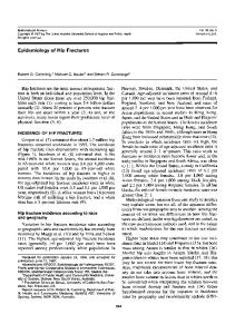
Fig. 12.1a–e. Pelvic fracture personality types. The management of a pelvic disruption depends on a clear evaluation of the personality of the fracture. The good personality types as noted in the drawing in a and the radiograph in b which demonstrates a relatively undisplaced stable fracture of the pelvis is different than the bad personality type as noted in the drawing in c and the radiographs in d and e. The anteroposterior radiograph (d) is that of a 21-year-old man who sustained a crush injury to the pelvis. The degree of instability was not recognized, and the patient was treated with bed rest while the extremities were attended. The final results (e) show severe shortening of the right hemipelvis with internal rotation. Note also the extremely high position of the right ischial tuberosity, which made sitting almost impossible (lower arrow). Marked shortening is indicated by the upper arrows above. Comparison of these two cases is like comparing apples to oranges or chalk to cheese
e
12.2
Understanding the Injury
12
241
Fractures of the Pelvis
a
b Fig. 12.2. a Radiograph of a patient with an apparently undisplaced fracture of the inferior and superior pubic ramus on the right side (white arrow). No lesion is seen posteriorly. The deformity of the left hemipelvis represents a malunion of an old left acetabular fracture. b Technetium polyphosphate bone scan of the same patient clearly showing the increased uptake of the superior and inferior pubic ramus fracture anteriorly, but also a massively increased uptake at the right sacroiliac joint, indicating a posterior lesion (black arrow). (From Tile 1984; courtesy of Dr. S.D. Gertzbein)
confirmed in a study by Bucholz (1981), in which posterior lesions at autopsy were found in all patients with pelvic trauma even when the radiograph had revealed only an anterior lesion.
12.2.2 Anatomical Lesions The anterior pelvic lesion may be a symphysis pubis disruption or overlap, or pubic rami fractures unilaterally or bilaterally. A symphysis disruption may also occur in combination with pubic rami fractures. The posterior lesion may be a fracture of the ilium, often in the coronal plane, a dislocation or fracture-dislocation of the sacroiliac joint, or a fracture through the sacrum (Fig. 12.3). The commonest lesion is a sacral fracture followed by a combined injury, i.e., a fracture-dislocation of the sacroiliac joint, usually with a portion of the ilium remaining attached to the main sacral fragment. Sacral fractures, in turn, may be classified as lateral, medial, or through the foramina or as complex types (H types).
Of greater importance than the site of the posterior lesion is the degree of displacement of the posterior sacroiliac complex. This can best be seen on the inlet radiograph showing posterior displacement of the so-called sacrogluteal line (Fig. 12.4) and is best confirmed by computed tomography (CT) scan. Therefore, the posterior lesion, although present, may be undisplaced and have intact posterior ligaments, often associated with a sacral crush, or may be displaced with a major ligamentous disruption of the posterior pelvic complex (Fig. 12.5).
12.2.3 Stability of the Pelvis The anatomical lesions are important for surgical management, but the stability factor is more important for overall decision making in the management of patients. Stability may be defined as the ability of the pelvis to withstand physiological forces without significant displacement. It is obvious that pelvic stability is dependent not only on the bony struc12.2
Understanding the Injury
242
M. Tile
a
Fig. 12.4. The dotted line on the right represents the sacrogluteal line on the inlet view of the pelvis. Any break in the continuity of this line, as shown on the left, represents displacement of the posterior complex, an ominous prognostic indicator. (From Tile 1984)
b
c Fig. 12.3a–c. Injuries to the posterior pelvic complex. The posterior injury may be a fracture through the ilium (a), a pure dislocation of the sacroiliac joint (b, straight arrow), or a fracture through the sacrum (c, straight arrow). A common pattern is a fracture dislocation through the sacroiliac joint, as shown by the small curved arrows in b and c
Fig. 12.5a,b. The posterior lesion may be stable or unstable. a The impacted right sacrum is clearly seen (white arrow). There is at least 1 cm of overlap between the two fragments. This posterior lesion is stable and cannot be moved. b The left sacral lesion is grossly unstable (black arrows). As well as the displacement at the fracture, all soft tissues are disrupted. (From Tile 1984)
12.2
Understanding the Injury
a
b
12
tures, but also on the strong ligamentous structures binding together the three bones of the pelvis, i.e., the two innominate bones and the sacrum. If these ligamentous structures are removed, the pelvis falls into its three component parts. Moreover, stability is a spectrum: at one end of the spectrum is the intact pelvic ring, at the other end a completely unstable pelvis, an internal hemipelvectomy. In our pelvic classification based on stability, the fractures at the stable end are type A, at the unstable end type C, and those with partial stability in the middle type B. The stability of the pelvic ring depends upon the integrity of the posterior weight-bearing sacroiliac complex (Fig. 12.6) and the pelvic floor. The major ligaments are the sacroiliac, the sacrotuberous, and the sacrospinous.
a
b
Fractures of the Pelvis
12.2.3.1 Sacroiliac Complex
The intricate posterior sacroiliac complex is a masterly biomechanical structure able to withstand the transference of the weight-bearing forces from the spine to the lower extremities. The ligaments have a major role as posterior stabilizers, because the sacrum, contrary to what is expected, does not form the shape of a keystone in a Roman arch, but is quite the reverse. Therefore, the strong posterior sacroiliac interosseous ligaments have been described as the strongest in the body, maintaining the sacrum in its normal position in the pelvic ring. Also, the iliolumbar ligaments join the transverse processes of L5 to the iliac crest, and the intervening transverse fibers of the interosseous sacroiliac ligaments further enhance
Fig. 12.6a,b. The major posterior stabilizing structures of the pelvic ring, as seen from the anteroposterior (a) and posterior view (b). The anteroposterior view (a) indicates the sacrospinous ligament as a strong triangular ligament lying anterior to the sacrotuberous ligament, a strong band extending from the lateral portion of the dorsum of the sacrum to the ischial tuberosity. These two ligaments form part of the pelvic floor, which is also supported by the pelvic floor muscles and fascia. The anterior sacroiliac ligament is flat and not as strong as the posterior sacroiliac ligamentous structures noted in the drawing (b). The posterior sacroiliac ligament, the sacrotuberous ligaments, and the sacrospinous ligaments are the major posterior stabilizing structures of the pelvic ring, that is, the posterior tension band of the pelvis. The ipsilateral sacroiliac complex often shows a compression through the sacrum. The pelvic floor integrity is usually maintained by the implosion force, thereby buckling the ligaments on the pelvic floor as noted. (From Tile 1984)
12.2
Understanding the Injury
243
244
M. Tile
the suspensory mechanism. The entire complex looks and functions like a suspension bridge (Fig. 12.7). The anterior sacroiliac ligaments are flat and strong and resist external rotation and shearing forces, although they do not have the strength of the posterior ligaments. 12.2.3.2 Pelvic Floor
The pelvic floor, with its muscular layer covered by investing fascia, also acts as a stabilizer of the pelvic ring. Two major ligaments also form part of the pelvic floor, namely the sacrospinous and sacrotuberous. The strong sacrospinous ligament, with fibers running transversely from the lateral edge of the sacrum to the ischial spine, resists external rotation of the pelvic ring (Fig. 12.8). The complex sacrotuberous ligament arises from most of the sacroiliac complex posterior to the sacrospinous ligament and extends to the ischial tuberosity. This strong ligament, positioned in the vertical plane, resists vertical shearing forces applied to the hemipelvis (Fig. 12.9). Therefore, these two supplementary ligaments, the sacrospinous and sacrotuberous, placed at 90° to each other, are well adapted to resist the two major forces
acting upon the pelvis, i.e., external rotation and vertical shear. In this way, they supplement the posterior sacroiliac ligaments.
12.2.4 Types of Injurious Forces Acting on the Pelvis Most forces acting on the pelvis are (a) external rotation, also called anteroposterior compression, (b) internal rotation (lateral compression), or (c) shearing or translational forces in the vertical plane. In the complex high-energy trauma seen in our society, some forces defy description, but, in general, the above are the three major force vectors acting upon the pelvic ring.
Fig. 12.8. The sacrospinous ligaments, joining the sacrum to the ischial spines, resist external rotatory forces (arrows). (From Tile 1984)
Fig. 12.7. The suspension bridge-like appearance of the ligaments binding the posterior sacroiliac complex. Note the vertical direction of the interosseous posterior sacroiliac ligaments, noted by Grant to be the strongest in the body, as well as the transverse component acting as the suspension, joining the pillars, represented by the posterior superior or iliac spines, to the sacrum. (From Tile 1984)
12.2
Understanding the Injury
Fig. 12.9. The sacrotuberous ligament, joining the sacrum to the ischial tuberosity, resists a shearing rotatory force (arrows). (From Tile 1984)
12
External rotation forces occur with a direct blow to the posterior superior spine or, more commonly, by forced external rotation through the hip joints unilaterally or bilaterally. This force usually produces an open book-type injury, i.e., the symphysis pubis disrupts and, as further force is applied, the sacrospinous ligament and the anterior ligaments of the sacroiliac joint may also open (Fig. 12.10). Eventually, impingement of the posterior ilium on the sacrum occurs. At this point, the posterior sacroiliac ligaments still confer stability to the ring, and translation vertically or posteriorly is not possible.
Fractures of the Pelvis
The force of internal rotation or lateral compression may be transmitted by a direct blow to the iliac crest, often causing an upward rotation of the hemipelvis or the so-called bucket-handle fracture, or through the femoral head, often causing an ipsilateral injury (Fig. 12.11). In this pattern, the anterior structures, usually the rami, break and the hemipelvis rotates internally. If the posterior ligaments remain intact, the anterior sacrum will compress. If the posterior ligaments tear, stability is still maintained by the pelvic floor. Shearing forces in the vertical plane cross the main trabecular pattern of the posterior sacroiliac
a b Fig. 12.10. a A direct blow to the posterior superior iliac spines will cause the symphysis pubis to spring open. b External rotation of the femora or direct compression against the anterior superior spines will also cause springing of the symphysis. (From Tile 1984)
a b Fig. 12.11. a A lateral compressive force directed against the iliac crest will cause the hemipelvis to rotate internally, crushing the anterior sacrum and displacing the anterior pubic rami. b Lateral compression injury may also be caused by a direct force against the greater trochanter. In that situation, the femoral head acts as a battering ram, dividing the pubic rami as shown, often through the anterior column of the acetabulum. The ipsilateral sacroiliac complex is also crushed in this injury. Note that the sacrospinous and sacrotuberous ligaments generally remain intact along with the pelvic floor in this lateral compressiontype injury. (From Tile 1995)
12.2
Understanding the Injury
245
246
M. Tile
complex, whereas a lateral compressive force causes impaction of the cancellous bone and usually allows retention of the ligament integrity. However, external rotation and lateral compression forces may be so great that they overcome the restraining effect of the ligament. Therefore, a completely unstable pelvic ring may be caused by complex forces acting on the pelvis. The term “shear” is synonymous with these complex forces. Shearing forces cause marked displacement of bone and gross disruption of the soft tissue structures (Fig. 12.12), including the pelvic floor. Continuation of these forces beyond the yield strength of the soft tissues produces an unstable pelvic ring with major anterior and posterior displacement. No finite point is reached with these shearing forces; therefore, the entire hemipelvis may be avulsed from the body, occasionally resulting in a traumatic hindquarter amputation.
12.2.5 Effect of Forces on Soft Tissue External rotation and shear forces tend to tear soft tissue; therefore, the injuries caused by these forces are usually major: tearing viscera and arteries and causing traction injuries to nerves. Lateral compression forces (internal rotation) tend to puncture viscera and compress nerves (Dalal et al. 1989).
12.3 Classification 12.3.1 Comprehensive Classification (from Tile 1988) 12.3.1.1 General Concepts
By combining the concepts of stability, force direction, and pathoanatomy, a meaningful classification may be developed to aid in patient management. No classification can answer all the questions regarding a specific injury. Since the first edition of this book, and since our publication in The Journal of Bone and Joint Surgery in January 1988 (Tile 1988), refinements have been made to our original classification to allow acceptance as the comprehensive classification of pelvic fractures. The basics of this classification stem from the concepts of George Pennal, who developed a classification based on force direction. The Young-Burgess classification has retained those basic principles. With members of the AO group, we have expanded the concept to include stability as well as force direction (Tile et al. 1988). All classifications should serve as guides to treatment and should allow centers to compare simi-
Fig. 12.12. A shearing force (arrows) crosses perpendicular to the main trabecular pattern of the posterior pelvic complex in the vertical plane. These forces cause marked displacement of bone and gross disruption of the soft tissues, resulting in major pelvic instability. (From Tile 1995)
12.2
Understanding the Injury
12
Fractures of the Pelvis
lar cases. The management of individual patients requires careful specific assessment, and the surgeon must be able to draw the fracture lines on a dry skeleton as well as determine the degree of soft tissue injury. The classification (Table 12.1) also follows the A,B,C nomenclature of the comprehensive classification (Müller et al. 1990), with increasing severity of injury from A to C. It must also be remembered that the types A, B, and C based on stability form a spectrum rather than a rigid black and white concept. For the purpose of this classification, the posterior pelvis is located posterior to the acetabulum, and the anterior arch anterior to it. The fracture type is based on the posterior lesion, which is more important for stability, and the anterior lesions are denoted by modifiers.
injury may be unstable in internal rotation or may be rigidly impacted, but neither is unstable in the vertical plane unless a force which disrupts the posterior ligamentous structures is present. Also, it is self-evident that unstable pelvic injuries may be produced by any force vector that overcomes the yield strength of the soft tissues. As well, there is a tendency in the relevant literature, especially from European centers, to group all type B injuries into one. Since the B1 external rotation type is vastly different from the B2 lateral compression, and has a different prognosis, these two types must be separated in any studies and reports.
Table 12.1. Classification of pelvic ring disruption ( from Tile 2002)
In the type A injury, the pelvic ring is stable and cannot, by definition, displace by physiological force. These injuries include type A1 avulsion fractures, which usually occur in adolescents and do not involve the pelvic ring. The type A2 fractures involve the iliac wing or the anterior arch without a posterior injury, a rare occurrence. The type A3 fractures are transverse fractures of the sacrum and coccyx and should more correctly be considered spinal injuries.
Type A: stable pelvic ring injury Type B: partially stable pelvic ring injury B1: Open book injury (AP compression, external rotation) B2: Latera; compression (internal rotation) B3 : Bilateral injuries Type C: completely unstable (allows all degrees of translational displacement)
Stability is defined as the ability to withstand physiological forces without deformation. Therefore, at one end of the stability scale, the type A pelvic lesions do not displace the ring, only involving the avulsions of the iliac wing or transverse sacral fractures, really spinal injuries. In all these cases the pelvis remains intact. At the other end of the spectrum, the type C fractures are unstable, with complete disruption of the posterior arch, the pelvic floor, and usually the anterior arch. The type B fractures retain some posterior stability and are therefore partially stable; they cannot, by definition, translate vertically or posteriorly. A and B types generally comprise about 70% of the total fractures, even in trauma centers; the remainder are unstable type C (Pohlemann and Tscherne 1995). The partially stable (type B) injuries are of two varieties: the open book or anteroposterior compression injury, caused by external rotation, and the lateral compression injury, caused by internal rotation. It should be remembered that the open book injury caused by an external rotatory force is unstable in external rotation, whereas the lateral compression
12.3.2 Type A Stable Fractures (Table 12.2)
12.3.3 Type B – Partially Stable Fractures (Table 12.2) 12.3.3.1 Open Book (Anteroposterior Compression) Fractures (B1, B3.1)
External rotatory forces applied to the pelvis usually cause a disruption of the symphysis pubis; however, they may also cause an avulsion fracture of the pubis adjacent to the symphysis or a fracture through the pubic rami, the symphysis avulsion or disruption being more common. Since the force is a continuum and may stop at any point, several possibilities exist. First, an opening of the symphysis pubis less than 2.5 cm permits stability to be retained in the pelvic ring, a situation not dissimilar to that observed during delivery of a baby. In the rare traumatic injury, the sacrospinous and anterior sacroiliac ligaments remain intact (Fig. 12.13). Therefore, a CT scan will show no opening of the sacroiliac joints. Second, continuation of the external rotatory force will reach a finite end point when the “book” opens to 12.3 Classification
247
248
M. Tile Table 12.2. Classification of pelvic ring disruption (from Tile 1988) Comprehensive Classification
Young and Burgess Classification
Type A: Stable pelvic ring injury
No equivalent
A1: Avulsion of the innominate bone
No equivalent
A2: Stable iliac wing fracture or stable minimally displaced ring fracture
No equivalent
A3: Transverse fractures of the sacrum and coccyx
No equivalent
Type B: Partially stable B1: Open-book injury
APC 1, APC 11
B2: The lateral compression injury
LC1, LC11, crescent fracture
B3: Bilateral B injuries
Windswept, complex
Type C: Complete unstable C1: Unilateral
APC 111, vertical shear
C2: Bilateral, one side B, one side C
Complex
C3: Bilateral C lesions
Complex
a
b Fig. 12.13a,b. The first stage of an open book injury (type B1) is a disruption of the symphysis pubis only with no involvement of the sacroiliac joints (a). The patient in b, a hockey player who sustained a direct blow to the posterior sacroiliac area bilaterally, noted immediate pain anteriorly at the symphysis pubis. His radiograph indicates a symphysis pubis separation of 1.5 cm with no opening of the sacroiliac joints posteriorly. (From Tile 1995)
the extent that the posterior iliac spines abut upon the sacrum. In this particular circumstance, the sacrospinous ligaments and the anterior sacroiliac ligaments are torn, but the strong posterior sacroiliac ligaments remain intact (Fig. 12.14). Occasionally, the posterior injury may be a fracture of the ilium or sacrum. 12.3 Classification
Therefore, this injury is unstable in external rotation, but as long as the force does not continue beyond the yield strength of the posterior ligaments, stability can be returned to the pelvic ring by internal rotation. It is extremely important to realize that the external rotatory force may in fact continue beyond the
12
Fractures of the Pelvis
Ultimately, with this type of force, a symphysis disruption as well as involvement of the pelvic soft tissues such as the vagina, the urethra, the bladder, or the rectum may occur. In the classification, the anterior lesion is designated by modifiers (Table 12.3). In the open book injury, typical varieties of open book fracture may occur with fractures anteriorly through the pubic rami unilaterally or bilaterally. These modifiers are descriptive of the injury, and are important for decision-making as well as clinical investigation.
a
b Fig. 12.14a,b. The second stage of an open book injury. a In this diagram note that the symphysis pubis has disrupted more than 2.5 cm. If that occurs, the sacrospinous ligaments tear or an equivalent avulsion of the adjacent sacrum or ischial spine occurs, as well as an avulsion of the anterior sacroiliac joints, causing a wide anterior opening of the sacroiliac joints. However, pelvic stability is maintained by the intact posterior ligamentous structures, indicated by the black lines. The endpoint is reached when the posterior iliac spines abut the sacrum. b A typical radiograph showing the disruption of the symphysis pubis and the markedly widened sacroiliac joints anteriorly (arrows). (From Tile 1984)
Fig. 12.15. The presence of a symphysis disruption does not imply a stable configuration; in fact, most symphysis disruptions are associated with unstable posterior lesions, as shown. Note the telltale avulsion fracture of the L5 transverse process, indicating instability and posterior displacement of this fracture. (From Tile 1984)
Table 12.3. Anterior pelvic qualifiers. The Qualifiers of the anterior arch lesions C1 to C9 are identical for all subgroups of Types B and C (in part from Tile 2003) C1)
yield strength of the posterior ligament, causing a complete avulsion of the hemipelvis. This is no longer an open book configuration but is now an unstable fracture of the worst variety (Fig. 12.15). In fact, as previously indicated, a complete traumatic hemipelvectomy may ensue. Therefore, the presence of a symphysis disruption does not always imply an open book fracture. Careful assessment is required to be certain that vertical instability is not also present.
Unilateral pubis / rami fx, ipsilateral
C2)
Unilateral pubis / rami fx, contralateral
C3)
Bilateral pubis / rami fx
C4)
Symphysis pubis disruption, m 2.5 cm
C5)
Symphysis pubis disruption, >2.5 cm
C6)
Symphysis pubis disruption, locked
C7)
Symphysis + ipsilateral pubis / rami fx (tilt) (yes)
C8)
Symphysis + contralateral pubis / rami fx
C9)
Symphysis + bilateral pubis / rami fx
C10)
No anterior lesion
12.3 Classification
249
250
M. Tile
12.3.4 Partially Stable Fractures (Type B2) 12.3.4.1 Lateral Compression Fractures (Tables 12.1, 12.2, 12.4)
There are several types of lateral compression injury depending upon the site of the anterior and posterior lesion (Table 12.4). The anterior and posterior lesions may be on the same side or ipsilateral (type B2.1), or they may be on opposite sides, producing the socalled bucket-handle type of injury (type B2.2).
Table 12.4. Lateral compression injury (from Tile 2003) B2 Lateral compression injury B 2-1 Ipsilateral B 2-2 Contralateral type (bucket-handle)
Type B2.1 – Ipsilateral Fractures
An internal rotation force applied to the ilium or, more commonly, a direct blow to the greater trochanter may cause a typical lateral compression or internal rotation fracture of the hemipelvis. The superior and inferior rami break, and a crush may then occur anteriorly at the sacroiliac joint or through the sacrum, but, commonly, the posterior ligamentous structures do not disrupt (Fig. 12.16a). The entire hemipelvis may be forced across to the opposite side, thereby rupturing the bladder or blood vessels within the pelvis. The elastic recoil of the tissues may deceive the examiner, and the fracture may appear undisplaced in the radiograph. However, the radiographs in Fig. 12.16b,c show the bladder being drawn back into the fracture site by the recoiling pelvis. If the bone is stronger than the ligaments, the posterior ligaments may disrupt, but stability may be retained by an intact pelvic floor, not disrupted by the implosion force.
Fig. 12.16a–c. Lateral compression fracture, type B2.1: ipsilateral. The diagram (a) shows a typical ipsilateral type of lateral compression injury. Note the anterior crush to the sacrum and the overlap of the pubic rami. In this particular case there is posterior disruption, but stability is afforded by the crush in the sacrum and the intact pelvic floor. The force necessary to produce this seemingly minimally displaced fracture is often underestimated because of the elastic recoil of the pelvis. This fracture, barely perceptible on the inlet radiograph (b, arrow), was obviously grossly displaced at the moment of injury, since the bladder was pulled back into it, as shown in the cystogram (c, arrow). (From Tile 1984)
a
b
c
12.3 Classification
12
The anterior injury designated by the modifiers may be as follows (see Table 12.3): - Fractures of both rami. This is the most common injury, with a spike of bone possibly penetrating the pelvic viscera. - Locked symphysis. This rare injury is a form of ipsilateral lateral compression type. As the hemipelvis internally rotates, the symphysis disrupts and locks, making reduction extremely difficult (Fig. 12.17). - Tilt fracture. The tilt fracture consists of a symphysis disruption and a fracture of the superior and/or the inferior pubic ramus, with possible impingement of the bone into the vagina of young females (Fig. 12.18).
251
Fractures of the Pelvis
Type B2.2 – Contralateral: Bucket-Handle Injuries
The bucket-handle type of injury is usually caused by a direct blow to the ilium. The anterior fracture may be on the opposite side to the posterior lesion (contralateral type), or all four rami may fracture anteriorly but the anterior displacement is on the side opposite the posterior lesion. Another combination might be a symphysis disruption with two rami fractures. This injury has particular characteristics that may seem confusing. The affected hemipelvis rotates anteriorly and superiorly like the handle of a bucket (Fig. 12.19). Therefore, even if the posterior structures are relatively intact, the patient may have a major leg
a Fig. 12.17a,b. Locked symphysis. a Diagram and b anteroposterior radiograph showing an unusual type of lateral compression injury where the symphysis becomes firmly locked anteriorly. (From Tile 1984)
b
a
b
Fig. 12.18. a A variant of the type I injury often seen in young women. The lateral compressive force fractures the superior ramus, often through the anterior column of the acetabulum. Continuing lateral compression rotates the distal fragment through the symphysis pubis, thereby disrupting it. This distal fragment assumes a vertical position and may impinge on the perineum, as demonstrated in the anteroposterior radiograph (b). (From Tile 1984)
12.3 Classification
252
M. Tile
a
b
c
d Fig. 12.19a–d. Type B2.2 lateral compression injury. The diagram (a) demonstrates a typical type B2.2 lateral compression injury, characterized by compression of the posterior sacroiliac complex associated with a straddle or butterfly fracture of the four pubic rami anteriorly. The anteroposterior (b), inlet (c), and outlet (d) views of this 19-year-old woman show this classic lesion with upward rotation and impaction of the right hemipelvis. Even under general anesthesia, this hemipelvis could not be moved on the third day following injury, indicating severe posterior impaction
length discrepancy. Very often, the posterior structures are firmly impacted, the deformity being clearly noted on physical examination. Reducing these fractures and thereby the leg length discrepancy requires derotation of the hemipelvis rather than pure traction in the vertical plane. With continued internal rotation, the posterior structures may yield, producing some instability. However, the anterior sacroiliac crush is usually so stable that reduction is difficult, and some stability is maintained by the intact pelvic floor and the sacrospinous and sacrotuberous ligaments. This is akin to the situation with a vertebral fracture, where the vertebral body may be crushed by 12.3 Classification
flexion forces but the posterior spinous ligament has ruptured. An excellent example of this is shown in Fig. 12.20a. The original radiograph of this 16year-old girl shows the internal rotation of the left hemipelvis and the posterior impaction. All four rami are broken anteriorly and the leg length discrepancy is seen. The CT scan (Fig. 12.20b) again shows the left hemipelvis to be internally rotated and the anterior portion of the sacroiliac joint crushed. Posteriorly, the arrow points to the avulsion of the posterior iliac apophysis (Fig. 12.20b,c). At surgery, the apophysis was clearly avulsed but the posterior sacroiliac ligaments were completely intact. After posterior reduction of the fracture and
12
253
Fractures of the Pelvis
a
b
c
d
e
Fig. 12.20. a Anteroposterior radiograph of a 16-year-old girl with a type B2.2 bucket-handle fracture. The fracture involves all four pubic rami and the left sacroiliac joint. b Computed tomography (CT) clearly outlines the essential features of this fracture. Note the anterior crush of the sacrum, the internal rotation of the left hemipelvis, and, in this case, the avulsion of the iliac apophysis, which had not yet fused to the ilium (arrow). c Clinical appearance at surgery of this apophysis avulsion (outlined by the probe, arrow). d Appearance after reduction and fixation with two lag screws crossing the sacroiliac joint. e Postoperative radiographic appearance
12.3 Classification
254
M. Tile
fixation by two screws, the pelvis is anatomically reduced (Fig. 12.20d,e). Type B3 – Bilateral Partially Stable Injuries
The B3 bilateral injuries may be the classical open book type (B3.1), or one side B1 and one side B2 (B3.2), or bilateral B2 (B3.3).
12.3.5 Type C – Unstable Fractures – Complete Disruption of the Posterior Arch (see Tables 12.1, 12.2)
Telltale radiographic signs of instability include avulsion of the transverse process of the L5 vertebra or of either attachment of the sacrospinous ligament (Fig. 12.22). Greater than 1 cm of posterior or vertical translation is noted. The CT scan shows the radiographic appearance of the unstable posterior complex better than the plain radiograph and should be obtained in all cases. A comparison of the CT scans (Fig. 12.23) shows clearly the difference between the impacted stable posterior complex and the grossly unstable complex of the vertical shear injury.
An unstable pelvic disruption implies disruption of the posterior sacroiliac arch as well as a rupture of the pelvic floor, including the posterior structures as well as the sacrospinous and sacrotuberous ligaments (Fig. 12.21). The unstable injury may be unilateral (type C1), affecting one posterior iliac complex, or may be bilateral (type C2 or C3), affecting both. The unilateral lesions may be fractures of the ilium (type C1.1) through the sacroiliac joint, or either a pure dislocation or a fracture-dislocation with involved ilium or sacrum (type C1.2), or a fracture of the sacrum (type C.1.3). The bilateral types C2 include one side unstable (C) and one side partially stable (B), while the C3 lesions include bilaterally unstable types.
a
b Fig. 12.21. Unilateral unstable vertical shear fracture. Shearing forces cause massive disruption of the pelvic ring, including the pelvic floor. Note the avulsion of the ischial spine and the tip of the transverse process of L5, both signs of pelvic instability. Note also the stretch of the lumbosacral plexus, commonly injured in this pattern of injury. (From Tile 1984)
12.3 Classification
Fig. 12.22a,b. Telltale signs of instability. a Avulsion of the ischial spine (black arrow) and posterior displacement of the ilium (white arrow). b Avulsion of the sacral end of the sacrospinous ligament (black arrow) and the tip of the transverse process of L5 on the opposite side (white arrow) in this bilateral injury. (From Tile 1984)
12
255
Fractures of the Pelvis
a
b Fig. 12.23. a CT scan showing marked disruption and instability of the left sacrum as a result of a shearing force. b CT scan showing impaction of the right sacrum from a lateral compression injury. This young woman sustained an acetabular fracture as well, confirming the mechanism of injury. Note the marked overriding of the sacral fragments on the fractured side as compared to the normal left side. Impaction was so rigid that no abnormal movement of the hemipelvis was detected on physical examination with image intensification. (From Tile 1984)
12.3.6 Unusual Types of Fracture
12.3.6.3 Pelvic Disruptions Associated with Acetabular Fractures
12.3.6.1 Complex Fractures
Many severe types of fracture dislocation of the pelvis defy precise classification because of the complex forces causing the injury. In these cases, the pelvic ring may be disrupted in a very bizarre fashion. Because of the high-energy forces involved, the pelvic ring is usually unstable; since most are bilateral, most will fall into the C3 classification. 12.3.6.2 Bilateral Sacroiliac Dislocation with an Intact Anterior Arch
This unusual injury is usually caused by hyperflexion of the legs (for example, two of our cases were in young women who were crushed in the hyperflexed position under a horse that reared and fell backwards). In this particular situation, the anterior complex remains intact but both sacroiliac joints dislocate posteriorly (C3).
If a pelvic ring disruption is associated with an acetabular fracture, the prognosis will clearly change and will be more dependent upon the acetabular component than upon the pelvic ring disruption. These complex injuries are relatively common. CT scanning of acetabular fractures has indicated a significant number of sacroiliac injuries and pelvic ring disruptions associated with acetabular fractures. In the comprehensive classification, the pelvic ring component is classified separately from the acetabulum (see Chap. 13).
12.4 Natural History In an attempt to further elucidate the incidence and severity of the early and late musculoskeletal complications of this injury, we undertook a clinical study in association with R. Lifeso, D. Dickinson, and R. McBroom (Dickinson et al. 1982). The purpose of this study was to place the management of this injury in perspective by determining which pelvic fractures 12.4 Natural History
256
M. Tile Table 12.5. Comparison of series A and series B patientsa (from Tile 1984) Series A (n=148)
Series B (n=100)
34.2 years
30.9 years
(15–81)
(14–85)
Male
91
55
Female
57
45
1. Age (range) 2. Sex
3. Injury types Motor vehicle accidents
89 (60%)
81
Fall
17 (11.5%)
11
Crush
34 (23%)
4
8 (5.5%)
4
43 (29%)
5
CNS
31 (21%)
38
Chest
19 (13%)
15
Gastrointestinal
10 (6.6%)
20
Bladder
17 (11%)
8
Miscellaneous 4. Workmen’s Compensation Board 5. Associated injuries
Urethra
Table 12.6. Factors resulting in unsatisfactory results (from Tile 1984)
6 (4%)
4
Nerve
12 (8%)
3
Musculoskeletal
63 (43%)
10
60 mo
2 years
6. Follow-up average
had the poorest prognosis. With the current trend to internal fixation of the pelvis, a study of the natural history of this injury is even more important, in order to place that trend in perspective, for without knowledge of the natural history, logical decision-making becomes impossible. The results of our review of 248 cases are shown in Tables 12.5–12.9. In this study, every patient was recalled, personally interviewed, examined, and radiographed using the inlet, outlet, and anteroposterior views. The conclusions may be summarized as follows: 1. Stable injuries gave few major long-term problems. Pain, if present, was usually mild or moderate. 2. By contrast, patients with unstable pelvic disruptions had many problems at review. Approximately 30% of this group had continuing pain, including 3% with nonunion of the posterior complex and 5% with malunion, defined as having a greater than 2.5-cm leg length discrepancy. In addition, 5% had permanent nerve damage, and 3% continuing urethral problems following urethral rupture.
a
In Tables 12.5 and 12.6, series A is a group of 148 cases of pelvic fracture managed in Toronto teaching hospitals and in nonteaching hospitals in Ontario, retrospectively reviewed; series B consists of the first 100 cases of pelvic fracture treated at the Sunnybrook Medical Center, Toronto, prospectively reviewed
Pain
Series A: 37/148
Series B: 35/100
37
32
Leg length discrepancy >2 cm
7
2
Nonunion
5
3
Permanent nerve damage
9
3
Urethral symptoms
5
1
Deaths
17
Table 12.7. Pain (moderate and severe) (from Tile 1984) Series A (n=148)
Series B (n=100)
No.
Nil
Moderate
Severe
No.
Nil
Moderate
Severe
53 (36%)
–
–
–
35
–
–
–
Posterior
47 (32%)
–
–
–
32
–
–
–
Anterior
6 (4%)
3
Anteroposterior compression
23
14
8
1
6
3
3
Lateral compression
86
47
35
4
69
53
16
Unstable (shear)
9
4
2
3
25
9
13
3
65
45
8
100
65
32
3
Incidence Location
Severity
Total a
118
a
Thirty cases with major acetabular involvement were not considered in this total
12.4 Natural History
12
The pain, when present, usually arose from the posterior sacroiliac joint area or from the lower lumbar spine. CT has shown lumbar spine involvement in significant numbers of patients with pelvic disruption. The pain in these cases was more severe, and usually associated with an unreduced sacroiliac dislocation or a nonunion. In summary, the natural history of pelvic trauma depends on the degree of violence, the type of injury, the method of treatment, and the presence or absence of complications such as a urethral tear, permanent nerve damage, malunion, malreduction of the sacroiliac joint, or nonunion. The unstable vertical shear injury results in a significant number of permanent problems resulting in posterior pain. Therefore, it is obvious that most of our energies should be directed to the management of the unstable vertical shear injury, especially if the sacroiliac joint is dislocated or subluxated, since more stable injuries achieve good to excellent results when managed by simple means, as will be described.
Table 12.8. Leg length discrepancy (malunion) (from Tile 1984) Amount (cm)
Series A (%)
Series B (%)
0
64
68
0–1
19.5
19
1–2
11.5
11
>2
5
2
Fractures of the Pelvis
12.5 Management of the Pelvic Disruption Management of a pelvic disruption depends on the “personality” of the injury as well as that of the associated injuries (see Fig. 12.1) and may be considered under the following four headings: assessment, resuscitation, provisional stabilization, and definitive stabilization, which, although considered separately, form a continuum of care.
12.5.1 Assessment 12.5.1.1 General Assessment
It is beyond the scope of this chapter to detail the general assessment of the polytraumatized patient. Suffice it to say that a polytraumatized patient with a pelvic fracture represents a therapeutic challenge to the treating surgeon because the mortality rate remains approximately 10%, and as high as 31% in the unstable pelvis (type C) (Pohlemann and Tscherne 1995). The necessity of a planned treatment protocol for the polytraumatized patient cannot be overemphasized. The patient must have immediate appropriate treatment from the time of injury until stabilization in an appropriate intensive care unit. The central theme of system management during resuscitation is simultaneous rather than sequential care. We recommend the treatment protocol of the American College of Surgeons in
Table 12.9. Results by fracture type (from Tile 1984) Series A (n=148) Total No.
Series B (n=100) Satisfactory
Unsatisfactory
(n)
%
(n)
%
Total No.
Satisfactory
Unsatisfactory
(n)
%
(n)
%
Anteroposterior compression
23
18
78
5
22
6
3
50
3
50
Lateral compression
114
79
69
35
31
69
53
77
16
23
Unstable (shear)
9
5
56
4
44
25
9
36
16
64
12.5
Management of the Pelvic Disruption
257
258
M. Tile
the Advanced Trauma Life Support (ATLS) Program (Aprahamian et al. 1981). In the primary survey, problems involving the airway, bleeding (shock), and the central nervous system have the highest priority. Immediate lifesaving resuscitation, therefore, must be directed to both the airway and the presence of shock. In pelvic trauma, shock may be profound due to retroperitoneal arterial or venous hemorrhage. The secondary survey following the primary resuscitation includes further examination of the airway, bleeding, the central nervous system, the digestive system, the excretory system, and, finally, the fracture. For further study in the management of polytrauma patients, we refer the reader to the excellent monograph on this subject by the American College of Surgeons mentioned above.
compression of both anterior iliac spines. Lateral compression injuries are usually in an anatomical recoiled position unless they have been impacted. Further internal rotation by compression of the iliac crests will displace the fracture. Finally, by applying one hand to the pelvic iliac crest and using the other to apply traction to the leg, displacement in the vertical plane can usually easily be diagnosed (Fig. 12.24b). This maneuver may require two examiners, one to apply traction and the other to palpate the iliac crests. If possible, these manipulations should be done under image intensification to verify the type of displacement and whether displacement in the vertical plane is present.
12.5.1.2 Specific Musculoskeletal Assessment
For the management of the musculoskeletal injury, assessment is directed to the determination of the stability of the pelvic ring. Clinical Assessment
As in all areas of clinical medicine, an accurate history is essential; patients who have sustained a high-energy injury from motor vehicle trauma or falls from a height are much more likely to have an unstable pelvic injury than are those who have sustained low-energy trauma. The physical examination is at least as important as the radiographs in determining pelvic stability. The essence of the physical examination is to inspect the patient for major bruising or bleeding from the urethral meatus, vagina, or rectum. If these latter two areas are not carefully inspected, occult lacerations may be overlooked, with dire consequences, since these lacerations always mean an open fracture of the pelvis. The pelvic area and the lower extremities should be examined with the patient undressed, so that displacement and limb shortening can be detected. In the absence of a lower extremity fracture, rotatory deformity or limb shortening usually implies an unstable pelvic injury. Determination of pelvic stability can simply be done by the physician applying his or her hands to the anterior superior spine and moving the affected hemipelvis (Fig. 12.24a). Open book injuries are maximally externally rotated and can be closed by 12.5
Management of the Pelvic Disruption
a
b Fig. 12.24. a Direct palpation of the iliac crest will reveal crepitus or abnormal motion, which, if present, is the best indicator of instability of the pelvis. b With one arm controlling the injured hemipelvis and the second arm applying traction, the amount of instability present can be determined. (From Tile 1984)
Radiographic Assessment
Plain Radiographs. As a routine in the acute situation, a single anteroposterior radiograph as commonly used in most trauma centers is usually sufficient to determine the presence or absence of pelvic ring instability. Although this radiograph will suffice in the acute injury during the resuscitative phase, a single anteroposterior radiograph may be mislead-
12
ing. Therefore, for accurate assessment of pelvic ring displacement, an inlet and an outlet view should be added (Fig. 12.25). The inlet view, taken by directing the X-ray beam 60° from the head to the midpelvis, is the best radiographic view to demonstrate posterior displacement. The outlet view, taken by directing the X-ray beam from the foot of the patient to the symphysis at an angle of 45°, demonstrates superior or inferior migration of the hemipelvis.
Fractures of the Pelvis
These views are now also used for radiographic control of iliosacral screw placement; therefore, knowledge of the skeletal landmarks is important. CT Scan. The CT scan is the best single investigative tool for determining pelvic instability, since the sacroiliac area is best visualized by this technique. Stable impacted fractures of the sacrum can be clearly differentiated from grossly unstable ones by
a
b
c
d
e
Fig. 12.25a–e. With the standard anteroposterior radiograph, inlet and outlet views may be very helpful. The outlet view as shown on the skeleton (a) and the radiograph (b) is the best view for visualizing the sacrum, the sacroiliac joints, and the sacral foramina, caudad and cephalad displacement is seen as well. The inlet view, as noted in the skeleton (c) and radiograph (d) best delineates posterior displacement of the hemipelvis. e Note the different information obtained from the above views compared to the anteroposterior view. (From Tile 1984)
12.5
Management of the Pelvic Disruption
259
260
M. Tile
this method (see Fig. 12.3). In pelvic ring trauma, three-dimensional CT is helpful in assessing the overall injury pattern, but not nearly as much as in acetabular trauma. 12.5.1.3 Diagnosis of Pelvic Instability
Careful clinical and radiographic assessment will allow the surgeon to determine the personality of the pelvic injury, i.e., whether the musculoskeletal injury is more to the stable or to the unstable end of the stability scale. The completely unstable (type C) can usually be diagnosed clinically by the lack of a firm endpoint in rotation or traction. Radiographically, a displacement or gap on plain X-ray or CT equivalent to 1 cm and the presence of avulsion fractures of the ischial spine or sacrum all suggest instability. Patients with partially stable (type B) have a firm endpoint on palpation, be it external rotation (B1) or internal rotation (B2). In the latter, the pelvis may be impacted in the internally rotated position (see Fig. 12.19).
12.5.2 Resuscitation Hemorrhage in pelvic trauma may be life-threatening. The site of bleeding is determined by peritoneal lavage, portable ultrasound, or CT. CT using contrast may give a rapid picture of arterial bleeding, and is being advocated by some authors (Fig. 12.26). In the resuscitative phase, control of hemorrhage must be rapid and may be lifesaving. Patients with an unstable pelvic disruption are at much greater general risk than those with a stable pelvis. In our first prospective study of 100 patients, 12 of the 15 mortalities were in this unstable group (McMurtry et al. 1980). Their blood transfusion requirements were three times greater (15.5 units vs. 5.5 units), their injury severity score was 37 (vs. 29 in those with a stable pelvis), and their overall complication rate was three times higher. Patients suffering this complication require massive fluid replacement, as outlined by the American College of Surgeons’ ATLS protocol. Early management of shock should include the pneumatic anti-
a
c
Fig. 12.26a–c. A 70-year-old female struck by a car sustained an unstable C-type pelvic fracture. She was in shock, with no obvious source. a: Skeletal traction was applied to her right leg. b: A contrast-enhanced CT showed extravasation that correlated with angiographic findings indicating bleeding from the obturator artery. c: 9 h post-injury shows successful angiographic embolization of obturator artery. (Courtesy of Dr. David Stephen). (From Tile 2003)
b
12.5
Management of the Pelvic Disruption
12
shock garment (PASG). The advantages and disadvantages of the PASG are listed in Table 12.10. In our opinion, the advantages outweigh the disadvantages, the only notable disadvantage being restriction of access to the abdomen. The garment must not be precipitously released. During gradual release of the garment, the blood pressure must be carefully monitored. Any drop greater than 10 mmHg in the systolic blood pressure is a contraindication to further deflation. Other guidelines of importance include inflation of the legs prior to the abdominal portion and reversing that order during deflation. In transfer situations, great care must be taken to prevent lengthy inflation periods, which may cause compartment syndromes in the lower extremity. Table 12.10. Pneumatic antishock garment (PSAG; from Tile 2003) P. 74 Advantages
Disadvantages
Simple
Short-lasting volume effect
Rapid
Compartment syndrome of lower extremity
Reversible
Decreased access to abdomen and lower extremity
Accessible and available
Decreased visibility of abdomen and lower extremities
Safe
Fracture of lower extremity Decreases lung compliance
Fracture stabilization belongs in the resuscitative phase of management. There is a growing body of evidence to suggest that the application of a simple anterior external frame will reduce retropelvic venous and bony bleeding to the extent that other intervention is rarely required. Therefore, pelvic stabilization should be performed early. Pelvic clamps which can be applied in the emergency room with direct skeletal fixation are now available and are useful in the patient with an unstable pelvic ring and severe bleeding (Ganz et al. 1991). It is hoped that this will reduce mortality by allowing the volume of the pelvis to decrease to its normal size, thereby restoring the tamponade effect of the bony pelvis and helping to stop the venous bleeding. The precise method for early fracture stabilization will be discussed in the next section. The role of embolization of the pelvic vessels has largely been clarified, but its use varies greatly from institution to institution, depending on local circumstances. In our trauma unit, we have narrowed its use to those patients who are bleeding mainly from a
Fractures of the Pelvis
small-bore artery such as the obturator or the superior gluteal arteries. Because of early diagnosis of arterial bleeding, using advanced imaging (including contrast-enhanced CT), the overall use and effectiveness of embolization have increased. Earlier arterial embolization for the right indication is desirable. Embolization is of little value in hemodynamically unstable patients in extremis with massive bleeding from the major vessels of the internal iliac system, because the emboli cannot control this type of hemorrhage, and the patient may die during the attempt. It is also, of course, of no value in venous or bony bleeding. Small-bore artery bleeding may be assumed if, although the patient can be well controlled using the above methods of fluid replacement, PASG, and fracture stabilization, he or she goes back into a shocked state each time the fluid is slowed down. A more precise diagnosis is made on contrastenhanced CT (see Fig. 12.26) or definitively on angiography. In those circumstances, after hemodynamic stability has been achieved, the patient is moved to the vascular suite, an arteriogram is performed, and if a small-bore artery is lacerated it is embolized with Gelfoam (Upjohn Pharmaceutical) or other embolic material. Direct surgical control is rarely indicated and is usually unsuccessful. However, urgent laparotomy and packing the pelvis in patients in extremis are becoming widely used, especially in Europe, and will be evaluated (Pohlemann et al. 1993; Ertel 2003). Open surgery is also indicated for open fractures. Very high mortality rates have been reported with open pelvic fractures (Richardson et al. 1982). However, the type of open pelvic injury, be it posterior or peroneal, is of great prognostic significance, and therefore all open pelvic fractures cannot be lumped together. It must be recognized that some pelvic fractures are actually traumatic hemipelvectomies, and, rarely, completing the hemipelvectomy may be lifesaving (Lipkowitz et al. 1982).
12.5.3 Provisional Stabilization Provisional stabilization is required only for those fractures that potentially increase the volume of the pelvis, i.e., the wide open book injury (B1, B3.1) or the unstable pelvic fracture (C). It is rarely required for lateral compression injuries (B2), which make up a large percentage of the total number of pelvic disruptions. 12.5
Management of the Pelvic Disruption
261
262
M. Tile
12.5.3.1 External Fixation or Pelvic Clamp
Provisional fixation can be obtained by a pelvic clamp or by an anterior external skeletal fixator. Whichever is used, it should be applied quickly. The AO Pelvic Clamp (Fig. 12.27a,b; Ganz et al. 1991, 2003) was designed to be used in the resuscitation room, to be applied quickly, to reduce the pelvic volume, and to impart some stability to the pelvis, thereby reducing bleeding. This clamp is designed to close the posterior aspect of the pelvic ring; therefore the concept is good, and in the right indication it has proved successful in the early resuscitation phase of treatment. The anterior frame will reduce the volume of the pelvis, thereby reducing venous
and bony bleeding. An added beneficial effect is a major reduction in pain and the ability to induce the upright position to better ventilate the patient in the intensive care unit. Since such patients are usually extremely ill, we believe that a simple configuration will suffice – two pins percutaneously placed in each ilium at approximately 45° to each other, one in the anterior superior spine and one in the iliac tubercle, joined by an anterior rectangular configuration (Fig. 12.27). Recently, especially in older patients, there has been a trend towards the use of one pin in the supraacetabular area (Fig. 12.27b). There is good bone in this area, but care must be taken to avoid penetration of the hip joint. The pin must be confirmed to be extra-articular on image intensification.
b
a
c
Fig. 12.27a–d. External fixation devices for the pelvis. a The AO pelvic clamp applied in the axis of the sacroiliac joints by hammering the spikes into the outer table of the ilium. b Note the clamp in place in a patient who had uncontrollable bleeding which stopped quickly following the application of the clamp. c Traditional fixation device on the pelvic ring. In this case, only two pins were used at 45° to each other during the resuscitation of a critically injured patient. d The use of supraacetabular pins which must be inserted with image intensification to avoid entering the hip joint
12.5
Management of the Pelvic Disruption
d
12
Biomechanical studies performed in our laboratory and elsewhere have shown that simple frames can give good stability in the open book fracture (Fig. 12.28a, from Tile et al. 2003). However, in the unstable pelvic disruption, even the most elaborate frames cannot fully stabilize the pelvic ring if the patient is to be ambulated (Fig. 12.28b, from Tile et al. 2003). In our opinion, sophisticated frames requiring dissection to the anterior inferior spine are contraindicated in the acute resuscitation period. They have some biomechanical advantage, but this advantage is so slight that the added risk of the operative procedure is not worth taking. However, the supra-acetabular pin is usually inserted by closed percutaneous techniques, and is generally safe and effective. 12.5.3.2 Role of Skeletal Traction
Skeletal traction has its major indication in the early phase of treatment, in stabilizing the unstable pelvis, in association with an external frame or clamp. Since the frames or clamps cannot restore stability to unstable type C fractures, a temporary traction pin
in the distal femur may be very helpful until definitive treatment is initiated. With the frame or clamp in place, 15–20 kg of traction will prevent the hemipelvis from shortening (Fig. 12.29, Tile et al. 2003), thus facilitating secondary internal fixation. In the event that the patient is extremely ill and internal fixation is undesirable or unsafe, this may become definitive and lead to a good outcome (Fig. 12.30). In most cases of unstable pelvic fractures, the frames or clamps should be applied rapidly and in such a way that they do not interfere with laparotomy. This is desirable in almost all instances, unless it is certain that the lifethreatening hemorrhage is intraperitoneal. 12.5.3.3 Early Internal Fixation
What is the role of internal fixation in the early resuscitative phase of treatment? Present literature reports show increased complications (McGowan et al. 1987; Schied et al. 1991). However, early internal fixation may be indicated as follows: a) Anterior stabilization of the symphysis and medial rami. If the patient is undergoing laparotomy and
Antero-Posterior Compression Injury Newtons 2000
Fractures of the Pelvis
Unstable Vertical Shear Injury (Anterior Fixation) Newtons
Dwyer
1000
Vertical Loads
Vertical Loads
> 1960 Newtons 2 Plates 300
2 Plates + Trapezoidal Frame
200
Rectangular (5mm pins)
Double Cluster Frame Rectangular Frame
Double Cluster 2 Plates 1 Plate Rectangular Trapezoidal
100
Trapezoidal Frame a
0
1 cm
Displacement
0
1 cm
Displacement
b
Fig. 12.28. a Results of the biomechanical tests in the typical anteroposterior (open-book) type injury produced in the laboratory by division of the symphysis pubis and anterior sacroiliac ligaments. The posterior tension band of the pelvis was intact. Of the external frames, the double-cluster frame was best, the trapezoidal the weakest. Because 1 kg equals approximately 10 N, both the rectangular and double-cluster frames gave suitable stable fixation for this type of injury. b Graph demonstrates the biomechanical results of the unstable vertical shear injury produced by complete division of the symphysis pubis anteriorly of the sacrospinous and sacrotuberous ligaments. Note that the vertical axis is measured in hundreds of newtons, as compared with thousands in the stable configuration. A 100-N load is equal to approximately 10 kg. From this graph, one can see that all forms of anterior fixation fail under 20 kg of load when used to stabilize an unstable vertical shear type pelvic disruption. The best frame tested was one anchored on 5-mm pins with a rectangular configuration and two side bars for triangulation. (From Tile 2002)
12.5
Management of the Pelvic Disruption
263
264
M. Tile
Fig. 12.29. Unstable pelvic ring (Type C) in 19-year-old man. Note the intramedullary nail in L femur and the external fixator. Traction was not used because transfer was contemplated, but was delayed 6 weeks because of his medical state. Skeletal traction would have avoided this extreme deformity. (From Tile, Helfet, and Kellam 2003)
a symphysis disruption is present, plating will greatly simplify the further treatment. This is also true for medial rami fractures. Lateral rami fractures at this time are better treated by an external frame. If a urologic procedure has been performed, the urologist should use a suction drain and catheter, not a suprapubic tube, which is a risk factor for sepsis. Two plates at 90° to each other will give excellent stability to the unstable pelvis when combined with an external frame, and may be definitive care in some very sick patients. b) Posterior stabilization. Early posterior fixation is risky and should only be done in pelvic centers. However, with improved imaging and guidance systems and using percutaneous minimally invasive techniques, many centers now proceed to fix the posterior pelvis acutely, if the general state of the patient allows.
a
b
c
d Fig. 12.30a–d. This 39-year-old patient sustained an unstable pelvic disruption with abdominal and head injuries. An external fixator was applied as a life-saving measure, as was a skeletal traction pin in the left femur. Note the wide posterior gap at the sacroiliac joint in spite of the external fixator and the traction. Attempts were made on three separate occasions to take this patient back for internal fixation of the posterior sacroiliac complex, but he was so medically unstable that on two occasions he had cardiac arrests in the intensive care unit prior to surgical intervention. Because of that it was decided to continue his pelvic treatment. At 6 weeks there was massive callus in the left sacroiliac joint. Traction was continued for 8 weeks. The left sacroiliac joint healed with no shortening and a good outcome. b CT showing a wide opening of the sacroiliac joint
12.5
Management of the Pelvic Disruption
12
12.5.4 Definitive Stabilization Definitive stabilization of the musculoskeletal injury depends upon a precise diagnosis of the fracture configuration. No matter what the configuration, if the pelvic ring is stable and undisplaced or minimally displaced, symptomatic treatment only is necessary. Patients with this injury may be mobilized quickly and the pelvic fracture, i.e., the musculoskeletal injury, largely ignored. 12.5.4.1 Stable Fractures (Type A)
Therefore, virtually all type A fracture can be managed symptomatically with the following exceptions. Avulsion fractures (A1) of the iliac crest, especially in young athletes, can be simply fixed with lag screws if widely displaced. Fractures of the iliac wing with wide displacement (A2) may, with full informed consent, be fixed with standard techniques, especially in young women, as this injury can leave a malalignment of the iliac crest. Transverse sacral fractures (A3) should be considered spinal injuries; therefore, with wide displacement and a sacral plexus neurological deficit, reduction of the fracture with or without decompression is usually required. Open Book (Anteroposterior Compression) Fractures (Type B1, B3.1)
This lesion may be unilateral or bilateral, but the treatment is more dependent on the extent of the injury overall. The anterior disruption is indicated in the classification by a modifier: α4 indicates a symphysis disruption of less than 2.5 cm, and α5 of more than 2.5 cm. In the open book fracture, with the symphysis pubis open less than 2.5 cm (α4), no specific treatment is indicated. Patients with this injury usually have no posterior disruption and have intact sacrospinous ligaments (see Fig. 12.13). Therefore, the situation is somewhat akin to the stretching of the symphysis pubis that takes place during pregnancy. With simple symptomatic treatment, i.e., bed rest until comfortable, healing is usually adequate and few patients complain of any symptoms. If the symphysis pubis is open more than 2.5 cm (see Fig. 12.14), several options are available to the surgeon. External Fixation. We prefer stabilization of the pelvis with a simple anterior external frame, as described
Fractures of the Pelvis
above (Fig. 12.27). The pins should remain in place for approximately 6–8 weeks; the frame should then be loosened and radiographs taken under stress to see whether healing has occurred and whether there is stability across the symphysis. If healing is adequate, the pins are removed at this stage. If not, the anterior frame is reattached for a further 4-week period. With no vertical displacement possible, the patients may be quickly ambulated. Reduction is best obtained in the lateral position or in the supine position with both legs fully internally rotated. Internal Fixation. If the patient has a visceral injury necessitating a paramedian midline or Pfannenstiel incision, or if preferred by the surgeon to external fixation, internal fixation using a 4.5-mm plate will restore stability. In this particular injury with partial stability, a single four-hole plate placed across the superior surface of the symphysis pubis will restore stability. The type of plate will vary with the specific injury: 3.5-mm low contact-dynamic compression (LC-DC) plate or curved reconstruction, occasionally 4.5 mm. This should be done immediately after the abdominal procedure prior to closure of the skin. In this instance a double plate, recommended for symphysis fixation of unstable fractures, is unnecessary, since the open book fracture is inherently stable. Spica or Sling. The patient with an open book fracture may also be treated with either a hip spica with both legs internally rotated or in a pelvic sling. These two methods are better suited to children and adolescents than to adults, and we much prefer external fixation as definitive treatment for this fracture configuration. Nursing care with these options is difficult, and long periods of bed rest are required, with the ensuing complications; therefore this method is not recommended at this time. Lateral Compression Fractures (Type B2)
Lateral compression fractures are usually partially stable, and therefore surgical stabilization is rarely required; it is called for only if reduction is necessary to correct malalignment or leg length discrepancy. Since these injuries often result in an impacted posterior complex, an intact pelvic floor, and hence a relatively stable pelvis, disimpaction and reduction should only be done if the clinical state of the patient warrants it. This will vary with the age of the patient, the general medical state, the degree of rotation of the hemipelvis, and the amount of leg length discrepancy. In a young individual, a leg length discrepancy of 12.5
Management of the Pelvic Disruption
265
266
M. Tile
more than 2.5 cm or marked internal rotation which cannot be compensated by external rotation of the hip are indications to reduce the lateral compression injury. This is especially true in bucket-handle injuries. However, we must stress again that the vast majority of lateral compression injuries may be treated with bed rest alone and do not require any external or internal fixation (Fig. 12.31). If reduction is desirable for the above reasons, it may be effected manually with external rotation (Fig. 12.32a) or with the aid of external skeletal pins placed in the hemipelvis (Fig. 12.32b,c). By placing a handle on the cross-rod and applying an external rotation force, the bucket-handle fracture may be reduced by derotation externally and posteriorly, allowing disimpaction of the posterior complex. In some instances, reduction is impossible and the surgeon must decide whether open reduction, the only remaining option, is necessary. Undue force applied to the pins may dislodge them from the bone; therefore, the major reduction force must be on the bone itself, not the pins. If external skeletal pins have been used to help with reduction, a simple rectangular anterior frame should be applied at the end of the maneuver to hold the hemipelvis in the external rotated position. In polytrauma patients, a simple external skeletal frame is indicated to relieve pain, to allow some movement in bed, and even the upright position, which in turn allows for easier nursing care. In this pattern, good stability is obtained with the anterior frame. Internal fixation of a lateral compression injury is rarely indicated except in the atypical type with bony protrusion into the perineum, especially in women. In that particular case, a short Pfannenstiel incision will allow derotation of the superior ramus, and fixation with a threaded pin is ample (Fig. 12.33). The pin may be removed at 6 weeks in the stable configuration. Rarely, if deformity is great and cannot be reduced closed, open reduction and internal fixation are indicated. Warning: Pelvic slings are contraindicated in lateral compression and unstable vertical shear injuries since they will cause further major displacement (Fig. 12.34). 12.5.4.2 Unstable Fractures (Type C)
In unstable shear fractures, simple anterior frames will not be adequate for definitive management, as an attempt to ambulate the patient will often result in redisplacement (Fig. 12.35). Therefore, the two options 12.5
Management of the Pelvic Disruption
a
b
c Fig. 12.31a–c. Stable lateral compression injury (type B2.2). a Anteroposterior X-ray of a 16-year-old girl with a stable lateral compression injury. Note the fracture in the left ilium (arrow) and all four pubic rami. b Cystogram of the same patient showing a ruptured bladder. c Final result at 1 year was excellent. Treatment consisted of 8 weeks of complete bed rest followed by ambulation with partial weight bearing for a further 4 weeks. Note that all fractures are healed and the position is good
12
267
Fractures of the Pelvis
a
a
b
b
c c Fig. 12.32. a Closed reduction of a lateral compression-type injury is performed by external rotation of the hip with the knee flexed and direct pressure on the hemipelvis, as shown. b The type of leverage that can be obtained by placing handles on the crossbars of the external fixation device to allow for both internal and external rotation of the unstable hemipelvis. c Diagrammatic representation on a CT scan indicating the type of direct leverage that can be obtained on the affected hemipelvis. (From Tile 1984)
Fig. 12.33. a The original radiograph demonstrates the rotated superior ramus of the left pubis through a disrupted symphysis pubis. b Since the posterior complex is stable, open reduction and internal fixation with a threaded Steinman pin restored stability. c Union occurred quickly, and the pin was removed at 6 weeks. (From Tile 1984)
12.5
Management of the Pelvic Disruption
268
M. Tile
a b Fig. 12.34. a Pelvic slings are illogical in patients with lateral compression or unstable injuries, as they will recreate the original force and cause displacement. b Note the amount of persistent displacement and impingement on the bladder. Note also that neither the superior ramus nor the fracture through the sacrum is united. (From Tile 1984)
a
b
c
d Fig. 12.35. a Anteroposterior cystogram showing an unstable fracture of the pelvic ring including a symphysis disruption and fracture through the left sacrum. Treatment consisted of a double-cluster frame. b The postreduction radiograph shows adequate position. c,d After ambulation, however, redisplacement occurred, as shown on the radiograph (c) and CT scan (d). Anterior frames do not afford sufficient stability to allow early ambulation in unstable pelvic ring disruptions. (Case courtesy of Dr. Ronald Rosenthal, Long Island, NY)
12.5
Management of the Pelvic Disruption
12
open to the surgeon are either the addition of femoral supracondylar skeletal traction or internal fixation. Skeletal Traction with External Fixation
Isolated, unstable shear injuries may be safely and adequately managed by the addition of a supracondylar femoral traction pin to a pelvis stabilized with an anterior external frame (Fig. 12.36). In our clinical review, patients managed in this fashion, especially those with fractures of the sacrum, fracture-dislocations of the sacroiliac joint, or fractures of the ilium had satisfactory long-term results. Redisplacement, if it occurred, was minimal and rarely clinically significant. Internal fixation may be a preferred option, but in many instances this may be undesirable because of the poor accessibility to a surgeon or center with expertise in pelvic surgery. In those circumstances, since internal fixation of the posterior pelvic complex is fraught with many complications, it is far safer for the general orthopedist to manage pelvic trauma, especially isolated pelvic trauma, in this manner, than attempt ill-advised open reductions (Fig. 12.37).
The traction must be maintained for 8–12 weeks and the patient monitored with anteroposterior and inlet radiographs as well as CT scans, where indicated. A major problem in the past has been too-early ambulation of these patients, who require a longer period of recumbency to allow for sound bony union. Open Reduction and Internal Fixation
Internal fixation of the pelvis, especially the posterior sacroiliac complex, was virtually unreported prior to 1980, with almost no literature on this subject except for sporadic case reports. There are reports of plating and wiring of the anterior symphysis complex but few of the posterior complex. The past two decades have brought a marked increase of internal fixation of the pelvis. Clearly, for the right patient and with a knowledgeable surgeon, the benefits outweigh the risks. The indications are strong in the unstable ring, much less so in the stable types. We have seen from our study of the natural history of pelvic fractures that the stable injuries, which comprise approximately 70% of the total number of cases, have few indica-
a
c
269
Fractures of the Pelvis
b Fig. 12.36a–c. Unstable pelvic disruption treatment with external frame and traction. a Anteroposterior intravenous pyelogram of a 59-year-old man who sustained a grossly unstable pelvic ring disruption in a motor vehicle accident. The white arrow indicates the marked disruption of the left sacrum with posterior displacement of 2.5 cm, causing an injury to the lumbosacral nerve plexus. The two black arrows show the avulsion of the rectus abdominus muscle anteriorly through the symphysis. b Restoration of alignment was possible with an anterior frame and 30 lb (about 13.5 kg) of supracondylar traction on the left leg (broad arrows). Note the distraction of the left hip joint (black arrow). c Final result showing healing of all fractures and adequate restoration of the pelvic ring. The clinical result was good except for permanent nerve damage in the left leg. (From Tile 1984)
12.5
Management of the Pelvic Disruption
270
M. Tile
a
b Fig. 12.37. Anterior posterior radiograph indicating an unstable left hemipelvis through a symphysis disruption and left sacroiliac disruption. Initial treatment with an external fixator and traction showed an acceptable reduction. An attempt made at open reduction and internal fixation using screw fixation redisplaced the fracture with the resulting significant rotational malunion and leg length discrepancy
tions for internal fixation. For unstable disruptions, many patients can be safely and adequately managed by external fixation and skeletal traction., but stable internal fixation offers many advantages, as outlined in Tables 12.11 and 12.12.
Risks. The risks include the following: 1. In general, early fracture stabilization is beneficial in the unstable pelvic injury, but prolonged surgery may bring its own problems in polytraumatized patients.
Table 12.11. Benefits of internal fixation (from Tile 2002) Obtain and maintain anatomic reduction Biomechanically more stable fixation Safer techniques (minimally invasive, guidance systems, image intensification) Early mobilization, shorter hospitalization, improve outcomes Table 12.12. Risks of internal fixation (from Tile 2002) General effects of surgery Complications to nerve, vessel and viscera Infection Failure of fixation
Benefits. The benefits of pelvic internal fixation are as follows: 1. Anatomical reduction and stable fixation would reduce the risk of late malunion. 2. Internal fixation is more biomechanically more stable (Fig. 12.38), allowing easier pain-free movement in the polytrauma patient. 3. Modern techniques of internal fixation include minimally invasive techniques, reducing the risk of soft tissue complications. 12.5
Management of the Pelvic Disruption
Fig. 12.38. a Results of the biomechanical testing show the superior stability afforded the pelvis by posterior fixation in the unstable vertical shear injury. (Adapted from Tile 1984). b Graph indicates the main stiffness of the pelvis (in newton meters) achieved with various forms of anterior fixation, noted by the bars; namely, one inferior plate on the symphysis pubis, one superior plate on the symphysis pubis, two plates, and external fixation. In each case, the anterior fixation was associated with anterior sacroiliac plates, lag screws, and transiliac (sacral bars). Note that no matter what form of posterior fixation was used, two plates across the symphysis (dark bars) yielded the highest values of overall ring stiffness, stressing the importance of stable anterior fixation in this model. (From Hearn et al. 1991). c Schematic representation of displacement transducers and target, aligned with three orthogonal sacroiliac axes. d Mean displacement, in micrometers per newton applied axial load (±SD), in the medial-lateral axis, corresponding to sacroiliac joint separation. Values are grouped by posterior fixation, showing anterior fixation within each group (n=8). e Mean anteroposterior displacement (±SD), in micrometers per unit of applied load, corresponding to interfragmentary shear in the direction of an axis aligned normal to the posterior sacral surface. Values are grouped by posterior fixation, showing anterior fixation within each group (n=8). f Mean vertical displacement (±SD), in micrometers per newton applied axial load, corresponding to shear displacements of the sacroiliac joint in the direction of the longitudinal axis of the sacrum. Values are grouped by posterior fixation, showing anterior fixation within each group (n=8). (From Hearn et al. 1991)
12
Fractures of the Pelvis
a
b
d c
e
f
12.5
Management of the Pelvic Disruption
271
272
M. Tile
Loss of tamponade and the possibility of massive hemorrhage because of clotting problems must be recognized. The superior gluteal artery is commonly injured during pelvic trauma, but the injury may be unrecognized because the artery may clot. With the massive blood transfusions required in these patients, the clotting mechanism may be defective on the fifth–tenth postoperative day when surgical exploration is performed. Reinjury
to the artery during exposure of the fracture may result in massive hemorrhage. 2. Sepsis. Posterior incisions in the acute trauma situation have resulted in an unacceptably high rate of skin necrosis (Kellam et al. 1987). Even without posterior incisions, we have seen skin breakdown in many of our patients with severe unstable vertical shear injuries (Fig. 12.39). At surgery, the gluteus maximus muscle is often torn from its
c a
d
b Fig. 12.39. a This 29-year-old man was struck by a motor vehicle, sustaining an unstable shear injury to the right hemipelvis. Note the marked displacement of the bladder by the pelvic hematoma and also the protrusion of the bladder through the symphysis on this intravenous pyelogram. b Application of an external skeletal fixator restored only partial stability. Note the deformity of the right hemipelvis with the fixator in place. c Stability was restored by dual plating of the symphysis pubis. d Ten days following injury, a large hematoma on the right sacroiliac joint spontaneously drained, indicating the marked soft tissue lesion of the posterior ligamentous complex. e The final result was good, with sound healing of the right sacroiliac joint and no displacement of the pelvic ring. (From Tile 1984)
12.5
Management of the Pelvic Disruption
e
12
insertion, leaving no underlying fascia to nourish the skin. Skin breakdown has been frequent despite meticulous technique, adequate nutrition, and preoperative antibiotics (Fig. 12.39). Percutaneous techniques have improved the risks of soft tissue breakdown. 3. Complications to nerves, vessels, and viscera may occur, and must be avoided to achieve satisfactory outcomes. 4. Neurological damage is especially important, because of the ensuing impairment. We have now seen several cases of neurological damage caused by screws entering the first sacral foramen or the spinal canal.These injuries have occurred in patients previously neurologically normal (Fig. 12.40). The insertion of screws posteriorly across the sacroiliac joint must be precise in order to avoid that complication. In a recent series at our institution, only one screw caused neurological damage (Cogley et al. 1998). Improved imaging techniques have made this complication less likely. 5. Failure of Fixation: improved understanding of pelvic biomechanics has improved the outlook
273
Fractures of the Pelvis
for these patients. It must be remembered that in the most unstable types, internal fixation cannot restore full stability, at most 1.5X body weight. Great care must be taken when ambulating the patient and when weight bearing is allowed. Each case must be managed individually, according to circumstance. Indications
The general indications for internal fixation of the pelvis are summarized in Table 12.13. 1. Anterior internal fixation a) Symphysis disruption. If the patient has a disrupted symphysis pubis and the general surgeons, urologists, or trauma surgeons are proceeding with a laparotomy or exploration of the bladder, then plating the symphysis pubis in a reduced position will greatly simplify the management of the case. If the fracture pattern is a stable open book variety, a short two- or fourhole plate can be placed on the superior surface of the symphysis pubis to restore stability.
a
b
c
d Fig. 12.40. a Anteroposterior radiograph showing a fracture dislocation of the right hip and a dislocation of the left sacroiliac joint. b This is better seen on the CT scan. Treatment consisted of open reduction and internal fixation of the right acetabular fracture, internal fixation of the left sacroiliac joint, and an anterior frame. c Note the position of the screws. d Postoperative CT showing the tip of the screw in the first sacral foramen (arrow)
12.5
Management of the Pelvic Disruption
274
M. Tile Table 12.13. General indications for internal fixation of the pelvis Indications for posterior fixation of pelvic ring disruption Unstable sacroiliac complex with more than 1 cm of displacement, especially if through the sacroiliac joint Open fractures with posterior (not perineal) wound Unstable posterior complex associated with acetabular fractures Indications for anterior fixation of symphysis and pubic rami fractures Disrupted symphysis pubis In unstable (Type C) pelvic disruption Symphysis open >2.5 cm Laparotomy being performed for visceral injury Locked symphysis Rami fractures Associated with femoral artery or nerve injury Tilt fracture with ramus protruding into vagina Marked displacement (unstable Type C disruption)
If the symphysis pubis disruption is part of an unstable pelvic ring pattern, then double plating to prevent displacement in the vertical and anteroposterior planes is preferable (Fig. 12.39). When combined with an external frame, stability will be restored, as shown in Fig. 12.38; therefore, it may be used as definitive treatment in some cases. This is especially true with dual plating, which is biomechanically superior to a single plate (Fig. 12.38). However, plates should not be used in the presence of fecal contamination or the proposed use of a suprapubic tube; in that situation, external fixation is usually the safer and preferred option. b) Displaced fracture in the perineum (see Fig. 12.18). In the atypical type of lateral compression injury, with rotation of the superior pubic ramus through the symphysis into the perineum, i.e., the tilt fracture, a limited Pfannenstiel approach, derotation of the fragment, and fixation with a threaded pin will maintain the fracture until healing has been completed, usually a period of 6 weeks for this stable fracture. c) Associated anterior acetabular fracture. If a fracture of the anterior column of the acetabulum or a transverse fracture is associated with a symphysis disruption, a displaced sacroiliac joint, or a fracture in the ilium, an ilioinguinal 12.5
Management of the Pelvic Disruption
approach can be used to fix both components of the fracture, as shown in Fig. 12.41. This case represents an unusual configuration of an open book injury with a massive symphysis disruption, an anterior opening of the right sacroiliac joint, and an external rotation injury to the left ilium extending into the anterior column of the acetabulum with major displacement. This fracture was approached through an ilioinguinal incision and all components fixed as shown. Rami Fixation. Our recent biomechanics studies have shown that 40% of pelvic stability comes from the anterior pelvis; therefore, if early ambulation is to be instituted, fixation of widely displaced rami is biomechanically more sound than external fixation. In minimally displaced rami fractures, internal fixation is not desirable, but several methods are available for the unstable rami. A standard anterior approach or a modified extraperitoneal Stoppa approach can be used with standard methods of internal fixation. Percutaneous techniques have recently been developed to fix these fractures retrograde from the symphysis (Routt et al. 1995); however, the technique has considerable risk of penetration of the hip and damage to the anterior vessels, and should only be done in expert centers (see Fig. 12.50). 2. Posterior internal fixation. a) Malreduction of the posterior sacroiliac complex. This is difficult to define, but greater than 1 cm of displacement of the posterior sacroiliac complex, especially in pure sacroiliac dislocation, may be an indication for posterior internal fixation. If posterior fracture with or without dislocation is noted, some displacement may be acceptable because healing of the fracture may lead to a satisfactory outcome. However, there may be instances where the fracture itself cannot be reduced, thus requiring open reduction, as shown in Fig. 12.42. In this particular case, the patient was injured in a collision between a motor bike and a motor vehicle. Note the external rotation of the left hemipelvis. This unusual injury caused by an external rotatory force on the left hemipelvis has fractured the ilium and driven the iliac portion of the sacroiliac joint anteriorly until it rested on the front of the sacrum. The lumbosacral plexus was injured but gradually recovered, except for the fifth nerve root, which was permanently damaged. When seen at 6 months following injury, the left hemipelvis was exter-
12
275
Fractures of the Pelvis
a
b
Fig. 12.41. a Anteroposterior radiograph demonstrating a left acetabular fracture associated with a pelvic ring disruption. Note the left acetabular fracture (long arrow) and the massive symphysis pubis disruption associated with a fracture of the left ilium and an anterior opening of the right sacroiliac joint, a variation of an open book fracture (short arrows). b The open right sacroiliac joint and left iliac fracture are best seen on the CT scan. c Open reduction and internal fixation were performed through an ilioinguinal approach with fixation as shown. (From Tile 1984)
c
nally rotated 45° and the fracture dislocation was not united. The patient had significant pain on sitting or standing. All four rami were fractured anteriorly, the right ones not being united but the left being united. Utilizing two teams of surgeons with the patient in the right lateral position, the sacroiliac joint was approached from both the inside and the outside of the pelvis. The fracture could not be reduced until the left anterior pubic rami fractures, which were healed, were osteotomized. At that point, the left hemipelvis could be reduced and held with three anterior plates placed across the sacroiliac joint. One long anterior plate across the symphysis pubis and the rami fractures fixed the anterior complex. b) Polytrauma treatment. Current surgical wisdom requires polytraumatized patients to be nursed
in the upright position to improve chest ventilation. If the pelvic fracture is so unstable that this becomes impossible, then open reduction may aid in the post-trauma care of the patient. Since stabilization with an anterior external frame will usually allow nursing in the upright position for the first few days with or without a traction pin in the femur, when this position is often life-saving, this indication would be relative rather than absolute. c) Open posterior fracture. In those uncommon instances when the posterior sacroiliac complex is disrupted and the posterior skin has been lacerated from within, the same principles applied to other open fractures can be applied here. With the wound already open, the surgeon should take the opportunity to stabilize the posterior complex in a manner described later 12.5
Management of the Pelvic Disruption
276
M. Tile
a
c Fig. 12.42. a Anteroposterior radiograph of an unstable pelvic ring disruption. The injury looks relatively innocuous; however, note that the left hemipelvis is externally rotated approximately 45° compared to the right. The left hemipelvis shows an iliac oblique view, the right an anteroposterior view. b This is more clearly seen on the CT scan. Note that the hemipelvis anterior to the iliac fracture has rotated externally and has compressed into the sacrum (arrow). The patient had a lumbosacral nerve plexus injury. c The pelvis failed to unite, and 7 months after the original injury, because of severe pain, a combined anterior and posterior approach was employed and resulted in reduction of the pelvis and fixation as shown. Stability was restored and the final result is good
b
in this chapter. In those instances, the wound may be left open and closed secondarily. However, if the open wound is in the perineum, then all forms of internal fixation are usually contraindicated. Both the rectum and the vagina must be examined carefully for lacerations to rule out occult open fractures of the pelvis. Open fractures into the perineum are dangerous injuries and result in a high mortality rate. Treatment for the open pelvic fracture should include cleansing and careful débridement of the wound followed by open wound care. The fracture should be stabilized in the first instance with an external skeletal frame. Both bowel and bladder diversion with a colostomy and cystostomy are essential. Occasionally, minimal internal fixation, especially across the anterior arch or symphysis, may be very helpful; however, this requires much experience and surgical judgment. d) Associated posterior acetabular fractures. Transverse or posterior fractures of the acetabulum associated with pelvic ring disruption are also an indication in some instances for posterior 12.5
Management of the Pelvic Disruption
fixation of the pelvic ring and acetabulum. This requires careful decision-making and preoperative planning. The acetabular fracture cannot be reduced anatomically until the pelvic portion is reduced. 12.5.4.3 Surgical Techniques General Aspects
Timing. In general, we prefer to wait with pelvic open reduction until the patient’s general state has improved, which is usually between the fifth and the seventh post-trauma day. During this initial period, relative stability is maintained with the external fixator and skeletal traction. Exceptions to this rule are instances when a laparotomy or bladder exploration has been carried out, so that the symphysis is already exposed; it should then be internally fixed primarily. Secondly, in the rare instance of a vascular injury to the femoral artery necessitating vascular repair, associated with a pelvic factor, the incisions may be carefully planned
12
with the vascular surgeon to allow stabilization of the anterior pubic rami. As previously mentioned, a posterior open fracture may also be a rare indication for immediate open reduction and internal fixation. In centers with the facilities and expertise in percutaneous techniques and with better imaging and guidance systems, earlier surgery is now a preferred option, if the patient’s general state allows (see Fig. 12.50). Antibiotics. Prophylactic antibiotics are routinely given for these major operative procedures. A firstgeneration cephalosporin is given intravenously just prior to surgery and continued for 48 h or longer if necessary. Antithrombotic Therapy. Recent epidemiological studies (Geerts et al. 1990) have shown that 60% of patients with pelvic trauma develop deep-vein thrombosis. The specific balance between clots and further pelvic bleeding is delicate; suffice it to say that some form of therapy is indicated when the threat of further hemorrhage is appreciably over. Implants
Plates. Because of the difficulty in contouring the standard plates in the several directions required, we recommend the 3.5-mm and 4.5-mm reconstruction plates for pelvic fixation (see, e.g., Figs. 12.42, 12.43). These plates can be contoured in two planes and are most useful. In general, the 3.5-mm plates are used on most women and smaller men, and the 4.5-mm plates on larger men. Preshaped reconstruction plates are available for the anterior column fractures. Screws. The 3.5-mm fully threaded cancellous screws and the 6.5-mm fully threaded cancellous screws are essential components of the fixation system, as well as all the standard lag screws in the two sizes (4.0 mm and 6.5 mm). Screws of exceptional length, up to 120 mm, are required in the pelvis. Instruments. Since reduction of the pelvic fragments is the most difficult part of the operation, special pelvic clamps are essential. These include the pointed fracture reduction clamps and the large pelvic reduction clamps held in place with two screws (see Fig. 13.36). Other specialized pelvic reduction clamps are also available. We also find the flexible drills and taps as well as the universal screwdriver to be essential
Fractures of the Pelvis
for pelvic open reduction and internal fixation. This allows the surgeon to work around corners, especially necessary when working on anterior fixation of the symphysis pubis in obese individuals. Open methods: Anterior Pelvic Fixation Symphysis Pubis Fixation
Surgical Approach. If the abdomen is already open through a midline or paramedian incision, then the symphysis can be simply fixed through that approach. If no incision has been made and the symphysis is being approached primarily, the Pfannenstiel incision transversely offers excellent visualization (Fig. 12.43). In the acute case, the rectus abdominis muscle has usually been avulsed and dissection is easy. The surgeon must stay on the skeletal plane to avoid injury to the bladder and urethra. Reduction. Reduction of the symphysis is usually easy in the acute case. The medial aspect of the obturator foramen should be exposed and the fracture reduction clamp inserted through the foramen (Fig. 12.43b). Reduction should be anatomical. Care must be taken to avoid catching the bladder or urethra in the symphysis when closing the clamp. Internal Fixation. In the stable open book configuration, a simple two- or four-hole 3.5-mm or 4.5-mm reconstruction plate applied to the superior surface of the symphysis will afford excellent stability. An external frame is not essential in this particular injury. In the symphysis disruption associated with an unstable pelvic disruption, we favor a double-plate technique (Fig. 12.43c). A two-hole plate, either a 3.5 or 4.5 mm, is fixed to the superior surface of the symphysis and fixed with the appropriate-sized cancellous screws (Fig. 12.43d) immediately adjacent to the symphysis pubis. To avoid displacement in the vertical plane, an anterior plate, usually a 3.5mm reconstruction plate fixed with the appropriate screws and applied anteriorly, will offer increased stability (see Fig. 12.38). Restoration of this anterior tension band will allow the previously inserted anterior frame to compress the posterior complex by externally rotating the hemipelvis at the time the clamps are closed. Good stability may be obtained and the patient may assume the upright position. In addition, if later fixation of the posterior injury is attempted, the position of the injury will be relatively reduced. 12.5
Management of the Pelvic Disruption
277
278
M. Tile
a
b
c
d
e
Fig. 12.43. a Open reduction of the symphysis pubis is done through a Pfannenstiel incision. b Deep dissection of the symphysis pubis. Usually, in a symphyseal disruption, the dissection is done by the injury; rarely, the rectus needs to be removed from the pubis. The bladder is retracted, as noted, and the inferior epigastric artery and the spermatic cord are protected. c A large fracture reduction clamp is inserted around the medial border of both obturator foramina. Closure of the clamp will easily restore the anatomy of the symphysis pubis. d Fixation is secured using two 4.5-mm DC plates placed at right angles to each other. e Fully threaded cancellous screws should be used to anchor the plates to the soft bone of the symphysis. (From Tile 1995)
Pubic Rami Fractures
Given the increased knowledge of pelvic biomechanics revealing that the anterior ring accounts for 40% of ring stability, fixation of rami fractures has been revisited. If the rami fractures are not gapped, or unstable, the anterior ring can be maintained by external fixation. However, many centers have chosen to internally fix the rami by open means (Hirvensalo et al. 1993) to allow earlier rehabilitation. In unstable patterns, biomechanics dictate both anterior and posterior stabili12.5
Management of the Pelvic Disruption
zation, preferably by internal fixation. Open reduction requires major dissection, possibly using an ilioinguinal approach or extraperitoneal Stoppa approach. If fractures of the pubic rami are grossly displaced and unstable, the indications are stronger. Open fixation may include plates and/or screws. Retrograde percutaneous superior ramus screws have now been introduced through the symphysis, but should only be used in expert hands and only for very limited indications (see Fig. 12.49). The potential hazard of penetrating the hip joint and/or the
12
femoral artery may be potentially greater than the benefits; however, better imaging and guidance have increased the safety factor. Posterior Pelvic Fixation
The posterior sacroiliac complex may be approached anterior or posterior to the sacroiliac joint. Decisionmaking is as always based on fracture and patient factors, especially the state of the posterior soft tissues. Because the complication rate for posterior incisions through crushed skin is high, posterior surgery in this situation must be avoided. The posterior skin is often in a precarious state and spontaneously breaks down even without surgery because of the avulsion of the underlying gluteus maximus fascia. In that situation, an anterior approach to the sacroiliac joint is preferred, but only if the fracture pattern allows. Posterior pelvic fixation cannot be used in sacral fractures, only sacroiliac dislocation or iliac fracture. Therefore, we favor the anterior approach for fixation of sacroiliac dislocations and some fracture dislocations, and the posterior approach for some iliac fractures and sacral crush. Percutaneous techniques are becoming more prevalent and, with better radiographic or wand control, also safer. Skin problems may be avoided, but reduction remains a problem. Anterior Fixation of the Sacroiliac Joint
Surgical Approach. A long incision is made from the posterior portion of the iliac crest to beyond the anterior superior spine. The iliac crest is exposed and the iliacus muscle swept by subperiosteal dissection posteriorly to expose the sacroiliac joint including the ala of the sacrum (Fig. 12.44a). If further exposure is required, the incision may be extended distally, as for the iliofemoral or Smith-Petersen approach to the hip joint. The greater sciatic notch should be clearly exposed to protect the sciatic nerve. The L5 nerve root exits from the intervertebral foramen between L5 and S1 and crosses the L5–S1 disc to the ala of the sacrum, where it joins the S1 nerve root as it exits from the S1 foramen (Fig. 12.44b,c). These nerves are in jeopardy in this approach, and care must be taken not to injure them by pointed reduction clamps or by plates that are longer than one screw on the sacral portion. This technique is not suitable for fractures of the sacrum because of the proximity to the nerves; therefore, it can only be used in sacroiliac dislocations or fractures of the ilium. Reduction may be difficult and
Fractures of the Pelvis
is aided by longitudinal traction and pointed fracture clamps in the anterior superior spine of the ilium, pulling anteriorly. The reduction should be checked anteriorly at the greater sciatic notch. Two two- or three-hole 3.5 or 4.5-mm plates held by the appropriate fully threaded cancellous screws afford excellent fixation (Fig. 12.44d,e). A rectangular external frame will supplement the posterior fixation if no fixation is present at the symphysis. The wound should be drained and closed. If the patient is young and good stability has been attained, the upright position may be assumed, but weight bearing should be restricted until healing progresses, a period of approximately 6 weeks. Posterior Fixation of the Sacroiliac Joint
As noted previously, the posterior approach to the sacroiliac joint is safe and straightforward, but the risk of complications such as wound breakdown and nerve damage is significant, and it should therefore be approached with considerable caution. The indications include an unreduced sacral crush, sacroiliac dislocation, and fracture-dislocation, especially the crescent fracture pattern (Fig. 12.46). Since no clear indications exist at this time for favoring either the anterior or the posterior approach to the sacroiliac joint, the surgeon’s choice of approach can often be guided by personal preference and the state of the soft tissues. Surgical Approach. The incision should be longitudinal just lateral to the posterior superior iliac spine over the belly of the gluteus maximus muscle (Fig. 12.45a). The subcutaneous border of any bone should always be avoided, especially in this area. The incision is opened to the posterior superior spine and iliac crest area. The gluteus maximus muscle, which is often avulsed, is further dissected by the subperiosteal route to expose the superior gluteal notch. The sciatic nerve must be protected as it exits through the notch. In the unstable fracture for which this incision is indicated, the examining finger can be placed through the notch to explore the anterior aspect of the sacrum (Fig. 12.45b). Anatomical reduction can be verified only by this maneuver. Image intensification is most desirable, especially if screws are to be used across the sacroiliac joint and the sacral foramina are to be avoided. Open Techniques of Posterior Fixation
Fractures of the Ilium: Screw and Plate Fixation. Posterior fractures of the ilium or fracture dislocations of the sacroiliac joint are best fixed with standard techniques of open reduction of the fracture and primary 12.5
Management of the Pelvic Disruption
279
280
M. Tile
c
a
d b Fig. 12.44a–e. Anterior fixation of the sacroiliac joint. a Incision just medial to the iliac crest. b Subperiosteal dissection of the iliacus muscle will expose the sacroiliac joint. Note the proximity of the L5 nerve root (arrow), which when joined by the S1 nerve exiting through the S1 foramen becomes the sciatic nerve, which leaves the pelvis through the greater sciatic notch, as shown. c Anatomical dissection showing the relationship of the L5 nerve root to the sacroiliac joint. The L5 nerve root (small white arrow) is seen crossing the ala of the sacrum and joining with the S1 nerve root exiting the first sacral foramen (broad white arrow). The sacroiliac joint is outlined by the black arrow. d Inlet view showing an unstable right hemipelvis with posterior displacement of the right sacroiliac joint and disruption of the symphysis. e Fixation of the right sacroiliac joint with two anterior plates and a single plate on the symphysis pubis
12.5
Management of the Pelvic Disruption
e
12
a
c Thoracolumbar fascia
Sacrospinalis Longissimus iliocostalis Fig. 12.45a–c. Posterior pelvic fixation. Note that the surgical incision should be made either 1 cm medial or lateral to the posterior iliac spine and not directly over the subcutaneous border (a). Depending on the amount of exposure required posteriorly, the insertion of the gluteus maximus may be dissected from the lateral aspect of the ilium into the greater sciatic notch with an elevator, as noted in the diagram. b Note also that the superior gluteal artery and nerve exit from the greater sciatic notch, and great care must be taken when dissecting in that area. If exposure to the posterior aspect of the sacrum is required because of sacral fracture, note the medial dissection and the sacral fracture. Exposure of the posterior foramina may help with reduction. c Dissection should allow the palpating finger to explore the anterior aspect of the joint to confirm reduction. (From Tile 1984)
Articular capsule
Square periosteal elevator
S1
Glut. med.
S2 P. Sl. S
b
281
Fractures of the Pelvis
Sup. glut. Art. & nerve Piriformis Glut. max. Sacrotuberous ligament with chewed remains ofglut. max. orign
internal fixation with lag screws across the fracture, followed by the application of a 4.5-mm or 3.5-mm reconstruction plate, as a neutralization plate. Usually two plates or one plate and a derotation screw are required to prevent displacement (Fig. 12.46). Sacroiliac Dislocations: Screw Fixation. Screw fixation across the sacroiliac joint affords excellent fixation. The screws can be used alone or through a small plate as a washer, especially in older individuals. The
technique of placement of these screws must be precise; otherwise, damage to the cauda equina by penetration of the spinal canal or to the S1 foramen will be unacceptably common. If this technique is to be used, image intensification is essential in two planes as well as a lateral view of the sacrum (Fig. 12.47). The superior screw should be placed in the ala of the sacrum and across into the area of the S1 body. A 2-mm Kirschner wire should be inserted first and checked on the image intensifier to confirm the posi12.5
Management of the Pelvic Disruption
282
M. Tile
c
a
d
b Fig. 12.46. a The use of lag screws to fix a fracture of the ilium. b The use of plates to fix the posterior ilium. c Anteroposterior radiograph showing a fracture through the ilium. d CT scan of the same patient. e A 4.5-mm reconstruction plate fixing the anatomically reduced fracture in the coronal plane. Note the lag screw fixation being used to compress this fracture. (From Tile 1984)
tion. Across the sacroiliac joint, 6.5-mm cancellous lag screws over washers (Fig. 12.47) or cannulated screws should be used. In a sacroiliac dislocation, a length of 40–45 mm will suffice. However, for a sacral fracture or sacral nonunion, the screw must penetrate the S1 body to cross the fracture line. In those circumstances, longer screws of 60–70 mm must be used, and therefore the 12.5
Management of the Pelvic Disruption
e
position of the screw is critical. The drill and guide wire must be inserted under image intensification. The second screw, again using image intensification, should be inserted distal to the S1 foramen. To avoid the nerve within the foramen, the final screw can be placed distal to the S1 foramen, although in this area it is extremely difficult because of the thinness of the bone. The foramen can be seen on the image intensi-
12
Fractures of the Pelvis
a b
c
e
d
Fig. 12.47a–f. Fixation of sacroiliac dislocation by 6.5-mm cancellous bone screws (according to Matta). a Entry points of cancellous bone screws. b Insertion of the index finger through the incisura ischiatica helps in aiming the drill. c Xray controls of screw position. d,e Correct position of cancellous bone screws. f Drilling directions to be avoided (sacral foramina, spinal canal, great vessels)
f
12.5
Management of the Pelvic Disruption
283
284
M. Tile
fier or may be seen directly by posterior disruption and dissection. Often two screws may be placed proximally and one distally. It is essential that the surgeon embarking on this technique learn it in a manual skills laboratory prior to inserting these screws into a patient. Sacral Fracture: Transiliac Bar Fixation. In the acute sacral crush requiring open reduction through a pos-
terior approach, the use of sacral bars is safe and adequate. Since the device does not penetrate the sacrum, the neural elements are not at risk unless compression across the fracture is excessive and traps the nerve in the foramen. In all open approaches to sacral fractures, the fracture should be exposed, all fragments removed from the neural foramina, and the reduction as anatomical as possible, lining up the neural
a b
c d Fig. 12.48a–g. Transiliac bar posterior fixation. a Two slightly curved, vertically placed incisions are made just lateral to the 컄컄 posterior superior spines. b After reduction of the fracture dislocation, Kirschner wires are placed across the sacroiliac joint as provisional stabilization. The posterior iliac crest in the region of the posterior spine is predrilled with a 0.25-inch (6.4-mm) drill bit to provide a gliding hole. c A second hole is predrilled for the second bar. The sacral bar is then inserted posterior to the sacrum and the sharp trocar point driven into the opposite posterior iliac spine. d A standard washer is used to prevent sinking of the sacral rod nut into the bone. Then the nuts are tightened, compressing the sacroiliac joint or the sacral fracture. e The two transiliac bars in place on a cadaveric specimen. The tips of the bars should of course be cut short so as not to interfere with the posterior soft tissues. f An anteroposterior radiograph of a case of pelvic disruption in a 32-year-old female, showing the two sacral bars stabilizing the posterior lesion and an anterior frame, the anterior lesion. g The final result after healing of the fracture and removal of the anterior frame. (From Tile 1995)
12.5
Management of the Pelvic Disruption
12
foramina. The insertion of two sacral bars will restore excellent stability to the posterior complex, as shown in Fig. 12.48. The addition of the anterior frame or anterior fixation will complete the stabilization. The incision on the side of injury is the same as previously mentioned, just lateral to the posterior superior iliac spine. The posterior spine is exposed, a gliding hole made, and the threaded sacral bar
Fractures of the Pelvis
driven through until it hits the opposite posterior iliac spine. The sharp point on the sacral bar is driven through the posterior spine until it emerges on the outer table of the iliac crest. Washers and nuts are inserted and the bars cut off flush at the nut. A second bar is inserted distally. An absolute contraindication is a fracture in the posterior superior spine area. If none exists, good compression may be obtained for the sacral crush without fear of damaging the neural elements. We favor this approach for the acute sacral crush, where necessary. Newer transiliac bars have been developed but are not in general use (Gorczyca et al. 1994; Schied et al. 1991). Iliosacral Screws. In experienced hands, iliosacral screws inserted as previously outlined offer excellent fixation for sacral fractures, if the anterior arch is controlled by either internal or external fixation.
e
Sacral Plates. Small sacral plates have been developed by the Hannover Group (Pohlemann et al. 1993), but their use has been limited. Other methods include plates across the entire posterior complex, acting as a posterior tension band. Bilateral Sacroiliac Injuries. In bilateral injuries, the sacral bars cannot be used unless supplemented with screw fixation into the sacrum on at least one side to prevent posterior displacement of the entire complex. Closed techniques
f
g
Anterior. Techniques have been recommended for percutaneous fixation of the symphysis (Mears and Rubash 1986) as well as the pubic rami (Routt et al 1995). Although percutaneous fixation of the symphysis is rarely done, rami fixation using retrograde percutaneous screws using real-time imaging (Routtet al. 1995; Starr) or guidance wands (Kahler et al. 2001) is more frequently done in select centers (Fig. 12.49). Advantages include better stabilization of the unstable ring, with a minimal incision; the risks include damage to hip, femoral nerve, or blood vessels Closed Percutaneous Iliosacral Screws. As previously stated, percutaneous techniques are becoming more common with more experienced pelvic surgeons, for the reasons given. Precise placement of the screw is essential to prevent neurological complications (McLaren 1995; Matta and Saucedo 1989), and reduction must be adequate (Fig 12.50). The patient may be in the supine position, another advantage in polytrauma care, and damage to soft tissues is minimal. 12.5
Management of the Pelvic Disruption
285
286
M. Tile
a
b
c
d
e Fig. 12.49a-e. The patient is positioned supine and elevated on a soft lumbosacral support, usually a folded blanket. The contralateral thigh may obstruct screw placement in obese patients. The C-arm is placed on the side of injury. The obturatoroutlet image is obtained by tilting the image intensifier approx. 30° toward the foot of the bed (outlet image) and then rotating it 30° toward the affected hip joint (obturator oblique image). This combination image demonstrates the safe zone for screw placement cephalad to the acetabulum. (b) This intraoperative image demonstrates the ideal starting point for the retrograde medullary screw. The inlet (c) and obturator-outlet (d) images guide drilling and screw placement. (e) The retrograde ramus screw is located medial to the acetabulum or in cases of lateral ramus fracture cephalad and beyond the acetabulum, exiting the lateral iliac cortical bone. (From Tile, Helfet, and Kellam 2003)
12.5
Management of the Pelvic Disruption
12
287
Fractures of the Pelvis
a
b
d Fig. 12.50. a The lateral sacral view: anatomic model. b A cross section of the pelvis at the upper level of the S1 vertebral body shows the safest path for a screw at this level between c the two dotted lines. Note the concavity of the sacral ala anteriorly. Screws should not cross any of the black boundaries, nor should they be aimed posteriorly, toward the spinal canal. c Case example: Anteroposterior pelvis radiograph showing a common iliac artery injury and an unstable left sacroiliac dislocation. d Anteroposterior pelvis radiograph showing the reduced left sacroiliac dislocation with callus formation seen at the inferior margin of the S1 joint 8 weeks postinjury. The external fixator is removed when callus is seen or after 3 months if there is no callus. (From Tile, Helfet and Kellam 2003)
12.5.4.4 Postoperative Care
12.5.4.5 Complications
Postoperative care depends entirely on the quality of the bone and the quality of the fixation. If the bone quality is good and fixation is stable anteriorly and posteriorly, ambulation with crutches is possible. However, each case must be individually managed, and in some, a period of postoperative traction is prudent and may prevent late displacement.
Early Complications
Complications of pelvic trauma occur as a result of the injury pattern and/or the operative intervention. The surgeon must ensure that the treatment modality chosen is safe. Prophylactic antibiotics are a necessity to reduce the incidence of sepsis. Wounds must be kept 12.5
Management of the Pelvic Disruption
288
M. Tile
away from areas of skin and soft tissue crush to limit the incidence of wound necrosis. Fixation devices must be carefully placed to avoid penetration of the great vessels or the neurological structures. During surgery, neurological monitoring is desirable. Pelvic vein thrombosis is common; therefore, antithrombotic prophylaxis is indicated. However, this is extremely difficult in the immediate post-trauma period because of the danger of further bleeding. Therefore, anticoagulants should be used as soon as possible when the threat of bleeding has diminished, often at 24 hours. Late Complications
Nonunion and Malunion. Since nonunion of the pelvis is not a rare event, occurring in 3% of cases, the above techniques may prove valuable in the management of these difficult problems. The surgeon must be familiar with all of the above techniques before embarking on the management of a nonunion, especially in the malreduced position. These complex problems require individualization and careful preoperative planning. Posterior iliac osteotomies may be required to correct vertical displacement. If major amounts of correction are necessary (more than 2.5 cm), we favor a staged procedure. The first operative procedure should include freeing up of the nonunion and corrective osteotomies posteriorly or anteriorly, as required. The patient should then be placed in supracondylar skeletal traction with a weight of 30–40 lb (14–18 kg) applied to the limb. With the patient awake, correction can be monitored radiographically. Problems with the sciatic nerve may be detected because the patient is awake. At 2–3 weeks following the primary operation, a secondary procedure to stabilize the pelvis may be performed (Fig. 12.51). Occasionally, the large, double cobra plate or a long, contoured DC plate may be helpful for these difficult cases.
12.6 Conclusions Disruption of the pelvic ring is a serious injury with a significant mortality. Early management is directed to the essentials of polytrauma care. Complications of this injury are many, including massive hemorrhage, rupture of a hollow viscus, especially bladder, urethra, and small bowel, and open wounds in the perineum. As the general aspects of the injury are dealt with, the musculoskeletal injury should not be 12.5
Management of the Pelvic Disruption
forgotten but should be managed concurrently with the other injuries. The trauma or orthopedic surgeon must carefully plan the early management to include stabilization of the pelvic fracture. Knowledge of the fracture types as well as a careful assessment of the hemodynamic state of the patient, that is, the personality of the injury, is essential for logical decision-making. Four possibilities exist: 1. Stable hemodynamics and a stable pelvic injury 2. Unstable hemodynamics and a stable pelvic injury 3. Stable hemodynamics and an unstable pelvic injury 4. Unstable hemodynamics and an unstable pelvic injury Each of the above scenarios requires a different plan of management, as has been described. The role of external skeletal fixation or a pelvic clamp may be life-saving in provisional fixation of the unstable pelvic disruption in a hemodynamically unstable patient. It should be applied quickly and simply. External skeletal fixation may also be used as definitive treatment in the stable open book (B1) fracture, in the occasional lateral compression injury (B2) which requires reduction by external rotation, or in a patient with polytrauma and in the unstable pelvic disruption (C) in association with supracondylar skeletal traction, or open reduction and internal fixation. In the acute situation, skeletal traction should be maintained until secondary internal fixation is completed. The role of internal fixation is becoming clearer, and has clear benefits that will outweigh the risks in carefully selected cases and in expert hands. Internal fixation does afford the advantage of excellent stability to the ring, thereby maintaining satisfactory reduction, resulting in easier nursing care. Therefore, the indications will widen with improved techniques, percutaneous or open, and improved guidance systems to improve safety. Anterior fixation of symphyseal disruption is desirable, and in some cases so is rami fixation. Using open techniques for posterior pelvic fixations, we favor the anterior approach to the sacroiliac joint for sacroiliac dislocations and iliac fractures, and the posterior approach for sacral fractures and some fracture dislocations of the sacroiliac joint, especially is there is any soft tissue compromise. For sacral disruptions, we favor the insertion of two transiliac bars posteriorly or iliosacral screws. Where possible, closed techniques are favored using percutaneous screw insertion (see Figs. 12.50, 12.51).
12
289
Fractures of the Pelvis
a
b
c
d Fig. 12.51. a Anteroposterior radiograph of a 35-year-old woman who sustained an unstable disruption of the pelvic ring 18 months prior to this radiograph. Note the marked internal rotation of the right hemipelvis as well as posterior displacement of greater than 2 cm. Note also the leg length discrepancy, as indicated by the position of the femoral heads. A major problem in these individuals with pelvic nonunion is their difficulty in sitting because of the different planes of the ischial tuberosities (white arrows). Note also the nonunion of the left superior pubic ramus. b The nonunion is confirmed on the CT scan. The fibrous tissue in the posterior nonunion site was divided and the patient placed in traction. c After 2 weeks in traction, note on the anteroposterior radiograph that the posterior displacement of the right hemipelvis has been corrected. At this time open reduction and internal fixation through a posterior approach restored stability to the pelvis. d Note that three of the long lag screws cross the nonunion site and enter the body of the sacrum. A bone graft was placed around the nonunion, which subsequently healed. e The appearance on the CT scan after healing
e
Above all, these fractures occur in very ill polytraumatized patients and are often extremely complex. Therefore, management must be individualized and cannot be doctrinaire, since the outcome of this injury, especially the sacral fracture, currently depends more
on the injury than on the treatment (Schied et al. 1991; Tornetta and Matta 1996). This is especially true if a nerve lesion is present, as it is in 40%–50% of unstable (C) sacral fractures (Schied et al. 1991). The final result in these patients may be disappointing. 12.6
Conclusions
290
J. Schatzker
Further study is thus still necessary, as are further refinements of technique, which in the future will be made, I believe, with percutaneous techniques. Also needed at this time is careful decision-making to be certain that patients are being helped with minimal risk.
References Aprahamian C, Carrico CJ, Collicott PE et al (1981) Advanced trauma life support course. Committee on Trauma, American College of Surgeons Park Ridge, IL Bucholz RW (1981) The pathological anatomy of Malgaigne fracture dislocations of the pelvis. J Bone Joint Surg 63A(l):400–404 Cogley D, Stephen DJG, Kreder (1998) Functional outcomes of pelvic fractures treated by iliosacral screw fixation. Presented at the Canadian Orthopaedic Association annual meeting, Ottawa, Canada, June 1998 Dalal SA, Burgess AR, Siegel JM, Young JW (1989) Pelvic fractures in multiple trauma. Classification by mechanism is key to pattern of organ injuries, resuscitation requirements and outcome. J Trauma 29:981 Ertel W (2003) General assessment and management of the polytrauma patient. In: Tile, M, Helfet D, Kellam, J (eds) Fractures of the Pelvis and Acetabulum, 3rd ed, Lippincott Williams and Williams pp. 61–79 Dickinson D, Lifeso R, McBroom R, Tile M (1982) Disruptions of the pelvic ring. J Bone Joint Surg 64B(5):635 Ganz R, Krushell R, Jacob R, Kuffer J (1991) The anti-shock pelvic clamp. Clin Orthop 267:71 Ganz R (2003) Surgical Techniques for Acetabular Fractures. In:Tile, M, Helfet D, Kellam, J (eds) Fractures of the Pelvis and Acetabulum, 3rd ed.,Lippincott Williams & Williams, pp 533–603 Geerts WH et al (1990) Venous thrombosis in trauma – a prospective study. Orthop Trans 14: 271 Gertzbein SD, Chenoweth DR (1977) Occult injuries of the pelvic ring. Clin Orthop 128:202–207 Gorczyca J, Varga E, Woodside T, Hearn T, Powell J, Tile M (1994) The strength of iliosacral lag screws and transiliac bars in the fixation of vertically unstable pelvic ring injuries with sacral fractures. Presented at the Orthopaedic Trauma Meeting, Los Angeles Gorczyca, J, Hearn T, Tile M (2003) Biomechanics and methods of pelvic fixation. In: Tile, M, Helfet, Kellam J, (eds), Fractures of the Pelvis and Acetabulum, 3rd edn, Lippincott Williams & Williams, pp 116–129 Helfet D, Beck M, Gautier E, Ellis T, Ganz R, Bartlett C, Siebenrock K (2002) Surgical techniques for acetabular fractures In: Tile M, Fractures of the Pelvis and Acetabulum. 3rd edn.,Lippincott Williams & Williams, pp 533–603 Hirvensalo S, Lindahl J, Partio E (1993) Technique for internal fixation of pelvic fractures. Presented at SICOT Meeting, Seoul, Korea, August 29–Sepember 3 Hearn TC et al (1991) Effects of ligamentous sectioning and internal fixation of bending stiffness of the pelvic ring. In: Proceedings of 13th International Conference on Biomechanics, Perth, W. Australia, 9–13 December, pp 518–520 Holdsworth FW (1948) Dislocation and fracture dislocations of the pelvis. J Bone Joint Surg 3OB:461–466
1.1
Introduction
Kahler DM, Mallik, Tadje J (2001) Computer-guided percutaneous iliosacral screw fixation of posterior pelvic ring disruption compared to conventional technique. Presented at the Fifth North American Program on Computer-Assisted Orthopedic Surgery, Pittsburgh, July 7, 2001 Kellam JF, McMurtry R, Paley D et al (1987) The unstable pelvic fracture: Operative treatment. Orthop Clin North America 18–25 Lipkowitz G, Phillips T, Coren C, Spero C, Glassberg K, Velcek FT (1982) Hemipelvectomy: a lifesaving operation in severe open pelvic injury in childhood. J Trauma 25 (9):823–827 Matta J, Saucedo T (1989) Internal fixation of pelvic ring fractures. Clin Orthop 242: 83–98 McGowan S, Kellam JF, Tile M (1987) Unstable pelvic ring disruptions – results of open reduction and internal fixation. Orthop Trans 11: 478 McLaren AC (1995) In: Tile M (ed) Fractures of the pelvis and acetabulum. Williams and Wilkins, Baltimore, p 150 McMurtry R, Walton D, Dickinson D, Kellam J, Tile M (1980) Pelvic disruption in the polytraumatized patient: a management protocol. Clin Orthop 151:22–30 Mears DC, Rubash HE (1986) Pelvis and Acetabular Fractures. Thorofare, NJ Monahan PR, Taylor RG (1975) Dislocation and fracture dislocation of the pelvis. Injury 6(4):325–333 Muller ME, Allgower M, Schneider R, Willenegger H (1990) Manual of internal fixation, 3rd edn. Springer, Berlin Heidelberg New York Pennal GF, Sutherland GO (1959) The use of external fixation. Paper presented at the Canadian Orthopaedic Association Annual Meeting Pohlemann T, Bosch U, Gansslen A, Tscherne H (1993) The Hannover experience in management of pelvic fractures. Clin Orthop 305:69–80 Pohlemann, T, Gansslen A, Schellwald O et al (1996) Outcome after pelvic ring injuries. Injury 27 (Suppl): B31-B 38 Pohlemann T, Tscherne H (1995) Fixation of sacral fractures. Techniques in Orthopaedics 9:315 Richardson JD, Harty J, Amin M et al (1982) Open pelvic fractures. J Trauma 22(7):533–538 Routt ML Jr., Simonian PT, Grujic L (1995) The retrograde medullary superior pubic ramus screw for the treatment of anterior pelvic ring disruptions: a new technique. J Orthop Trauma 9(1):35–44 Schied DK, Kellam JF, Tile M (1991) Open reduction and internal fixation of pelvic ring fractures. J Orthop Trauma 5:226 Schopfer A, Hearn TC, D’Angelo D, Tile M (1994) Biomechanical comparison of fixation methods of vertically unstable pelvic ring disruption. Int Orthop 18 (2):96–101 Slatis P, Huittinen VM (1972) Double vertical fractures of the pelvis: a report on 163 patients. Acta Chir Scand 138:799–807 Tile M (1984) Fractures of the pelvis and acetabulum, 1st edn. Williams and Wilkins, Baltimore Tile M (1988) Pelvic fractures: should they be fixed? J Bone Joint Surg. 70B:l Tile M (1995) Fractures of the pelvis and acetabulum, 2nd edn. Williams and Wilkins, Baltimore Tile M (2003) Management of Pelvic Ring Injuries. In Tile, M, Helfet D, Kellam J, Fractures of the Pelvis and Acetabulum, 3rd edn., Lippincott Williams and Williams, pp 168–202 Tornetta P 3rd, Matta JM (1996) Outcome of operatively treated unstable posterior pelvic ring disruptions. Clin Orthop 329:186–193