FOLIA MEDICA CRACOVIENSIA Vol. LVI, 1, 2016: 27–32 PL ISSN 0015-5616
Vascular density, angiogenesis and pro-angiogenic factors in uterine fibroids Marek Sajewicz1, Monika Konarska2, Anna Natalia Wrona3, Veronika Aleksandrovych4, Tomasz Bereza2, Kinga Komnata2, Bernard Solewski2, Aleksandra Maleszka5, Paweł Depukat2, Łukasz Warchoł2 Clinic of Obstetrics and Perinatology, The University Hospital ul. Kopernika 23, 31-501 Kraków, Poland 2 Department of Anatomy, Jagiellonian University, Medical College ul. Kopernika 12, 31-034 Kraków, Poland 3 Szpital Specjalistyczny im. J. Śniadeckiego w Nowym Sączu, Oddział Ginekologiczno-Położniczy ul. Młyńska 10, 33-300 Nowy Sącz, Poland 4 Department of Pathophysiology, Jagiellonian University, Medical College ul. Czysta 18, 31-121 Kraków, Poland 5 Department of Diagnostics, Jagiellonian University Medical College ul. Kopernika 15b, 31-501 Kraków, Poland 1
Corresponding author: Tomasz Bereza, MD, PhD; Department of Anatomy, Jagiellonian University, Medical College; ul. Kopernika 12, 31-034 Kraków, Poland Phone/Fax: +48 12 422 95 11; E-mail:
[email protected]
Abstract: Angiogenesis is a process of development of new vessels from the preexisting vascular network of a host. This is process which is seen in many physiological situations but it accompanies also a development of different lesions, i.e. neoplasms. Uterine fibroids are one of the most frequent lesions which affect human internal female genital tracts. Authors briefly review most important pro-angiogenic factors, based on their own observation as well as reviewing current literature. They pay much attention to vascular density which is significantly changed in the uterine tumors. Key words: uterine fibroids, vascular density, angiogenesis.
Introduction Analysis of corrosion casts by scanning electron microscopy enables first of all to evaluate the quality of vascular density of uterine leiomyomata. The depth of the acuity, given by SEM, possible obtaining quasi three-dimensional pictures owing to stereoscopic observation
28
Marek Sajewicz, Monika Konarska, et al.
(stereopairs) [1–3] allow to measure in some specimens certain parameters necessary i.e. to calculate the values of the pressure within the vascular network. Promising method is computer microtomography [4–6], which enables evaluation of the spatial organization of the vascular networks even in large [7–9] and also in frozen specimens [10]. Unfortunately currently used microtomography does not allow observation of the vessels with caliber less than 40 μm. The studies carried out on the vessel microdensity of uterine fibroids were carried out using light microscopic studies of the specimens of leiomyomatous uteri in which immunohistochemical markers of endothelial cells, i.e. von Willebrand’s factor [11], CD 31, CD 34 or vascular endothelial growth factor (VEGF) were visualized. Such material may be useful for evaluation of essential parameters of vascular system, i.e. microvessel density, surface of vascular bed, caliber of the vessels. Results of such measurements deliver trustworthy statistical data. Vascular density of different uterine tissues under the normal and pathological conditions (endometrium, myometrium, uterine fibroids) does not alter so significantly during a cycle. It is important that index of vascular density of the uterine fibroids does not change even as a result of preoperative treatment using agonists of gonadotropin releasing hormone (GnRH) [12]. The vascular density of the endometrium remains unchanged in the presence of the uterine fibroids while the density of the myometrium is highly increased in myomatous uteri in comparison to the control group [13]. Most authors postulate that microvessel density is significantly lower in the fibroids than in the regular myometrium [14–17]. We can never rule out that microvessel density of the fibroids and myometrium is comparable, if it is calculated with respect to the number of smooth muscle cells. It results from the fact that most of the mass of uterine fibroid is consisted of intercellular matrix [14]. Results of morphometric studies may also differ a little depending on the type of the marker used to visualize endothelial cells, as shown by Poncelet et al. [18], which observed lower density of the microvessels in fibroids during immunohistochemical reactions for factor VIII or CD34. However they did not find significant differences in microvessel density between fibroids and normal myometrium, using anti-CD31 and anti-VEGF. Following the studies carried out by the same authors [19], uterine fibroids have smaller surface of vascular bed and a greater caliber of the vessel (sinuses) comparing to normal myometrial microvessels.
Material and methods Studies were carried out on 37 human uteri obtained during necropsy of women aged 27–58 years, deceased due to causes not related to disorders of the reproductive system. The material was collected 6–24 h after death. Each uterus together with ovaries and cervical portion of the vagina was removed in such a way that relatively long fragments
Vascular density, angiogenesis and pro-angiogenic factors in uterine fibroids
29
of uterine and ovarian vessels (arteries and veins) were retained. Immediately after removal, sixteen uteri were perfused via the afferent arteries with prewarmed (37oC), heparinized saline (12.5 IU/ml heparin; Polfa, Poland, containing 3% dextrane (70kDa) and 0.025% lidocaine (Lignocaine; Polfa, Poland), until the fluid outflowing via the veins was completely transparent (~5 min). Next perfusion was continued using a solution of 0.66% paraformaldehyde/0.08% glutaraldehyde (Sigma, Germany) in 0.1 mol/l cacodylate buffer, pH 7.4 supplemented with 0.2% lidocaine. Finally, the vascular system was injected with 60–80 ml of Mercox CL-2R resin (Vilene Comp. Ltd. Japan) containing 0.0625 mg/ml methyl acrylate polymerization initiator (Vilene Comp. Ltd., Japan) and the uteri were left in a warm water bath (56oC) for several hours to allow polymerization and tempering of the resin. When the polymerization was completed, the uterine tissues were macerated for 5–6 days by repeated baths in 10% potassium hydroxide at 37oC followed by washing with warm (50–55oC) running tap water. The obtained vascular casts were washed for the next 4–5 days in multiple changes of distilled water under mild vacuum conditions, cleaned in 5% trichloroacetic acid for 1–2 days, washed again in distilled water for 2–3 days and freeze-dried in a lyophilizer (Liovag G2; Aqua Fina, Germany). Next the casts were embedded in a mixture of poliethylenoglycols [20]. The casts were gently dissected to expose the vasculature of myomata and stored in an exiccator containing phosphorus pentoxide until the microscopic examination. They were then mounted onto copper plates using colloidal silver and ‘conductive bridges’ and coated with gold. The casts were examined using a JEOL SEM 35-CF scanning electron microscope at 20–25 kV.
Results and discussion Immunohistochemical/morphometric studies of Casey et al. [14] proved significantly increased microvessel density of the myometrium located in the direct vicinity of the fibroids. These observations confirm the existence of a dense ‘vascular capsule’ surrounding the fibroid, which was found also in the studies carried out by SEM [21]. Such peripherally located zone characterized by higher density seems to be a common feature of all fibroids (seen in this study, too), except for these smallest, almost avascular fibroids, which did not manage to develop their own vascular network yet. Vascular growth factors cause sprouting of capillaries, what leads to revascularization of growing tumors. From another hand however, uterine fibroids may show decreased angiogenic response or show increase of angiogenesis inhibiting factors. In such case the myometrium may react with a growth of vessels around the tumor, and increase of the blood flow in the region of the lesion as compensation [14]. Such phenomenon induces formation of rich vascular plexus around the myoma. From clinical point of view such plexus has a diagnostic value because in about 90% of myomas it has been observed during transvaginal Doppler sonography [22] and may be the source of bleeding during myomectomy.
30
Marek Sajewicz, Monika Konarska, et al.
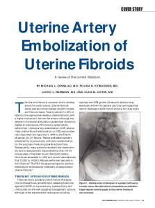
Fig. 1. Corrosion cast. SEM. Uterus of 40-years old female. Corpus, myometrium – coronal section. Avascular gap (↑) between fibroid (below) and regular myometrium (above). The area marked (ООО) is seen on Fig. 2. Bar = 1000 μm.
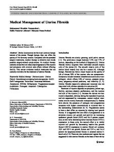
Fig. 2. Corrosion cast. SEM. Uterus of 40-years old female. On the left large fibroid growing within myometrium, to the right unchanged myometrium. In the depth of the gab visible minute vessels penetrating the lesion (↑). Bar = 1000 μm.
Vascular density, angiogenesis and pro-angiogenic factors in uterine fibroids
31
It is still believed that reports considered to the differences in microvessel density between the uterine fibroids and uterine fibrosarcomas are sparse and unclear. Hata et al. [23] observed significantly lower microvessel density in the fibroids, while Chou et al. [24] did not find such differences. In our study relatively dense capsule was observed (Fig. 1, 2), penetrated by minute vessels which run across the gap. It agreed both with the Doppler sonography findings as well as with the observations of most authors cited.
Conflict of interest None declared.
References 11. Malkusch W., Konerding M.A., Klapthor B., Bruch J.: A simple and accurate method for 3-D measurements in microcorrosion casts illustrated with tumour vascularization. Anal Cell Pathol. 1995; 9, 1: 69–81. 12. Minnich B., Bartel H., Lametschwandtner A.: How a highly complex three-dimensional network of blood vessels regresses: the gill blood vascular system of tadpoles of Xenopus during metamorphosis. A SEM study on microvascular corrosion casts. Microvasc Res. 2002; 64, 3: 425–437. 13. Minnich B., Leeb H., Bernroider E.W.N., Lametschwandtner A.: Three-dimensional morphometry in scanning electron microscopy: a technique for accurate dimensional and angular measurements of microstructures using stereopaired digitized images and digital image analysis. J Microsc. 1999; 195, Pt 1: 23–33. 14. Bentley M.D., Ortiz M.C., Ritman E.L., Romero J.C.: The use of microcomputed tomography to study microvasculature in small rodents. Am J Physiol Regul Integr Comp Physiol. 2002; 282, 5: R1267–1279. 15. Kozerska M., Skrzat J., Walocha J., Wróbel A., Leszczyński B.: Imaging of the wormian bones using microcomputed tomography. Folia Med Crac. 2013; 53 (4): 21–28. 16. Leszczyński B., Skrzat J., Kozerska M., Wróbel A., Walocha J.: Three dimensional visualisation and morphometry of bone samples studied in microcomputed tomography (micro-CT). Folia Morphol. 2014; 73 (4): 422–428. 17. Jorgensen S.M., Demirkaya O., Ritman E.L.: Three-dimensional imaging of vasculature and parenchyma in intact rodent organs with X-ray micro-CT. Am J Physiol. 1998; 275, 3, Pt 2: H1103–1114. 18. Kantor B., Jorgensen S.M., Lund P.E., Chmelik M.S., Reyes D.A., Ritman E.L.: Cryostatic micro-computed tomography imaging of arterial wall perfusion. Scanning. 2002; 24, 4: 186–190. 19. Fay P.J.: Factor VIII structure and function. Thromb Haemostat. 1993; 70: 63–67. 10. Gossl M., Rosol M., Malyar N.M., Fitzpatrick L.A., Beighley P.E., Zamir M., Ritman E.L.: Functional anatomy and hemodynamic characteristic of vasa vasorum in the walls of porcine coronary arteries. Anat Rec A Discov Mol Cell Evol Biol. 2003; 272, 2: 526–537. 11. Lerman A., Ritman E.L.: Evaluation of microvascular anatomy by micro-CT. Herz. 1999; 24, 7: 531–533. 12. Abulafia O., Kleinhaus K., Levi G., Lee Y.C., Sherer D.M.: Effect of gonadotropin-releasing hormone agonist treatment upon angiogenesis in uterine leiomyoma. Gynecol Obstet Invest. 2001; 52: 108–113. 13. Hague S., Zhang L., Oehler M.K., Manek S., MacKenzie I.Z., Bicknell R., Rees M.C.P.: Expression of hypoxically regulated angiogenic factor adrenomedullin correlates with uterine leiomyoma vascular density. Clin Cancer Res. 2000; 6: 2808–2814.
32
Marek Sajewicz, Monika Konarska, et al.
14. Casey R., Rogers P.A.W., Vollenhoven B.J.: An immunohistochemical analysis of fibroid vasculature. Hum Reprod. 2000; 15: 1469–1475. 15. Poncelet C., Madelenat P., Feldmann G., Walker F., Darai E.: Expression of von Willebrand’s factor, CD34, CD31, and vascular endothelial growth factor in uterine leiomyomas. Fertil Steril. 2002; 78: 581–586. 16. Poncelet C., Fauvet R., Feldmann G., Walker F., Madelenat P., Darai E.: Prognostic value of von Willebrand factor, CD34, CD31, and vascular endothelial growth factor expression in women with uterine leiomyosarcomas. J Surg Oncol. 2004; 86: 84–90. 17. Weston G., Trajstman A.C., Gargett C.E., Manuelpillai U., Vollenhoven B.J., Rogers P.A.W.: Fibroids display an anti-angiogenic gene expression profile compared with adjacent myometrium. Mol Hum Reprod. 2003; 9: 541–549. 18. Walocha J.A., Szczepański W., Miodoński A.J., Gorczyca J., Skrzat J., Bereza T., Ceranowicz P., Lorkowski J., Stachura J.: Application of acrylic emulsion Liquitex R (Binney and Smith) for the preparation of injection specimens and immunohistochemical studies — an observation. Folia Morphol. 2003; 62 (2): 157–161. 19. Poncelet C., Madelenat P., Feldmann G., Walker F., Darai E.: Expression of von Willebrand’s factor, CD34, CD31, and vascular endothelial growth factor in uterine leiomyomas. Fertil Steril. 2002; 78: 581–586. 20. Walocha J.A., Miodoński A.J., Nowogrodzka-Zagórska M., Kuciel R., Gorczyca J.: Application of a mixture of glycol polyethylenes for the preparation of microcorrosion casts — an observation. Folia Morphol. 2002; 61 (4): 313–316. 21. Walocha J.A., Miodoński A.J., Szczepański W., Skrzat J., Stachura J.: Two types of vascularisation of intramural uterine leiomyomata revealed by corrosion casting and immunohistochemical study. Folia Morphol (Warsz). 2004; 63, 1: 37–41. 22. Chiang C.H., Chang M.Y., Hsu J.J., Chiu T.H., Lee K.F., Hsieh T.T., Soong Y.K.: Tumor vascular pattern and blood flow impedance in the differential diagnosis of leiomyoma and adenomyosis by color Doppler sonography. J Assist Reprod Genet. 1999; 16: 268–275. 23. Hata K., Hata T., Iida K., Miyazaki K.: Expression of thymidine phosphorylase in uterine sarcoma and uterine leiomyoma: association with microvessel density and Doppler blood flow analysis. Ultrasound Obstet Gynecol. 1997; 10: 54–58. 24. Chou C.Y., Huang S.C., Tsai Y.C., Hsu K.F., Liu C.H., Huang K.E.: Uterine leiomyosarcoma has deregulated cell proliferation, but not increased microvessel density compared with uterine leiomyoma. Gynecol Oncol. 1997; 65: 225–231.