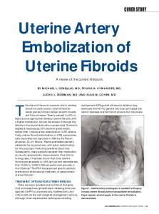
Removing Uterine Fibroids Laparoscopically Published on Physicians Practice (http://www.physicianspractice.com)
Removing Uterine Fibroids Laparoscopically July 07, 2011 By Herbert A. Goldfarb, MD [1] Hysterectomy is no longer the only treatment for uterine fibroids. Now, GnRH agonists and laparoscopic coagulation with lasers and bipolar needles are treatment options.
Before the advent of laparoscopy, uterine fibroids were often removed by surgical hysterectomy. The author describes a number of alternative treatments, including GnRH agonists and laparoscopic coagulation with the Nd:YAG laser and bipolar needles.
Over 80% of women between 30 and 50 years of age have uterine fibroids. Depending on their size and location, fibroids can be completely asymptomatic or can cause pelvic pain, dyspareunia, pressure, urinary problems, and recurrent menorrhagia. In general, the larger the fibroid, the more severe the symptoms. Abnormal bleeding is usually caused by fibroids adjacent to the uterine cavity. Patients who have smaller serosal fibroids may be completely asymptomatic or report only one symptom. Less than 1% of fibroids are malignant, and, unless they affect the patient's quality of life, there's no need for treatment. Current Approaches Medical therapy may be the best way to treat fibroids causing symptoms in premenopausal women. Synthetic gonadotropin-releasing hormone (GnRH) agonists inhibit estrogen production, causing, the uterine cavity to shrink by 36% within a few months. Fibroids may be reduced by 38% to 90% of their original size. Although these results are impressive, long-term estrogen suppression can cause Page 1 of 9
Removing Uterine Fibroids Laparoscopically Published on Physicians Practice (http://www.physicianspractice.com) bone loss. Also, GnRH agonists cannot be taken orally; a long-acting version of the drug requires injection. The drug is expensive and there's no guarantee of continued protection after treatment is stopped. Indeed, fibroids usually return to their original size within 4 months. Surgical therapy usually involves myomectomy or total abdominal or vaginal hysterectomy (Fig. 2). Some investigators, such as Camran Nezhat, MD, Mercer University, Atlanta, and Harry Reich, MD, The Graduate Hospital, Philadelphia, recommend laparoscopic myomectomy, which requires advanced pelviscopic skill. Selecting the appropriate treatment is controversial. Abdominal or vaginal myomectomy is recommended for women under 40 years of age who wish to have children, and total vaginal or abdominal hysterectomy is suggested for women who have completed their families. Fibroid uterus is the most frequent diagnosis leading to hysterectomy; this accounts for 37% to 62% of all US hysterectomies performed annually. Out of the estimated half-million hysterectomies performed annually in the US, the overall mortality rate is 12 per 10,000. Major complications, often include wound infection; postoperative bleeding; transfusion; ureteral, bladder, and bowel injuries. There are several new operative techniques that don't require major surgery. Laparoscopic myoma coagulation - myolysis is an alternative to myomectomy. This procedure can be combined with endometrial ablation for patients who have persistent uterine bleeding. Laparoscopic myomectomy is another alternative to hysterectomy. Laparoscopic Coagulation
Laparoscopic coagulation uses the Nd:YAG laser or bipolar needles (Reznik Instrument Co., Skokie, Ill., and J.E.M. Davis, Hicksville, N.Y.) to degrade myometrial stroma, denature protein, destroy vascularity, and substantially shrink fibroids. We use 50 to 70 W of continuous, pure cutting power with the Nd:YAG laser to repeatedly pierce and coagulate the fibroid, desiccating a cone of vasculature approximately 3 to 5 mm in diameter around each puncture point. Repeated puncturing effectively destroys the fibroids. We've tried using monopolar electrodes, but consider bipolar current safer. Unipolar energy may increase the size of the holes, but it doesn't increase the efficiency of the energy delivered to the fibroids. Future research may show that this technique may be used in women who want to maintain fertility, but current experience is based only on women who have completed their families. Anecdotal evidence suggests that myometrial necrosis is counterproductive to childbearing. In the hundreds of procedures I've performed, and in those performed in Europe, there have been no side effects. Compared with hysterectomy, which requires 5 to 6 weeks recovery, patients are discharged the same day and can return to work within 4 or 5 days. Because the uterus is left intact, hormonal and sexual function is preserved. After following these patients for up to 8 years, we have seen no regrowth of fibroid tumors. This treatment is successful for both serosal and subserosal fibroids and is recommended for women who have symptomatic fibroids that measure 10 cm or less. Imaging Techniques for Diagnosis Hysteroscopy, endovaginal ultrasonography, computerized axial tomography, and magnetic resonance imaging will allow you to visualize the interior of the uterine cavity accurately. By Page 2 of 9
Removing Uterine Fibroids Laparoscopically Published on Physicians Practice (http://www.physicianspractice.com) evaluating the uterine cavity, you can determine the size, shape, and position of any fibroids. If a myoma is asymptomatic and measures less than 5 cm in diameter, I recommend watchful waiting. However, when a fibroid measures 5 cm or more and continues to grow, I usually recommend a course of action. Click here for Decision-Making in treating fibroids Treatment Candidates Before treatment, always perform a pelvic examination including ultrasound examination to determine the size and location of the fibroid. If it measures 10 cm or less and responds to treatment with GnRH agonists, you may consider laparoscopic coagulation. Hysteroscopy with endometrial biopsy will help you identify submucosal and intrauterine fibroids, and rule out endometrial hyperplasia and premalignant or malignant conditions. Because the procedure shrinks the uterine cavity and may leave adhesions, it should be performed only on women who are no longer interested in conception. To maximize fibroid shrinkage before laparoscopic surgery, I suggest pretreating patients with depot leuprolide acetate, a synthetic pituitary hormone. A monthly injection (3.75 mg) given at the onset of menses for at least 3 months will reduce estrogen production and promote fibroid atrophy. Coagulation with Md:YAG Laser
Laser coagulation proceeds as follows: To begin, inject 5 mL to 10 mL of 10% vasopressin solution directly into the serosa of the uterine fibroid to eliminate hemorrhagic diathesis. It is critical to measure the desired depth of penetration that will thoroughly coagulate the myoma. By placing the Nd:YAG laser fiber against the fibroid, you will be able to visualize it in relation to the surrounding tissue. After determining the desired depth of penetration, puncture the fibroid contiguously using a drilling technique. To totally coagulate the fibroid, repeat drilling with the bare fiber tip at multiple, concentric sites. Fluid may bi used to cool the fiber tip. Because each puncture coagulates a diameter of 5 mm surrounding the puncture, it takes ten punctures per diameter, or a total of 50 to 75 punctures, to totally coagulate a 5-cm fibroid. The coagulation dispersion of the Nd:YAG laser fiber at 5-mm intervals creates a contiguous core of coagulation. To protect the rectum, bladder, and uterine blood vessels, it is extremely important to visualize the fibroid's posterior surface during the entire procedure. A uterine grasper inserted through the suprapubic puncture will elevate the uterus and help protect against posterior perforation of the rectum. I suggest a suprapubic steerable laser probe to obtain right-angle punctures for an anterior fibroid; for a posterior fibroid, you can achieve a horizontal approach by using an operative laparoscope with a 50-cm guide. The entire procedure takes about 25 to 30 minutes. In my experience, minimal - if any-bleeding - has occurred and transfusions haven't been necessary. After 4 hours of recovery from anesthesia, the patient is discharged. We've performed over 800 procedures without any serious complications. One patient developed symptoms of fibroid degeneration and was treated successfully with analgesics, bed rest, and tetracycline. We follow up all patients with endovaginal ultrasound. There has been no residual fibroid exceeding 5 cm, and, since October 1990, no patient has exhibited any fibroid regeneration nor required a second procedure. Coagulation with Bipolar Needles
Page 3 of 9
Removing Uterine Fibroids Laparoscopically Published on Physicians Practice (http://www.physicianspractice.com)
Because many hospitals do not have access to an Nd:YAG laser, we have developed a bipolar needle instrument. Laparoscopic coagulation of subserosal fibroids using a newly developed bipolar needle produces the same effects as the Nd:YAG laser. The bipolar instrument has two, 5-cm steel needles connected to a standard bipolar generator that procluces 70 to 120 W. Two forms of bipolar needles have been used to puncture uterine myomas and obtain multiple cores of coagulation: a 30-cm instrument with a 5-mm probe, and a 45-cm probe attached to the operating laparoscope, which provides a horizontal approach for posterior fibroids. When placed through a 5-mm suprapubic puncture, the bipolar needles develop a core of coagulation and effectively destroy the vasculature of the uterine myoma (Figure 4). After the tissues are lavaged, there is minimal bleeding. Using this technique, a 7-cm myoma can be thoroughly coagulated in 20 minutes. After 18 months, we have seen fibroid reduction comparable with that achieved by laser laparoscopic coagulation. When patients are treated with depot leuprolide, fibroids are reduced to about 20% to 50% of pretreatment size. Laparoscopic coagulation reduces fibroid size by another 50%. For example, an 8-cm myoma reduced to 4 cm before the procedure, typically measures 2 cm at 2 months after myoma coagulation (Tables 1 and 2). We perform vaginal ultrasound after 6 months, 1 year, and 2 years and have seen no uterine or myoma regrowth. Endometrial Ablation A majority of our patients afflicted with fibroids also have persistent uterine bleeding. We treat these patients with a combination of myoma coagulation and endometrial ablation. Good candidates for endometrial ablation are women who have persistent abnormal uterine bleeding whose preoperative uterine size measures between 5 cm and 10 cm. You can use the Nd:YAG laser for endometrial ablation, but it has a small field of focus, requires at least I hour to ablate a small uterus, and it costs the hospital more than $100,000. Ablation can also be performed with a continuous flow resectoscope fitted with a roller-bar electrode at 75 to 100 W of coagulating current (some physicians recommend cutting current). Results with the roller electrode are comparable with those achieved by the Nd:YAG laser. To dilate the cervix, insert laminaria tents the night before surgery. This dilation prevents cervical spasm during the procedure, facilitates the insertion of a 27F-continuous flow resectoscope, and Page 4 of 9
Removing Uterine Fibroids Laparoscopically Published on Physicians Practice (http://www.physicianspractice.com) allows fluid to circulate easily, thus preventing excessive intrauterine pressure and fluid absorption. The loop electrode is used to shave intercavity or submucosal fibroids, Once this is accomplished, use the roller-bar electrode to coagulate the entire uterine cavity. Combining this technique with myoma coagulation allows you to treat serosal and submucosal fibroids simultaneously. The results of endometrial ablation with the Nd:YAG laser or roller-bar electrode show that uterine hemorrhage is controlled in 95% of patients and uterine size is reduced more than 50%. Amenorrhea rates vary, depending on the technique used and the size of the uterine cavity. In patients whose preoperative uterine cavity measures 7 cm or less, amenorrhea rates are about 60%. This rate decreases to 30% when the uterine cavity measures between 10 cm and 15 cm (Tables 3 and 4). Since October 1990, when we performed the first myoma coagulation procedure in the US, we have seen no recurrence of abnormal uterine bleeding nor has any patient required a second procedure. Although evaluation is necessary to confirm long-term fibroid suppression (more than 2 years), the immediate benefits of the Nd:YAG laser and bipolar needles include same-day surgery, avoidance of hysterectomy, and elimination of abnormal bleeding.
Laparoscopic Myomectomy Abdominal myomectomy is considered a complicated procedure because of technical difficulties inherent in removing large fibroids from the abdominal cavity, controlling bleeding, and suturing the uterine defect. GnRH analogs are used preoperatively to reduce fibroid size and vascularity, and eliminate the need for transfusion. Laparoscopic myomectomy is a relatively new technique that treats symptomatic fibroids and avoids the complications associated with laparotomy and hysterectomy. This technique should be performed by advanced pelviscopic surgeons who have mastered techniques of laparoscopic suturing and tissue removal. When fertility is an issue, we restrict laparoscopic myomectomy to pedunculated and superficial serosal fibroids, and those in which the uterine cavity is not compressed. Because of the potentially Page 5 of 9
Removing Uterine Fibroids Laparoscopically Published on Physicians Practice (http://www.physicianspractice.com) destructive impact on the uterine cavity, I don't recommend laparoscopic myomectomy for deep intramural fibroids in women who want to have children. The experience of the operator, blood loss, and safety are the criteria that determine the appropriateness of the procedure when fertility isn't an issue. Before laparoscopic myomectomy, perform an endometrial biopsy and a diagnostic hysteroscopy to evaluate the uterine cavity. Exclude women with malignant or premalignant conditions. Pretreatment with leuprolide therapy for 3 to 5 months will shrink fibroids by 30% to 50%. First, inject the fibroid with 5 mL to 10 mL 10% vasopressin solution of vasoconstriction. We inject the fluid immediately under the serosa. We incise the fundus of the uterus in the anterior-posterior plance, using a knife electrode at 100W of cutting power. A suction lavage instrument helps evacuate smoke as well as prode adequate lavage. A 1-mm corkscrew is inserted into the fundus of the the myoma to aid in the dissection process. As soon as a bleeding point is found, we coagulate it with the knife electrode, using 100 W cutting power. This power level provides effective cutting as well as excellent coagulation. The combination of the knife electrode, the suction lavage instrument, and the corkscrew allow adequate dissection of the fibroid. Be careful to keep the knife electrode away from the intestines, and use caution when cutting. We repeat this dissection procedure until the root of the fibroid is exposed and can be incised. After the excess blood is suctioned and the tissue thoroughly lavaged, we coagulate oozing points, and then place the myoma in the cul-de-sac.
Suturing In the past, we performed suturing intracorporeally by stitching the wound inside the abdominal cavity. Because this was time consuming and awkward, we now use an extracorporeal suturing technique, which shortens the procedure considerably. Using a .00 PDS CT.2 needle placed through a 10-mm trocar, we position the needle inside the Page 6 of 9
Removing Uterine Fibroids Laparoscopically Published on Physicians Practice (http://www.physicianspractice.com) abdomen with the help of two needle-holders. One needle-holder (Wolf Castroviejo) is placed through the 10-mm trocar and is used to grasp the needle; the other needle-holder is used for manipulation. After suturing, we withdraw the needle through the 10-rnm trocar. We tie the knots extracorporeally in a double surgeon's knot and use a knot-pusher to secure them. Three knots are made for each suture before cutting. This process is repeated as often as necessary to adequately close the uterine defect. Bleeding is minimal and there has been no need for transfusion. The area is then thoroughly lavaged. Removing the Myoma
To remove the myoma from the abdominal cavity, we expand the trocar to 20 mm, using the Semm tissue removal system. The procedure requires 30 to 40 minutes for a large myoma, and is an alternative to colpotomy, which is also an effective method of removal. Once the myoma is completely removed, tissues are thoroughly lavaged with 2,000 mL to 8,000 mL of warm lactated Ringer's solution. Another 1,500 mL of Ringer's irrigation are left in the pelvic cavity for hydroflotation and adhesion prevention. It is very important to close the fascia and peritoneal surfaces of the trocar sites of 10 mm or more so that a loop of intestine cannot isolate itself. This entire procedure can be successfully completed in approximately 2 hours and provides an excellent, but highly technical, alternative to open abdominal surgery. Hysterectomy Unacceptable The use of hysterectomy to treat myomatous conditions in post-reproductive women is no longer acceptable to increasing numbers of patients. Learning these alternative laparoscopic options requires diligence and dedication, but allows us to provide patients with alternatives to major surgery.
Page 7 of 9
Removing Uterine Fibroids Laparoscopically Published on Physicians Practice (http://www.physicianspractice.com)
References: Dr. Herbert A. Goldfarb M.D. F.A.C.O.G. F.A.C.S. The Montclair Reproductive Center 29 The Crescent Montclair, New Jersey 07042 973-744-7470 For the New York Office call (212) 585-4400 Please send emails to Dr. Goldfarb The Montclair Reproductive Center is a full-service gynecological and reproductive center specializing in the treatment of women's disorders where caring goes hand-in-hand with treatment. Queries may be faxed to the Montclair Reproductive Center at 973-744-1274 or phone us at 973-744-7470.
Source URL: http://www.physicianspractice.com/fibroids/removing-uterine-fibroids-laparoscopically Links: [1] http://www.physicianspractice.com/authors/herbert-goldfarb-md Page 8 of 9
Removing Uterine Fibroids Laparoscopically Published on Physicians Practice (http://www.physicianspractice.com)
Page 9 of 9