Abnormal Uterine Bleeding and Fibroids AGuideforPatients IN T R O D U C T I O N Menstruation is considered normal when uterine bleeding occurs every 21 to 35 days and is not excessive in amount or duration. The normal duration of menstrual bleeding is between two and seven days and the normal amount is less than 80 milliliters (about 3 ounces). Abnormal uterine bleeding occurs when either the frequency or the quantity of uterine bleeding differs from that mentioned above or the woman has breakthrough spotting between her normal menstrual periods. Abnormal uterine bleeding is caused by a variety of factors. The two main causes are structural abnormalities of the reproductive system and o v u l a t i o n disorders. Abnormal uterine bleeding often causes a women to become anemic, chronically. This may raise her risk for illness by reducing her immune status. In addition, AUB can be embarrassing, can cause loss of work, family time and can impact quality of life. Finally, AUB can impact sexual relationships.
NO R M A L OVA R I A N FU N C T I O N In women of reproductive age, the ovary secretes estrogen and progesterone into the bloodstream. These two hormones prepare the e n d o m e t r i u m (the lining of the uterus) for implantation of a fertilized egg. The pituitary gland, located at the base of the brain, influences ovarian hormone production and ovulation by secreting two hormones, follicle stimulating hormone (FSH) and luteinizing hormone (LH) . Following stimulation by FSH and LH, a follicle containing an immature egg begins to develop within the ovary. As the follicle enlarges, it secretes increasing amounts of estrogen. When a sufficient amount of estrogen is secreted, the pituitary gland releases a large amount of LH which causes the follicle to release its egg (ovulation). If the egg does not become fertilized or does not implant in the endometrium, the egg leaves the womb with the menstrual cycle.
Menstrual Cycle Comparisons Cyclic Hormone Production Ovarian Function
Endometrial Uterine Changes
1
The secretion of estrogen and progesterone declines approximately two weeks after ovulation if pregnancy hasn’t occurred. With declining levels of estrogen and progesterone, the lining of the uterus is shed as the menstrual period and the unfertilized egg is shed. The cyclical release of FSH and LH from the pituitary gland is tightly regulated and easily disrupted. When the pituitary gland does not release appropriate quantities of FSH or LH, ovulation may not occur and the cycle may disappear or be irregular. In some women who do not ovulate, the endometrium is stimulated by continuous exposure to estrogen without sufficient levels of progesterone to allow for complete shedding of the endometrial lining. This may eventually result in irregular or heavy bleeding. If estrogen exposure is continuous, abnormal cells may develop within the endometrium (hyperplasia) that could eventually form endometrial cancer.
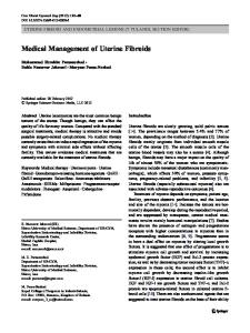
CA U S E S O F AB N O R M A L UT E R I N E BL E E D I N G Abnormal uterine bleeding (AUB) has many causes. It may be due to structural abnormalities of the uterus. Some of the more common structural causes of abnormal uterine bleeding include benign (noncancerous) l e s i o n s of the uterus such as p o l y p s , f i b ro i d s ( m y o m a s ), and a d e n o m y o s i s Other causes include bleeding associated with early pregnancy, including miscarriage and ectopic pre g n a n c y, and bleeding disorders which affect the ability of the blood to clot normally. Lesions of the cervix or vagina (benign and cancerous), chronic infections of the endometrial lining (e n d o m e t r i t i s), scar tissue (a d h e s i o n s) in the endometrium, and the use of an intrauterine device (IUD) may also be associated with abnormal uterine bleeding. Additional causes of abnormal bleeding include medications which can affect the normal release of estrogen and progesterone, chronic medical problems such as diabetes mellitus or disorders of the liver, kidney, t h y roid gland, or a d renal glands, or other medical problems which can affect the production and metabolism of estrogen and progesterone. Emotional or physical stress as well as significant changes in body weight may disrupt the pituitary’s release of FSH and LH and prevent ovulation.
Causes of Abnormal Uterine Bleeding 1,2: 30% of reproductive-age women worldwide Biochemical
Structural
Prostaglandin
Fibroids/polyp Adenomyosis Infection Hyperplasia Malignancy
Endocrinology
Ovulation Dysfunction “Hormone imbalance”
Hematologic
Clotting diseases Leukemia
Iatrogenic
Anticoagulant Exogenous hormones Intrauterine devices
2
Terms Used to Describe Abnormal Uterine Bleeding Term
Abnormal uterine bleeding pattern
Oligomenorrhea
Bleeding occurs at intervals of > 35 days and usually is caused by a prolonged follicular phase.
Polymenorrhea
Bleeding occurs at intervals of < 21 days and may be caused by a lutealphase defect.
Menorrhagia
Bleeding occurs at normal intervals (21 to 35 days) but with heavy flow (>=80 mL) or duration (>=7 days).
Menometrorrhagia
Bleeding occurs at irregular, noncyclic intervals and with heavy flow (>=80 mL) or duration (>=7 days).
Amenorrhea
Bleeding is absent for 6 months or more in a nonmenopausal woman.
Metrorrhagia or bleeding Irregular bleeding occurs between ovulatory cycles; causes to consider intermenstrual include cervical disease, intrauterine device, endometritis, polyps, submucous myomas, endometrial hyperplasia, and cancer. Midcycle spotting
Spotting occurs just before ovulation, usually because of a decline in the estrogen level.
Postmenopausal bleeding
Bleeding recurs in a menopausal woman at least 1 year after cessation of cycles.
Acute abnormal bleeding Dysfunctional bleeding
emergent Bleeding is characterized by significant blood loss that results in uterine hypovolemia (hypotension or tachycardia) or shock. uterine
This ovulatory or anovulatory bleeding is diagnosed after the exclusion of pregnancy or pregnancy-related disorders, medications, iatrogenic causes, obvious genital tract pathology, and systemic conditions.
Dysfunctional Uterine Bleeding Dysfunctional uterine bleeding is the occurrence of uterine bleeding unrelated to structural abnormalities of the uterus or the endometrial lining. The diagnosis is made after structural causes of bleeding and chronic medical diseases have been excluded. Other causes of abnormal bleeding must also be disproved, including pregnancy complications and medications which influence hormonal action or affect clotting. DUB occurs most commonly in the first five years after a woman starts menstruating and as during the perimenopause (4 years prior to the cessation of menses). The main cause of DUB is the absence of ovulation and the orderly secretion of estrogen and progesterone. DUB may alert the woman and her physician to the fact that she is no longer ovulating normally. Occasionally a woman may have ovulatory DUB.
3
DI A G N O S I S Women who experience abnormal uterine bleeding should be evaluated by a physician. A medical history, discussion of possible contributing factors, and a detailed physical exam are usually necessary. A variety of diagnostic techniques are available for determining the cause of abnormal uterine bleeding.
Diagnostic Procedures The following is a list of labs and procedures that may be performed to help clarify the problem.
Laboratory Evaluation Necessary Tests • • •
Complete Blood Count (CBC) (checks for anemia) HCG (Human Chorionic Gonadotropin) (pregnancy test) TSH (Thyroid Stimulating Hormone)
Tests that might be done based on Physician judgment • • • • • •
PT (Prothrombin Time) (checks for clotting disorder) PTT (Partial Thromboplastin Time) (checks for clotting disorder) Bleeding Time (checks for platelet function) FSH (Follicle Stimulating Hormone) (checks for menopause status) Prolactin (test of pituitary function) Testosterone (test associated with polycystic ovarian disorder)
Additional tests of the liver, kidney, pancreas, and other major organs may be useful, depending upon each woman’s medical history. Laboratory studies for abnormal uterine bleeding will be based on the physician’s clinical judgment as to the underlying cause of the bleeding.
Imaging Evaluation • Ultrasound Evaluation Ultrasound uses ultrasound energy to view internal organs in the body. In Gynecology you will get a vaginal ultrasound which allows the physician to closely inspect the uterus, cervix, tubes, and ovaries. Other tests may be performed based on the findings of the ultrasound examination •
Saline Infused Sonography: saline is infused into the cavity of the uterus and an ultrasound is done. This particular test outlines the endometrium (the lining of the uterus where bleeding occurs) well and helps determine if any structural abnormalities (fibroids, polyps) are present and responsible for bleeding.
4
Endometrial Polyps
•
MRI (Magnetic Resonance Imaging) Occasionally, a pelvic MRI may be ordered. This test evaluates fibroids well and helps locate them better than ultrasound. In addition, the MRI can be helpful locating adenomyosis (glandular endometrial cells that grow into the muscle wall of the uterus). MRI scan from the side showing a uterus with extensive adenomyosis (black arrows), which appears as a darker shade of gray on this scan when compared to the normal portions of the uterine wall (white arrows).
•
Office Hysteroscopy
This test is done in the office under local anesthesia and is well tolerated. A small telescope is inserted into the lining of the uterus (through the vaginalcanal) and the cavity of the uterus is visualized. Very specific pathology can be outlined and further treatment options can be clarified. At the same time a biopsy of the endometrial lining will be done to determine if any abnormalities of the cells are present.
A telescope is placed in the uterine cavity and in this case the patient has a uterine septum (a wall of tissue dividing the cavity).
5
TR E AT M E N T It is important to treat AUB (Abnormal Uterine Bleeding) for the following reasons: • • • • • •
embarrassment, fear (staining clothes in public) lost work days/time lost family time impact on sexuality impact on health Impact on quality of life
The individual therapy recommended will be tailored to the specific cause of abnormal bleeding. In general, the therapies fall into one of the following categories and can be used to treat abnormal uterine bleeding without structural causes as well as AUB with structural causes (i.e. fibroids, etc).
• Do Nothing This approach can be used for mild abnormal uterine bleeding not causing anemia and quality of life disruption. It is important to keep a menstrual calendar to document worsening of the condition. A person should be seen in 3-6 month intervals to evaluate for worsening symptoms.
• Iron Therapy (Hemax, Repliva, Ferrosequels) if anemia Iron therapy is used to treat anemia. This will usually raise a persons energy level by improving the delivery of oxygen to the cells of the body. This treatment does not affect the bleeding levels, however.
• Other Medical Treatments used to affect bleeding levels (hormonal and nonhormonal) Pharmacotherapy
Efficacy
Oral Contraceptive (Birth control pills)
Up to 50%improvement
Progestogens (Provera, Aygestin, Cycrin, Prometrium)
Up to 20% improvement
NSAIDS (Therapeutic dosing) (Advil, Aleve, Celebrex, Motrin) Up to 30% improvement
•
Danazol (Androgen) (200-400mgs/day)
Up to 75% improvement
GnRH agonists Injections (Lupron, Goserelin) (about $1500/3 month)
Up to 100% improvement
Progesterone IUDs (Mirena) (5 years) (about $500 and usually not covered by insurance)
Up to 97% improvement
Anti-Progestogens
Research only
Pain management: A variety of NSAIDS (motrin, advil, ultram er, aleve, ponstel) available with variable efficacies 6
Abnormal Uterine Bleeding without Structural Abnormalities (Dysfunctional Uterine Bleeding): Other options for Treatment Endometrial Ablation (Reduction in menstrual flow effective in 80-90% of cases) o Endometrial Ablation (Hysteroscopic): Place an electrode device through a telescope that is inserted into the cavity of the uterus and serially destroys the lining of the uterus (requires operator skill and takes about 30 minutes to perform) 1) Hysteroscopic Rollerball Endometrial Ablation 2) Hysteroscopic Loop Resection Endometrial Ablation
o Endometrial Ablation (Global): Place a device in the cavity of the uterus and destroy the lining all at once (requires very little operator skill, is quicker than the Hysteroscopic methods, and can be performed in the gynecology office) 1) ThermaChoice Global Endometrial Ablation: A balloon device containing the hot water that conforms to the cavity of the uterus (Takes 8 minutes)
7
2) HTA Global Endometrial Ablation: Free flowing hot saline instilled through a device into the cavity of the uterus. (takes 10 minutes)
3) Novasure Global Endometrial Ablation: An electrical device that burns the lining of the uterus (takes 2 minutes to perform)
4) Cryogen Global Endometrial Ablation: Uses freezing technology to destroy the lining of the uterus (Takes 10 minutes)
8
5) MicroWave Global Endometrial Ablation: Uses Microwave energy through a probe device inserted into the cavity of the uterus to destroy the lining. (Takes 3-4 minutes to perform)
Comparisons of Global Technologies
Therma
HTA
Cryogen
Novasure
choice
Micro wave
Success
80%
68%
67%
78%
87%
Amenorrhea
15%
35%
22%
36%
55%
10
10
4
3.5
Local Anesthesia 39%
45%
39%
73%
62%
Patient Satisfied
96%
N/A
86%
92%
98%
Uterus Size (cm)
66-10
10.5
9
Generation
third
Fibroid success
80%
?
?
?
Dysmenorrhea
70/78%
?
?
35%
55%
Avoid 2nd Therapy
83%
?
?
?
?
Time (min)
8
First
First
66-10 First
66-14 First
68%
9
o Uterine Artery Embolization (rarely used for abnormal bleeding unless in the setting of fibroids) A catheter is introduced into each uterine artery via the femoral artery (located in the groin). Polyvinyl particles are inserted into the artery which blocks the flow of blood to the fibroid and also, in some cases, the uterus.
Hysterectomy for the Treatment of Abnormal Uterine Bleeding without Structural Abnormalities (such as fibroids)
Hysterectomy A Hysterectomy is an operation to remove a woman’s uterus (womb). The uterus is where a baby grows when a woman is pregnant. Sometimes the fallopian tubes, ovaries, and cervix are removed at the same time the uterus is removed. These organs are located in a woman’s lower abdomen. The cervix is the lower end of the uterus (the opening). The ovaries are organs that produce eggs and hormones (estrogen, progesterone, and testosterone). The fallopian tubes carry eggs from the ovaries to the uterus. A hysterectomy will stop your monthly period and you will not be menopausal unless ovaries are removed. There are several types of hysterectomy: •
A complete or total hysterectomy: removes the
cervix and uterus (the most common type). A women will be menopausal if the ovaries are removed also. This type of hysterectomy can be performed through the vagina or with the laparoscope. With many physicians it is done through large incisions on the abdomen (either vertical or horizontal based on surgical judgment). This should be 10
the last resort type of hysterectomy not the most common. •
A partial hysterectomy: removes uterus and cervix
•
Supracervical hysterectomy: removes uterus (Womb) and leaves the cervix. This should be done through the belly button (laparoscopically)
•
Radical Hysterectomy: removes uterus, cervix, upper part of the vagina, and supporting tissues (usually done when cervical cancer is found)
Often one or both ovaries and fallopian tubes are removed at the same time a hysterectomy is done. When both ovaries and both tubes are removed, it is called a bilateral salpingooophorectomy. If they are removed before the natural menopause, then one enters the menopause suddenly (surgical menopause). This can cause more severe menopause symptoms. The core symptoms are hot flushes (night sweats), insomnia, memory and concentration loss, and reduced libido (sex drive). Hysterectomy is the second most common major surgery among women in the United States (the most common major surgery that women have is cesarean section delivery). Each year, more than 600,000 hysterectomies are done. About one third of women in the United States have had a hysterectomy by age 60.
How is hysterectomy performed? Hysterectomies are done either through a cut (vertical or horizontal) in the abdomen (abdominal Hysterectomy); or through minimally invasive approaches (minimal or no incisions). These approaches include laparoscopy and vaginal. Most hysterectomies can be performed vaginally or laparoscopically (minimally invasive). The abdominal approached is more painful, involves more scarring, and requires a longer recovery. While the minimally invasive approaches have less scarring, less risk, faster recovery, and can be done as outpatient. With Laparoscopic Supracervical hysterectomy the womb is removed while conserving the cervix. This maintains support to the upper vagina and bladder (less risk of prolapse) and often women report no change in their sexual response. This type of minimally invasive hysterectomy can be done with many types of pathologies of the uterus and involves 3 small incisions, requires no hospital stay, has a 1-3 week recovery, very little scarring, and, finally, reduced surgical risk.
11
With Total Hysterectomy the womb and the cervix are removed. Total Hysterectomy can be performed through the vagina or with laparoscopy (minimally invasive approach) or through a vertical or horizontal incision on the abdomen. The approach to this type of hysterectomy is based on many factors and involves good surgical judgement. If done minimally invasive, this hysterectomy involves 4 small incisions (laparoscopically), no incision (vaginally), requires no hospital stay, has a 1-3 week recovery, very little scarring, and, finally, reduced surgical risk. If done through large abdominal incisions, this hysterectomy requires 1-2 day hospital stay, 4-6 weeks recovery, has a large scar, more blood loss and is riskier overall.
Typical hysterectomy percentages in the United States are 80% Abdominal Hysterectomy, 10% Vaginal Hysterectomy, 10% Laparoscopic. These numbers are based upon surgeon competence, surgeon training and familiarity, and surgeon interest. Most residency training institutions train in the above percentages.
Hysterectomies in the United States (600,000/year)
Hysterectomy in the US* Prolapse 15%
(Pre) cancer 10%
Chronic Pelvic Pain 10%
Endometriosis Adenomyosis 20%
Fibroids 30% Dysfunctional uterine bleeding 20%20%-30%
50-60%
150,000 *KJ Carlson et al. NEJM, 328:856, 1993
Uterine Fibroids are common, benign (non-cancerous) tumors that grow in the muscle of the uterus. More hysterectomies are done for fibroids than any other problem of the uterus. Fibroids require treatment when they cause pain, bleeding abnormalities, urinary frequency, or grow rapidly. There are alternatives to hysterectomy to treat fibroids. Medicinal therapy to shrink fibroids are temporary and include a shot (Lupron) or anti-progestational agents (under investigation). Myomectomy removes just the fibroids and maintains future fertility. A relatively new procedure to treat symptomatic fibroids is call Uterine Artery Embolization. It involves placing small plastic particles in the uterine artery as it feeds the fibroids. This stops the blood supply to the fibroid and causes it to degenerate. Recently, the FDA 12
approved the MRI guided Ultrasound Ablation (ExAblate 2000). This is a completely noninvasive approach to uterine fibroids. For most alternatives, more studies need to be performed to help clarify the benefits (long and short-term), and the risks. Abnormal Uterine bleeding (AUB) is the second most common reason for hysterectomy. If no cause for the bleeding can be determined it is called Dysfunctional Uterine Bleeding (DUB). Alternatives for the management of DUB are medicines such as oral contraceptive pills and a minor outpatient surgery called Endometrial Ablation. Endometrial ablation destroys the lining of the uterus (the endometrium) and reduces the menstrual blood loss in 80-90% of women who have it done. Endometriosis is the third leading cause of hysterectomy. It occurs when endometrial tissue (the inside lining of the uterus) begins to grow on the outside of the uterus and on nearby organs. This condition may cause painful menstrual periods, abnormal vaginal bleeding, and sometimes loss of fertility (ability to get pregnant). Endometriosis is usually not a problem for women after menopause. Alternatives for the management of endometriosis include hormones, surgical excision of the endometrial implants usually done laparoscopically. Hysterectomy is generally done after other treatments have failed.
Endometriosis
Uterine prolapse is a benign condition in which the uterus moves from its usual place down into the vagina. Uterine prolapse is due to weak and stretched pelvic ligaments and tissues. The bladder and colon can also be affected and prolapse into the vagina (cystocele and rectocele). Alternative treatments for this condition include pelvic floor exercises, estrogen therapy, pessary (a silicon device inserted into the vagina to help support it), Laparoscopy can be used to suspend the uterus without removing it in some cases. Usually there is little loss of blood when a Hysterectomy is done by a surgeon with extensive experience in the procedure. There are a number of ways to reduce blood loss. Before the incision, medicines are injected appropriately to shrink blood vessels near the operative site. In addition, vessel sealing technology is used which minimizes blood loss. As a result, our average blood loss during Hysterectomy is about a tablespoon. Bloodless surgery requires skill and experience. It is important to find a surgeon with experience in bloodless surgery.
13
Structural Problems (Fibroids) may occur within the cavity, in the muscle wall of the uterus, on the outside surface of the uterus
Fibroids are some of the most common benign tumor of the uterus and are classified by name and by number (location in relation to amount of fibroid within the uterine cavity (Type O or 1 and some type II) The fibroids affecting the uterine cavity are often treated with Operative Hysteroscopy. A telescope is inserted into the cavity of the uterus via the vagina. Instruments are then placed through the telescope and the abnormalities are surgically removed and sent to the pathologist for evaluation. This is outpatient surgery, requiring light anesthesia, has minimal risk, and the recovery is usually 1-3 days. There is usually very little discomfort associated with this surgery.
Fibroids within the muscle wall of the uterus (Intramural) and on the outside of the uterus (Serosal) are usually treated other ways depending on special circumstances. • Desire to maintain the ability to get pregnant or the desire to keep the uterus within the body
Myomectomy (removal of fibroids and reconstructing the uterus) When a person wishes to preserve her uterus for childbearing or for personal reasons surgery (myomectomy) is the only avenue used to remove these structural abnormalities.
14
This surgery may be performed laparoscopically (minimally invasive) or through an incision on the abdomen (laparotomy). The surgeon’s experience and judgment determines the best route of surgery. One main limitation with surgical myomectomy (removal of fibroids) is recurrence. It is reported in the medical literature that the recurrence of fibroids is at least 30% (and that’s if all of them were removed at the time of surgery).
o Laparoscopy
Laparoscopy involves 4 small incisions on the abdomen (the size of a pinky fingernail). The first incision is directly within the belly button (umbilicus). A telescope is placed through this incision to visualize the abdominal cavity. Visualization is much improved comparing laparoscopy and traditional abdominal surgery. The other small incisions are place depending on the architecture of the uterus and its problems. Occasionally it is physically impossible to perform laparoscopic myomectomy. This is usually determined by the location of the fibroids within the uterus. Inability to remove all of the fibroids is the main limitation to performing laparoscopic myomectomy (fibroid removal). Fibroids that are in the muscle wall usually requiring the surgeon to feel with his/her fingers for location. In laparoscopy there is very little tactile sensation for localization of a fibroid within the muscle wall of the uterus. The benefits of laparoscopy are well known. These include shorter hospital stays (possibly outpatient surgery), less scarring, faster recovery, quicker return to normal life (and work), and less risky overall. The overall recovery for laparoscopic myomectomy is 1 week.
o Abdominal Myomectomy This approach often is best when there are large numbers of fibroids (greater than 5), and when the uterus approaches a size > 18 weeks. With this approach all fibroids may be removed and a plastic reconstruction of the uterus can be accomplished. The limitations of this approach are also well known. These include more scarring, longer recovery, slower return to normal life (and work). The hospital stay is usually 23 hours and the risks are the same in either approach. The average recovery for this procedure is 2-4 weeks
Usually there is little loss of blood when a myomectomy is done by a surgeon with extensive experience in the procedure. There are a number of ways to reduce blood loss. . Before the 15
incision, medicines (diluted pitressin) injected into the uterus to shrink blood vessels. In addition, a tourniquet is placed around the uterine arteries reducing blood flow to the uterus. As a result, our average blood loss during myomectomy is about a tablespoon. Bloodless surgery requires skill and experience. It is important to find a surgeon with experience in bloodless surgery.
Fibroids within the muscle wall of the uterus (Intramural) and on the outside of the uterus (Serosal) are usually treated other ways depending on special circumstances. •
Doesn’t wish to retain fertility or keep the uterus
In this setting the hysterectomy options are similar as previously noted above.
What are fibroids? Uterine fibroids are tumors or growths, made up of muscle cells and other tissues that grow within the wall of the uterus (or womb). Although fibroids are sometimes called tumors, they are almost always benign (not cancerous). The medical term for fibroids is uterine leiomyomata (you-ter-in lieoh-my-oh-mah-tah). Fibroids can grow as a single growth or in clusters (or groups). Their size can vary from small, like an apple seed (or less than one inch), to even larger than a grapefruit, or eight inches across or more.
16
Why should women know about fibroids? Uterine fibroids are the most common, benign tumors in women of childbearing age, but no one knows exactly what causes them. They can be frustrating to live with when they cause symptoms. Not all women with fibroids have symptoms, but some have pain and heavy menstrual bleeding. Fibroids also can put pressure on the bladder, causing frequent urination.
Who gets fibroids? More research is being done to figure out who is at risk for fibroids. But it is known that: •
Most of the time, fibroids grow in women of childbearing age.
•
African American women are more likely to get them than women of other racial groups.
•
African American women tend to get fibroids at a younger age than do other women.
•
Women who are overweight or obese also are at a slightly higher risk for fibroids than women who are not overweight.
•
Women who have given birth appear to be at a lower risk for fibroids.
Where can fibroids grow? Doctors put fibroids into three groups based on where they grow, such as just underneath the lining of the uterus, in between the muscles of the uterus, or on the outside of the uterus. Most fibroids grow within the wall of the uterus. Some fibroids grow on stalks (called peduncles) that grow out from the surface of the uterus, or into the cavity of the uterus.
17
What are the symptoms of fibroids? Most fibroids do not cause any symptoms, but some women with fibroids can have: • • • • • • •
heavy bleeding or painful periods bleeding between periods feeling of fullness in the pelvic area (lower abdomen) urinating often pain during sex lower back pain reproductive problems, such as infertility, having more than one miscarriage, or having early onset of labor during pregnancy
What causes fibroids? No one knows for sure what causes fibroids. Researchers have some theories, but most likely, fibroids are the result of many factors interacting with each other. These factors could be hormonal (affected by estrogen levels), genetic (running in families), environmental, or a combination of all three. Because no one knows for sure what causes fibroids, we also don't know what causes them to grow or shrink. For the most part, fibroids stop growing or shrink after menopause. But, this is not true for all women with fibroids.
Can fibroids turn into cancer? Fibroids are almost always benign, or not cancerous, and they rarely turn into cancer (less than 0.1 percent of cases). Having fibroids does not increase a woman's chances of getting cancer of the uterus.
How do I know for sure that I have fibroids? Your doctor may find that you have fibroids when you see her or him for a regular pelvic exam to check your uterus, ovaries, and vagina. Often, a doctor will describe how small or how large the fibroids are by comparing their size to the size your uterus would be if you were pregnant. For example, you may be told that your fibroids have made your uterus the size it would be if you were 8 weeks pregnant. Your doctor can do imaging tests, or tests that create a "picture" of the inside of your body without surgery, in order to confirm that you have fibroids. These tests might include: •
ultrasound - uses sounds waves to produce the picture.
•
magnetic resonance imaging or MRI - uses magnets and radio waves to produce the picture.
Besides imaging tests, you also might need a surgery to know for sure if you have fibroids. These could include: •
laparoscopy - surgery with general anesthesia in which your doctor makes a small cut in the abdomen and places a small tube with a light inside to see any fibroids. 18
•
hysteroscopy - surgery in which your doctor inserts a long tube with a camera into the vagina and directly into the uterus to see any fibroids. It also shows any growths or problems inside the uterus.
What is the treatment for fibroids? Talk with your doctor about the best way to treat your fibroids. She or he will consider a number of things before helping you choose a treatment. Some of these things include: •
whether or not you are having symptoms from the fibroids
•
if you might want to become pregnant
•
the size of the fibroids
•
the location of the fibroids
•
your age
If you have fibroids, but do not have any symptoms, you may not need any treatment. But your doctor will check during your regular exams to see if they have grown. Medications If you have fibroids and have mild symptoms, your doctor might only suggest pain medication. Overthe-counter anti-inflammatory drugs, such as ibuprofen, or other painkillers such as acetaminophen can be used for mild pain. If pain becomes worse, your doctor can prescribe a stronger painkiller. Other drugs used to treat fibroids are called gonadotropin releasing hormone agonists (GnRHa). These drugs may decrease the size of the fibroids. Side effects can include hot flushes, depression, not being able to sleep, decreased sex drive, and joint pain. Anti-hormonal agents, such as a drug called mifepristone, also can stop or slow the growth of fibroids. These drugs only offer temporary relief from the symptoms of fibroids; once you stop the therapy, the fibroids often grow back. Surgery If you have fibroids with moderate or severe symptoms, surgery may be the best way to treat them. Here are the options: •
Myomectomy - a surgery to remove fibroids without taking out the healthy tissue of the uterus. There are many ways a surgeon can perform this procedure. It can be major surgery (with an abdominal incision) or minor surgery. The type, size, and location of the fibroids will determine what type of procedure will be done. Talk with your doctor about the different types of this surgery.
•
Hysterectomy - a surgery to remove the uterus. This surgery is the only sure way to cure uterine fibroids. This surgery is used when a woman's fibroids are large, or if she has heavy bleeding, and is either near or past menopause and does not want children. There are various types of hysterectomy that differ in how invasive they are. Sometimes, if the fibroids are large, a woman might need a hysterectomy that involves cutting into the abdomen to 19
remove the uterus. If the fibroids are smaller, the surgeon might be able to reach the uterus through the vagina, instead of making a cut in the abdomen. •
Endometrial ablation – the endometrial lining of the uterus is destroyed. This surgery controls very heavy bleeding, but afterwards a woman cannot have children.
•
Myolysis – a procedure in which an electrical needle or freezing probe is inserted into the uterus through a small incision in the abdomen to destroy the blood vessels feeding the fibroids. This is not widely available and generally has low success rates.
Uterine Fibroid Embolization (UFE) Uterine fibroid embolization (UFE) is a treatment that cuts off the blood supply to the uterus and the fibroids so they shrink. UFE is proving to be an alternative to hysterectomy and myomectomy. The recovery time is also shorter, and there is a much lower risk of needing a blood transfusion than for these surgeries. Many women can have UFE and go home the same day. There is a small risk of infection in the treated fibroid, but these are usually managed with antibiotics. Recent studies also suggest that most fibroid tumors are not likely to re-grow after UFE, although more long-term data is needed. Not all fibroids can be treated with UFE. All patients must first be evaluated with ultrasound or MRI to make sure the fibroids will respond well to this treatment. Doctors called interventional radiologists perform UFE. The best candidates for UFE are women who: •
have fibroid tumors that are causing heavy bleeding
•
have fibroid tumors that are causing pain or pressing on the bladder or rectum
•
don’t want to have a hysterectomy
•
don’t want to have more children
Sometimes after UFE, the particles that are put into the fibroids to cut off their blood supply have traveled to the ovaries. In a few women, the ovaries then stop working for a short time or permanently. Although researchers know that UFE may affect how ovaries function, they are unsure of how exactly UFE affects fertility. If you want to have children in the future, you should talk with your doctors about the small, but definite risk of UFE causing you to go into early menopause. Too few women have gotten pregnant after UFE for researchers to know if there is an increased risk of pregnancy complications.
How does uterine fibroid embolization work? Fibroids have a large blood supply that makes them grow. Fibroids may shrink if the blood supply is stopped. Embolization means to stop or block the blood flow. So, uterine fibroid embolization is a way to stop the blood flow that makes fibroids grow. The procedure works even if you have several fibroids.
How is uterine fibroid embolization done? Uterine fibroid embolization is not surgery, but it's done at a hospital. You will be given medicine to make you sleepy and relaxed. The procedure doesn't cause pain but can be associated with 20
significant pain after the procedure. The doctor (an interventional radiologist who is specially trained to do this procedure) will make a tiny cut in the skin in your groin area. Next, the doctor will pass a tiny tube called a catheter through an artery to your uterus. When the catheter is in place, the doctor will inject tiny particles into the catheter. These particles, made of plastic or gelatin sponge, are about the size of grains of sand. These particles move through the catheter into the arteries that send blood to the fibroid. The particles will stop the blood flow to the fibroid. Over time, most (but not all) fibroids will shrink in size.
How successful is uterine fibroid embolization? About 85 percent of women have a reduction in bleeding associated with fibroids. Typically a fibroid may shrink 1-2 centimeters in size. The fibroids, however, will always be present.
What are the advantages of uterine fibroid embolization? UFE offers no advantage over laparoscopic management of Abnormal Uterine Bleeding and Fibroids. It does, however, offer significant advantage over Total Abdominal Hysterectomy (the typical hysterectomy done through a large incision on the abdomen).
21
Are there any side effects from uterine fibroid embolization? Uterine fibroid embolization is very safe, but there are some side effects. Most women have moderate to severe lower abdominal pain for up to 2 weeks after the procedure. Some women have nausea and fever. Medicine can help with these symptoms. A few women get an infection after the procedure. Antibiotics can control the infection. About 1 percent of women have an injury to the uterus from the procedure. This could make a hysterectomy necessary. A few women have started menopause after uterine fibroid embolization. Studies about getting pregnant after having this procedure are not complete but case series suggest no particular harm to pregnancy (long term studies need to be performed in order to make recommendations).
How do I know if this procedure is right for me? You, your gynecologist and the interventional radiologist will help you decide if uterine fibroid embolization is right for you. This procedure is not recommended for women with Intracavitary fibroids (fibroids in the cavity of the uterus) nor for fibroids greater than 18 weeks size. Most insurance companies will pay for this procedure. You will want to talk with your insurance company and your doctors before having this procedure. This procedure requires about 1 week recovery time.
Where can I get more information? For more information on fibroids and their treatment and to find interventional radiologists in your area, you can call the Society of Cardiovascular & Interventional Radiology (SCVIR) at their toll-free telephone number: 1-800-488-7284. You can also access their Web site at: www.scvir.org.
ExAblate® 2000 System (MRI Guided Ultrasound Ablation of Fibroids) ExAblate® 2000 is a medical device that uses magnetic resonance image guided focused ultrasound to target and destroy uterine fibroids. The device is intended to treat women who have completed child bearing or do not intend to become pregnant. ExAblate® 2000 is non-invasive surgery. It spares the uterus and is an alternative to myomectomy, hysterectomy, watchful waiting, hormone therapy, or uterine fibroid embolization. ExAblate combines two systems – a magnetic resonance imaging (MRI) machine to visualize patient anatomy, map the volume of fibroid tissue to be treated, and monitor the temperature of the uterine tissue after heating, and a focused ultrasound beam that heats and destroys the fibroid tissue using high frequency, high-energy sound waves. The treatment requires repeated targeting and heating of fibroid tissue while the patient lies inside the MRI machine. The procedure can last as long as four hours. The new device can be used to treat some – but not all – fibroids. Fibroids close to sensitive organs such as the bowel or bladder and those outside the image area cannot be treated. This procedure may have successes in the 70% range for selected fibroids. 22
InSightec, the manufacturer, is conducting more research to assess the long-term safety and effectiveness of the treatment.
As can be seen in the diagram above, highly focused ultrasound waves are directed into the body that raise the temperature of the tissue leading to the fibroid destruction. Using the thermal imaging capabilities of MR equipment, real time feedback on the temperature achieved at the target tissue during treatment ensures a successful outcome.
Is MRgFUS Right For Me? MRgFUS is the treatment of choice for the busy woman who cannot afford to take several weeks off for recuperation, the patient who prefers non-invasive treatment over surgery, and the woman who is concerned with the effects surgery may have on her anatomy and hormonal balance in the future. This non-invasive procedure offers the following benefits: Incisionless thermal ablation of soft tissue. No hospitalization. Return to work or normal activity within 1-2 days. Low rate of complications. Real time visualization, monitoring, and control of treatment. Limited conscious sedation. Integrated MR imaging for treatment planning. MRgFUS is the only focused ultrasound system that uses Magnetic Resonance images for control and guidance.
23
However, MRgFUS is not for everyone and a full examination and diagnosis by a medical professional is required before the possibility of MRgFUS treatment can be determined. Common Exclusion Criteria Weight over 250 pounds Abnormal PAP smear or uterine cancer Acute pelvic infection Pregnant or desire for future pregnancy Anemia lab result for Hemoglobin of under or equal to 25% Uterine size larger than 24 weeks
For more Information . . . You can find out more about uterine fibroids by contacting the National Women's Health Information Center (NWHIC) at 800-994-9662 or the following organizations: National Institute of Child Health and Human Development Clearinghouse Phone Number(s): (800) 370-2943 Internet Address: http://www.nichd.nih.gov/publications/pubs.cfm American College of Obstetricians and Gynecologists (ACOG) Resource Center Phone Number(s): (800) 762-2264 x192 (Publications Requests Only) Internet Address: http://www.acog.org/ National Uterine Fibroids Foundation Phone Number(s): (719) 633-3454 or (800) 874-7247 Internet Address: http://www.nuff.org/ REFERENCES 1. Cramer, SF, Patel, A. The frequency of uterine leiomyomas. Am J Clin Pathol 1990; 94:435. 2. Parazzini, R, La Vecchia, C, Negri, E, et al. Epidemiologic characteristics of women with uterine fibroids: a case-control study. Obstet Gynecol 1988; 72:853. 3. Stewart, EA, Nowak, RA. New concepts in the treatment of uterine leiomyomas. Obstet Gynecol 1998; 92:624. 4. American College of Obstetricians and Gynecologists. Surgical alternatives to hysterectomy in the management of leiomyomas. ACOG practice bulletin #16, American College of Obstetricians and Gynecologists, Washington, DC 2000. 5. Iverson, RE Jr, Chelmow, D, Strohbehn, K, et al Relative morbidity of abdominal hysterectomy and myomectomy for management of uterine leiomyomas. Obstet Gynecol 1996; 88:415. 6. Spies, JB, Spector, A, Roth, AR, et al. Complications after uterine artery embolization for leiomyomas. Obstet Gynecol 2002; 100:873. 7. Pron, G, Bennett, J, Common, A, Wal,l J, Asch M, Sniderman K. The Ontario uterine fibroid embolization trial part 2. Uterine fibroid reduction and sympton relief after uterine artery embolization for fibroids. Fertil Steril 2003; 79:120. 8. Marshall, LM, Spiegelman D, Goldman, MB, Manson JE, Colditz GA, Barbieri RL, et al. A 24
prospective study of reproductive factors and oral contraceptive use in relation to the risk of uterine leiomyomata. Fertil Steril 1998; 70:432. 9. ACOG Committee Opinion. Uterine artery embolization. Obstet Gynecol 2004; 103:403.
Uterine Artery Embolization Dr. Oscar Sosa Medical Director Board Certified Interventional Radiologist Vascular and Spine Institute 7887 N. Kendall Dr. Suite 210 Miami Florida 33156 Tel 305-598-1555 www.vascularandspine.com Dr. Benenati Baptist Hospital 8900 North Kendall Dr Miami, FL 33176 tel: 786-596-7050 www.miamivascular.com
Exablate 2000 3848 FAU Boulevard, Suite 200, Boca Raton, Florida 33431 Tel 561-826-1274 www.exablateofsouthflorida.com
Research Performed by the Center •
“Does Cesarean Section Increase the Prevalence of Adenomyosis”, Verma U, Whitted RW, poster presentation ACOG, 2000; supplement to Obstetrics and Gynecology, 97(4); April 2000 (Abstract)
•
“Endometrial Ablation for Refractory, Life-Threatening Uterine Bleeding: A Case Report”. Felicia Cohen, Whitted RW poster presentation American Association of Gynecologic Laparoscopists, Annual Meeting. Supplement, Journal of the American Association of Gynecologic Laparoscopists,Orlando,2000 (Abstract)
•
“The Accuracy of Endometrial Pipelle Sampling in Diagnosing Endometrial Polyps”. Deborah Chong, Whitted RW, P. Pietro. Poster Presentation, ACOG 2002. Supplement Obstetrics and Gynecology 99(4): April 2002 (Abstract). In manuscript formation 25
•
“A Retrospective Study Evlauating the impact of Formal Laparoscopic Training on Dermoid Management in Residency”. Benezra V, Whitted RW, Pietro PA. Poster presentation, ASRM 2003; Supplement, Fertility and Sterility, October 2003.
•
“A Retrospective Study Evaluating the Impact of Formal Laparoscopic Training on Patient Outcomes in a Residency Training Program”. Whitted RW, Pietro PA, Martin G, etal. Oral Presentation, SLS, 2001; Accepted for publication in the Journal of The American Association of Gynecologic Laparoscopists, November 2003.
•
“Can A Commitment to Minimally Invasive Surgery Change the Resident Gynecologic Surgical Experience?”. Paul A. Pietro, Whitted RW. Supplement, Journal of The American Association of Gynecologic Laparoscopists, November 2003, (Abstract)
•
“The Preoperative Pelvic Organ Prolapse Quantified is not a Reliable Predictor of Intraoperative Findings or Success of Vaginal Hysterectomy”. Whitted RW, Pietro PA. Oral presentation, South Atlantic Association of Obstetricians and Gynecologists. Meeting Abstract publication January 2004.
•
Whitted RW. “Resident Endoscopic Skills Development Workshops…The Future of Gynecologic Surgical Training”. Gynetrends ., April 2004.
•
“Comparison of Laparoscopy versus Laparotomy for the Surgical treatment of Ovarian Dermoid Cysts”. Benezra V, Verma U, Whitted RW. Gynecol Surg (2005) 2:89-92.
•
“Quantification of Pseudo-Uterine Prolapse. Whitted RW, Pietro PA. Supplement Journal of Minimally Invasive Gynecology, September/October 2005:12(5), Abstract.
•
“Surgical Treatments Outcomes Project for Dysfunctional Uterine Bleeding (STOP-DUB): design and methods.” Kay Dickerson, Malcolm Munro, Patricia Langenberg, Roberta Scherer, Kevin Frick, Anne Weber, Alan Johns, Jeffrey Peipert, Melissa Clark, and the STOP-DUB Research Group. Controlled Clinical Trials 24 92003) 591-609.
•
“Gynecological Laparoscopy Improving Outcomes without Trocar Site Fascial Closure: McCarus, Zimberg, Porter, Wentworth, Cane, Shaban, Nieves, Scarborough, Whitted, Hidlebaugh, Murrmann, Ferland. Poster Presentation, AAGL 2005, Supplement Journal of Minimally Invasive Gynecology, September/October 2005: Abstract.
•
“Clinical Evlauation of GYNECARE THERMACHOICE III uterine balloon therapy system for menorrhagia”. Whitted, principle investigator
26