Endocrine-Related Cancer (2003) 10 193–202
International Congress on Hormonal Steroids and Hormones and Cancer
The expression and function of estrogen receptor ␣ and  in human breast cancer and its clinical application S-I Hayashi, H Eguchi, K Tanimoto, T Yoshida, Y Omoto, A Inoue, N Yosida and Y Yamaguchi Division of Endocrinology, Saitama Cancer Center Research Institute, 818 Komuro, Ina-machi, Saitama 362-0806, Japan (Requests for offprints should be addressed to S-I Hayashi; Email:
[email protected])
Abstract The overexpression of estrogen receptor α (ERα) is frequently observed in the early stage of breast cancer. We previously reported that the specific promoter of the ERα gene is responsible for this enhanced transcription of the gene, and identified the cis-acting elements which play an important role in its transcription. Furthermore, methylation of the ERα gene promoters also contribute to the regulation of gene transcription. Elucidation of these mechanisms of ERα gene expression may provide useful information for the early detection and chemoprevention of breast cancer. On the other hand, the expression of ERβ has been reported in breast cancer. We have also assessed the significance and function of ERβ and its variant types in breast cancer, and suggest that ERβ and ERβcx specifically suppress the function of ERα through different mechanisms. ERβ isoforms may be important functional modulators of the estrogen-signaling pathway in breast cancer cells, and might affect the clinical outcome of patients. Moreover, to address the role of these ERs on the estrogen-dependent growth of breast cancer cells and to develop a diagnostic tool, we have analyzed the gene expression profiles of estrogen-responsive genes using cDNA microarray. Based on these results, the expression of several candidate genes in breast cancer tissues were analyzed by real-time RT-PCR and by immunohistochemical techniques, in order to discover new predictive factors for the endocrine therapy of patients with breast cancer. These studies could provide new clues for the elucidation of the estrogen-dependent mechanisms of cancer and the clinical benefits for patients. Endocrine-Related Cancer (2003) 10 193–202
Introduction Estrogen and its receptor (ER) play important roles in the genesis and malignant progression of breast cancer. ERα regulates the transcription of various genes as a transcription factor, which binds to estrogen response elements (ERE) upstream of the target genes. The expression of ERα is closely associated with breast cancer biology, especially the development of tumors; for example, breast carcinomas which lack ERα expression often reveal more aggressive phenotypes. Furthermore, ERα expression in tumor tissues is a favorable predictor of prognosis in endocrine treatment. Studying the mechanisms that regulate the transcription of the ERα gene may therefore provide a new insight into the understanding of breast carcinogenesis. We have been investigating the molecular mechanisms of carcinogenesis and development of human breast cancer,
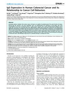
in terms of the cancer-specific modulation of ER expression and function, for clinical application. As mentioned above, the regulation of ERα gene expression is an important issue in breast cancer, and the overexpression of ERα is an initial significant event in its genesis. We have previously reported that the distal promoter (promoter B) of the ERα gene is responsible for this enhanced transcription of the gene (Hayashi et al. 1997a), and identified an important cis-acting element, which is located downstream of the transcription start site and plays an important role in its transcription (Tanimoto et al. 1999). Furthermore, methylation of the ERα gene promoters also contributes to the regulation of gene transcription (Yoshida et al. 2000). Elucidation of these mechanisms of ERα gene expression is important to develop a new tool for the early detection of breast cancer along with chemoprevention targeting breast cancer-specific promoters of the ERα gene (Fig. 1).
Endocrine-Related Cancer (2003) 10 193–202 1351-0088/03/010–193 2003 Society for Endocrinology Printed in Great Britain
Online version via http://www.endocrinology.org
Hayashi et al.: ERα and β in breast cancer
estrogen synthesis (aromatase)
? Growth factors
ERα gene ?
overexpression
coactivators phosphorylation signal
ERβ AIB1
ERα
overexpression
ROI
hypoxia
ERE
redox
P53 MDM2
E2
estrogen ablation LH-RH agonist aromatase inhibitor
anti-hormones target genes
TAM ICI raloxifene
anti-cancer drugs inflammatory cytokines Figure 1 ERs in the microenvironment of breast cancer. Breast cancer-specific phenomena are illustrated; for example, overexpression of ERα and its coactivators, alteration of intracellular signaling pathways triggered by various stimuli from outside the cancer cells, and the modulation of ERα function by ERβ or cancer-related genes. ROI, reactive oxygen intermediate; TAM, tamofixen; ICI, ICI182780.
Modulation of the ER function might be a promising tool with which to control breast cancer. In fact, anti-estrogens are widely used for its therapy. From this aspect, we have so far examined the cancer-specific modulation of ERα function, such as redox regulation (Hayashi et al. 1997b) and interaction with cancer-related genes (Saji et al. 2001), while clinically available tools have not been established hitherto. On the other hand, another ER, ERβ, was recently identified (Kuiper et al. 1996). Subsequently, numerous studies have reported on the expression of ERβ in various cancers, including our observations in breast (Omoto et al. 2002), lung (Omoto et al. 2001), and stomach (Matsuyama et al. 2002). Immunohistochemical studies suggest that ERβ tends to be expressed in ERα-positive breast cancers, and that there are ERα and ERβ co-expressing cells in human breast cancer. Furthermore, the existence of various variant forms of ERβ has been reported in breast cancer cells (Leygue et al. 1999). We then assessed the significance and function of ERβ and its variant types in breast cancer. Various observations obtained from experiments using ERβ-expressing stable transformant breast cancer cell lines suggested that ERβ and ERβcx truncated at the C-terminal region but has extra 26 amino acids (Ogawa et al. 1998) specifically suppress the function of ERα through different mechanisms. These ERβ isoforms could be important functional modulators of estrogen-signaling pathways in breast cancer cells, and might affect the clinical outcome of patients with primary breast cancer.
194
There are many reports concerning the target genes transcriptionally activated by ERα, but the entire mechanism of the pathway from ERα leading to the proliferation and progression of mammary tumors is far from being completely clarified. In order to elucidate the scheme of estrogen signaling and improvement of clinical decisions, expression profiling analysis using cDNA microarray technology should be one of the most effective procedures. Several laboratories have performed cDNA microarray analysis of breast cancer from patients, and novel genes whose expression status was highly correlated with the prognosis of the patients have been identified (Finlin et al. 2001, Sørlie et al. 2001, Veer et al. 2002). There has also been a report concerning gene expression profiling in human ZR-75-1 breast cancer cells in the presence of estrogen or estrogen antagonists using oligonucleotide microarray and several novel estrogen-responsive genes have been identified (Soulez & Parker 2001). Nonetheless, there is little information on how many markers are sufficient and which markers are suitable for accurate prognosis and diagnosis of breast tumors, especially regarding sensitivity to anti-hormone therapy. We first analyzed the estrogen-responsive gene expression profile in ER-positive breast cancer cells using largescale cDNA microarray (Inoue et al. 2002). Based on the results, the custom-made cDNA microarray, on which only estrogen-responsive genes were loaded, was produced. Using this microarray consisting of the narrowed gene subset, we analyzed the estrogen responsiveness of various cell lines and
www.endocrinology.org
Endocrine-Related Cancer (2003) 10 193–202 the effect of estrogen antagonists. Several candidate genes selected from the contents on the custom-made microarray were also analyzed by real-time RT-PCR and by an immunohistochemical technique using breast cancer tissues, in order to find new predictive factors for the responsiveness to hormone therapy of patients with primary breast cancer.
Regulation of ER␣ gene expression We previously analyzed promoter usage of ERα gene in human breast cancer tissues, and found that promoter B, one of the distal promoters, may be responsible for cancerspecific overexpression of ERα. We next analyzed the promoter B region using luciferase reporter plasmids containing several deleted fragments of this region and gel mobility shift assay. These experiments identified an important cis-element in the downstream region of the transcription initiation site, which contains the nucleotide sequence CTGGAAAG. This element was capable of transactivating a heterogeneous SV40 promoter in MCF-7 cells, confirming that the element is a transcriptional enhancer. The trans-acting factor binding to the element (named ERBF-1) was exclusively expressed in cells expressing ERα mRNA transcribed from promoter B. Furthermore, we also attempted to elucidate the mechanisms of suppression of ERα gene expression in advanced breast cancer. In the development of breast cancer, the loss of ERα gene expression is one of the most important steps in acquiring hormone resistance, although the mechanisms are poorly understood. We assessed the effects of methylation in the promoter region of ERα using breast cancer cell lines and tissues. The methylation status of promoter B as well as that of promoter A was inversely associated with ERα gene expression. In vitro methylation of ERα directly decreased the transcription of the ERα gene in a reporter assay. Demethylating treatment induced transcription of ERα from promoter B in ZR-75-1 cells which, by their nature, show no transcription from promoter B despite weak ERBF-1 expression. On the contrary, ERα-negative cells, such as MDA-MB-231 and BT-20 cells, which lack ERBF-1, did not show any induced expression with demethylation. Moreover, ZR-75-1 cells showed ERα promoter activity equal to that of MCF-7 cells when luciferase reporter plasmid was transiently transfected. These observations indicated that the methylation of promoters can directly regulate ERα gene expression, and loss of critical transcription factors such as ERBF-1 may also be involved in the negative regulation of ERα gene expression. As summarized in Fig. 2, down-regulation of ERα gene expression could be controlled by two distinct mechanisms: DNA methylation of the promoter region or loss of transcriptional activators.
Function of ER in breast cancer cells In order to assess the function of ERβ in ERα-positive breast cancer cells, we established cell lines expressing ERβ and
www.endocrinology.org
ERβcx by stable transfection of each expression plasmid into MCF-7 cells. This constitutive expression of ERβ and ERβcx significantly reduced cell growth, cell cycle, and colony formation in anchorage-independent situation. ERE activity in these cells was also strongly diminished compared with parental MCF-7 cells. Furthermore, endogenous expression of known ERα target genes, such as cathepsin D (Augereau et al. 1994), in these cells responded more weakly to estrogen. These observations indicated that both ERβ and ERβcx inhibit ERα function, resulting in growth inhibition of ERαpositive breast cancer cells. However, functional differences between ERβ and ERβcx were observed in the electrophoretic mobility shift assay and gene expression profiling using microarray. Although nuclear extracts prepared from ERβexpressing MCF-7 cells formed a DNA–protein complex despite different patterns from parental MCF-7 cells, ERβcx did not show any clear complexes in this assay. We further analyzed expression profiles of estrogen-responsive genes in these cells using our custom-made cDNA microarray as described in the next section. The estrogen-responsive expression profile of the genes in the ERβcx-expressing cells was clearly distinct from the ERβ-expressing cells. These results suggest that ERβ and ERβcx attenuate ERα function through distinct mechanisms as shown in Fig. 3. We reported very recently that ERβcx expression may affect the clinical outcome of breast cancer, such as the response to tamoxifen (Saji et al. 2002).
Expression profiling of estrogen-responsive genes To understand the estrogen-signaling pathway in breast cancer cells and to find the molecular markers which reflect the physiological status of ER-positive breast tumors for clinical application, such as the diagnosis of estrogen- and anti-estrogen responsiveness of mammary tumors, we analyzed the estrogen-responsive gene expression profiles in human MCF-7 breast cancer cells using a human UniGEMTM V 2.0 microarray system (IncyteGenomics, CA, USA) consisting of 9128 human cDNA clones covering 8502 unique gene/EST clusters. After MCF-7 cells were cultured in estrogen-starved medium for 5 days, the cells were treated with 10 nM 17β-estradiol, which is considered to be a saturating estrogen-rich condition for the MCF-7 cells but physiologically relevant and also a possible condition in mammary glands. From a total of 9128 clones, the data for 1846 clones were cut off because of low signal intensities as defined by the manufacturer. Among the remaining 7282 clones, 181 genes showed differential expression ratios equal to or more than 2.0, and 105 genes showed differential expression ratios equal to or less than 0.5; the remaining 96% of the genes revealed no significant differences in their expression levels. In a total of 286 genes which proved to be potentially estrogen-responsive genes by this analysis, there
195
Hayashi et al.: ERα and β in breast cancer
◊
transcriptional activators (ex. ERBF-1)
repression ◊ (1) methylated promoter
expression
HDAC1
unmethylated promoter
◊ ◊
HDAC2
◊
(2) loss of trancriptional activators
◊
◊
mSin3A
MeCP2
transcriptional activators (ex. ERBF-1)
repression
histone deacetylation
◊
repression
unmethylated promoter
chromatin remodeling
Figure 2 Proposed model of silencing ERα gene expression in breast cancer cells. HDAC, histone deacetylase.
A
AF-1
DBD
Hinge
LBD/AF-2
D
EE
ERβ
A/B
CC
β cx
A/B
CC
F
X EE splicing variants in the C-terminal region
D
B heterodimer
ERα ERE
>
ERE
ERβ
>
ERE
βcx
ERβ Target genes PgR etc
Clinical outcome response to tamoxifen
Figure 3 (A) Structure of wild-type ERβ and ERβcx and (B) how they modulate the transcription activity of ERα through the distinct mechanisms. AF-1, activation function 1; DBD, DNA binding domain; LBD, ligand binding domain; PgR, progesterone receptor.
196
www.endocrinology.org
Endocrine-Related Cancer (2003) 10 193–202 Table 1 Expression ratios of 148 genes loaded on the custom-made cDNA microarray Accession no.
Gene name Group)
Ratio of expression at 72 hrsa
Group A: early-responsive estrogen-induced genes M80244 solute carrier family 7, member 5 AJ011972 histone deacetylase 6 AB018010 antigen identified by monoclonal antibodies 4F2, TRA1.10, TROP4, and T43 X84373 nuclear receptor interacting protein 1 M62403 insulin-like growth factor-building protein 4 X05030 trefoil factor 1 U44427 tumor protein D52-like 1 D10495 protein kinase C, delta AB028974 KIAA1051 protein X16706 FOS-like antigen 2 D13643 KIAA0018 M63138 cathepsin D L06237 microtubule-associated protein 1B
8.34 4.75 4.70 3.78 3.58 3.34 2.85 2.78 2.71 2.20 2.02 1.88 1.84
Group B: late-responsive oestrogen-induced genes AF012281 PDZ domain containing 1 AA587912 ESTs, Highly similar to phosphoserine aminotransferase U72066 retinoblastoma-binding protein 8 N3555 EST L02785 down-regulated in adenoma X16396 methylene tetrahydrofolate dehydrogenase (NAD+ dependent), methenyltetrahydrofolate cyclohydrolase X62570 tryptophanyl-tRNA synthetase U29343 hyaluronan-mediated motility receptor (RHAMM) AI767533 EST AA430241 pituitary tumor-transforming 1 AI660571 argininosuccinate synthetase D30658 glycyl-tRNA synthetase X62585 EST X72875 B-factor, properdin AB018265 unc-51 (C. elegans)-like kinase 1 L35946 asparagine synthetase X92720 phosphoenolpyruvate carboxykinase 2 (mitochondrial) AF039022 exportin, tNRA (nuclear export receptor for tRNAs) U66838 cyclin A1 AI880413 Incyte EST AA629308 ESTs, Highly similar to BimL AI151190 S100 calcium-binding protein B AI878886 heat shock 70kD protein 5 (glucose-regulated protein, 78kD) X89773 interferon-stimulated gene (20kD) D90070 phorbol-12-myristate-13-acetate-induced protein 1 N39944 activating transcription factor 3 AF050110 TGFB inducible early growth response AB011123 KIAA0551 X74837 mannosidase, alpha, class 1A, member 1 U23143 serine hydroxymethyltransferase 2 (mitochondrial) AB012664 stanniocalcin 2 M37400 glutamic-oxaloacetic transaminase 1, soluble (aspartate aminotransferase 1) AA216685 prostate differentiation factor M90516 glutamine-fructose-6-phosphate transaminase 1 L14595 solute carrier family 1 (glutamate/neutral amino acid transporter), member 4 AB005047 SH3-domain-binding protein 5 (BTK-associated) AA102267 ferritin, heavy polypeptide 1 D42073 reticulocalbin 1, EF-hand calcium-binding domain AI1185199 EST U82984 ESTs, Weakly similar to oligophrenin-1 like protein N28312 HS1 binding protein AB011159 KIAA0587
6.64 5.54 5.33 4.94 4.16 4.13 4.01 3.76 3.70 3.53 3.53 3.42 3.38 3.24 3.21 3.18 3.02 2.89 2.87 2.79 2.76 2.75 2.65 2.61 2.56 2.53 2.52 2.51 2.47 2.47 2.44 2.42 2.32 2.26 2.24 2.24 2.14 2.13 2.07 2.04 2.03 2.03
Group C: oestrogen-repressed genes X76220 mal, T-cell differentiation protein U59325 cadherin 18 AI700706 general transcription factor II, i, pseudogene 1 D87993 paired basic amino acid cleaving system 4 U29091 selenium binding protein 1 AJ002308 synaptogyrin 2 U96136 catenin (cadherin-associated protein), delta 2 (neural plakophilin-related arm-repeat protein) AB002305 KIAA0307 gene product X13425 membrane component, chromosome 1, surface marker 1 (40kD glycoprotein, identified by monoclonal antibody GA733) AC002984 calpain, small polypeptide M19922 fructose-bisphosphatase 1 M59828 heat shock 70kD protein 1 X69433 isocitrate dehydrogenase 2 (NADP+ ), mitochondrial X69550 Rho GDP dissociation inhibitor (GDI) alpha AA374325 insulin-like growth factor binding protein 5 U03877 EGF-containing fibulin-like extracellular matrix protein 1 U97276 quiescin Q6 X56832 enolase 3, (beta, muscle) X51956 enolase 2, (gamma, neuronal) AA482422 MYC promoter-binding protein 1
www.endocrinology.org
− 2.00 − 2.04 − 2.21 − 2.34 − 2.37 − 2.48 − 2.60 − 2.65 − 2.65 − 2.69 − 2.76 − 2.94 − 3.23 − 3.38 − 3.46 − 3.94 − 4.13 − 5.24 − 5.42 − 5.79
197
Hayashi et al.: ERα and β in breast cancer Table 1 Continued Accession no. U30246 L27560
Gene name (Group)
Ratio of expression at 72 hrsa
solute carrier family 12 (sodium/potassium/chloride transporters), member 2 Human insulin-like growth factor binding protein 5 (IGFBP5)
Others: genes without significant response to estrogen M84755 Neuropeptide Y receptor Y1 D28473 Isoleucine-tRNA synthetase AI949781 ESTs, Weakly similar to phosphoprotein AF105826 Solute carrier family 1 (neutral amino acid transporter), member 5 X15187 Tumor rejection antigen (gp96) 1 AI492976 ESTs, Moderately similar to KIAA0400 AA557306 CCAAT/enhancer binding protein (C/EBP), beta U08316 Ribosomal protein S6 kinase, 90kD, polypeptide 3 AI332415 EST U89436 Tyrosyl-tRNA synthetase L20815 Corneodesmosin AI261366 DKFZP566G223 protein AA481712 Protein geranylgeranyltransferase type I, beta subunit AI188401 ESTs, highly similar to CGI-82 protein AI683760 Sema domain, immunoglobulin domain (Ig), short basic domain, secreted, (semaphorin) 3B U21858 TATA box-binding protein (TBP)-associated factor, RNA polymerase II, G, 32kD X64I16 Poliovirus receptor AI088306 FOS-like antigen 2 AI598150 v-jun avian sarcoma virus 17 oncogene homolog J05068 Transcobalamin I (vitamin B12-binding protein, R binder family) Z48950 H3 histone, family 3B (H3 3B) X06233 S100 calcium-binding protein A9 (calgranulin B) AF065388 Tetraspan 1 T73188 Aldo-keto reductase family 1, member C4 (chlorodecone reductase; 3-alpha-hydroxysteroid dehydrogenase, type I; dihydrodiol dehydrogenase 4) D32050 Alanyl-tRNA synthetase AF022109 CDC6 (cell division cycle 6, S. cerevisiae) homolog AA975298 EST AA484893 Matrix Gla protein AI342303 Solute carrier family 29 (nucleoside transporters), member 2 Y18448 Bassoon (presynaptic cytomatrix protein) AA143530 stearoyl-CoA desaturase (delta 9-desaturase) AA682502 EST AF055008 granulin AL022726 inhibitor of DNA binding 4, dominant negative helix-loop-helix protein Z80998 solute carrier family 5 (sodium/glucose cotransporter), member 1 X55122 GATA-binding protein 3 X63741 early growth response 3 L35546 glutamate-cysteine ligase (gamma-glutamylcysteine synthetase), regulatory (30.8kD) D21205 zinc finger protein 147 (oestrogen-responsive finger protein) AA994925 tumor necrosis factor receptor superfamily, member 7 AI239815 general transcription factor IIH, polypeptide 2 (44kD subunit) Z46973 phosphoinositide-3-kinase, class 3 L42611 keratin 6B AI679881 thrombospondin 3 X15393 motilin U07361 sorbitol dehydrogenase X74929 keratin 8 X00947 alpha-1-antichymotrypsin AL046741 chromobox homolog 1 (Drosophila HP1 beta) U79302 Human clone 23855 mRNA, partial cds AA173870 nucleophosmin (nuclear phosphoprotein B23, numatrin) U78525 eukaryotic translation initiation factor 3, subunit 8 (eta, 116kD) D21163 U5 snRNP-specific protein, 116kD) M30704 amphiregulin (schwannoma-derived growth factor) X00351 actin, beta L39211 carnitine palmitoyltransferase I, liver X14723 clusterin (complement lysis inhibitor, SP-40,40, sulfated glycoprotein 2, testosterone-repressed prostate message 2, apolipoprotein J) AI815757 ribosomal protein L35 D21064 KIAA0123 Y17977 fucosyltransferase 8 (alpha (1,6) fucosyltransferase) AL036958 KIAA0058 AF0544996 Homo sapiens clone 23783 mRNA AC004030 Homo sapiens DNA from chromosome 19, cosmid F21856 U83115 absent in melanoma 1 AF014807 CDP-diacylglycerol – inositol 3-phosphatidyltransferase (phosphatidylinositol synthase) U87939 aconitase 2, mitochondrial D83780 KIAA0196 X79568 protein tyrosine phosphatase, non-receptor type 18 (brain-derived) X03635 estrogen receptor 1 L25081 ras homolog gene family, member C J03075 protein kinase C substrate 80K-H
− 5.84 − 8.42 1.97 1.95 1.95 1.94 1.91 1.91 1.89 1.86 1.85 1.85 1.84 1.81 1.76 1.75 1.74 1.74 1.71 1.65 1.60 1.59 1.59 1.59 1.56 1.54 1.53 1.52 1.52 1.47 1.36 1.33 1.31 1.29 1.27 1.23 1.22 1.21 1.21 1.18 1.17 1.08 1.07 1.07 1.05 1.02 1.02 − 1.02 − 1.11 − 1.14 − 1.19 − 1.20 − 1.22 − 1.28 − 1.31 − 1.38 − 1.43 − 1.44 − 1.45 − 1.45 − 1.47 − 1.49 − 1.57 − 1.58 − 1.61 − 1.62 − 1.66 − 1.71 − 1.76 − 1.83 − 1.84 − 1.93 − 1.96
a ‘Ratio of expression at 72 hrs’ means the ratio of expression levels for each gene in MCF-7 cells treated with (E2+) and without (E2−) 17β-estradiol for 72 hrs, which examined by custom-made microarray analysis. For E2 -upregulated or E2 -downregulated genes, the values are shown as the ratios E2+/E2 – or – (E2-/E2+) respectively.
198
www.endocrinology.org
Endocrine-Related Cancer (2003) 10 193–202 were some genes which had previously been reported to be induced by estrogen such as pS2 (Brown et al. 1984), PDZK1 (PDZ domain-containing protein) (Ghosh et al. 2000), and insulin-like growth factor-binding protein (IGFBP)-4 (Qin et al. 1999), indicating the reliability of this analysis. From the 286 potentially estrogen-responsive genes mentioned above, a total of 138 genes were selected for production of a prototype of the custom-made microarray,
removing the genes which showed relatively low ratios of estrogen-responsive expression (near or equal to 2.0 or − 2.0) and which were inferred to have little relation to the nature of breast cancer according to the background information on the genes. Using this customized microarray prototype, we analyzed the time-course of estrogen response of the genes in MCF-7 cells. The estrogen-starved MCF-7 cells were treated with ethanol (E2 − ) (Cy5-labelled) or 10 nM 17β-estradiol (E2 + ) for 6, 12, 24 or 72 h (Cy3-labelled),
Fold Expression (E2+ / E2-)
A 9 trefoil factor 1 (pS2)
8 7
IGF-binding protein 4
6
KIAA1051 protein
5
solute carrier family 7, member 5
4 histone deacetylase 6
3 2
cathepsin D
1
tumor protein D52-like 1
0 0
12
24
36
48
60
72
Time after estrogen addition (h)
B Fold Expression (E2+ / E2-)
retinoblastoma-binding protein 8
8 asparagine synthetase
7 6
EST
5
ESTs, Highly similar to phosphoserine aminotransferase
4
cyclin A1
3
down-regulated in adenoma
2 pituitary tumor-transforming 1
1 tryptophanyl-tRNA synthetase
0 0
12
24
36
48
60
72
Time after estrogen addition (h)
C Fold Expression (-E2- / E2+)
0
IGF-binding protein 5
-1
MYC promoter-binding protein 1
-2
enolase 2 (gamma, neuronal)
-3
KIAA0307
-4
quiescin Q6 solute carrier family 12, member 2
-5
selenium binding protein 1
-6
EGF-containing fibulin-like extracellular matrix protein 1
-7 0
12
24
36
48
60
72
Time after estrogen addition (h) Figure 4 Expression profiles of several genes in MCF-7 cells at different times after estrogen stimulation analyzed by customized microarrays. Several representative genes in each group were selected and the time-courses of the ratios of expression levels between the cells treated with E2 + and E2 − are plotted. (A), (B) and (C) show the expression patterns of early-responsive estrogen-induced genes, late-responsive estrogen-induced genes and estrogen-repressed genes respectively.
www.endocrinology.org
199
Hayashi et al.: ERα and β in breast cancer and mRNA isolated from each culture of the cells was used for the microarray analysis. The mRNA was also isolated from the estrogen-starved cells but without any treatment (shown as 0 h). On the customized microarray, cDNA fragments for each gene were spotted in two blocks, yielding a pair of data sets of expression ratios from one hybridization. In the case of estradiol treatment for 0 h (without estradiol treatment), Cy3- and Cy5-labeled cDNA were produced from the same preparation of mRNA and hybridized together into one microarray glass slide, for the purpose of examining the level of technical variation in fluorescent labeling and/or hybridization. As a result, all the Cy3/Cy5 ratios for the genes were between 0.5 and 2.0. Hierarchical clustering clearly highlighted the gene clusters of estrogen-induced and estrogen-repressed genes, and made it possible to subdivide the cluster of estrogen-induced genes into two subgroups; one group containing the genes which showed expression ratios above 2.0 after 12 h of estrogen treatment (early-responsive estrogen-induced genes, group A) and the other group containing the genes which showed significant induction after 24 h or, in most cases, 72 h of estrogen treatment (late-responsive estrogen-induced genes, group B). On the other hand, the cluster of estrogen-repressed genes (group C) did not show any apparent subgroups. The expression ratios after estradiol treatment for 72 h of all the 148 genes loaded on the custom-made microarray are listed in Table 1, being divided into four groups including groups A, B and C.
Figure 4 shows the time-course profile of estrogenresponsive expression of several genes selected from groups A, B and C. As expected, pS2, IGFBP-4 and cathepsin D, which were reported to be the target genes of ERα, were found in the group of early-responsive estrogen-induced genes (Fig. 4A). IGFBP-5, which was reported to be downregulated by estrogen (Huynh et al. 1996), was found in the group of estrogen-repressed genes (Fig. 4C). Among these genes listed in Table 1, we performed Northern blot analysis for several of them to confirm the results from microarray analysis, and all the examined genes exhibited similar expression patterns in Northern analysis to that from microarray analysis (data not shown). We have developed a second version of the custom-made estrogen-responsive array (estrogen-responsive gene (ERG) chip in Fig. 5), and are currently applying it to various studies such as basic research for estrogen signaling and clinical application for the diagnosis of estrogen-dependent cancer, as summarized in Fig. 5.
Identification of predictive factors for endocrine therapy Based on the results obtained from the microarray analysis, we selected 11 genes as candidate factors for predicting the efficacy of endocrine therapy. We assessed their expression levels in breast cancer tissues by real-time RT-PCR, and the
Microarray analysis of 10000 genes
Identification of estrogen regulated genes (Estrogen Responsive Genes) Production of custom-made cDNA microarray
ERG chip (200 genes)
Basic research for estrogen signaling pathway
Diagnosis of hormonedependent cancers
Screening of novel prognostic factors
•Prediction of hormone therapy •Screening of ER-target genes •Classification of patients for individualized therapy •Functional analyses of ERα, ERβ •Monitoring of hormone therapy •Evaluation of SERMs •Mechanisms of anti-hormone resistance Figure 5 Development of the custom-made estrogen-responsive cDNA microarray and its application to basic and clinical studies. SERMS; selective estrogen receptor modulators.
200
www.endocrinology.org
Endocrine-Related Cancer (2003) 10 193–202
Gene B
Gene A 1
Low (n=47)
.8
.6 High (n=18)
.4
.2
p=0.02 (Logrank (Mantel-Cox) test) p=0.01 (Breslow-Gehan-Wilcoxon test)
Disease-free survival rate
Disease-free survival rate
1
Low (n=15)
.8
.6
High (n=50)
.4
.2
p=0.04 (Logrank (Mantel-Cox) test) p=0.06 (Breslow-Gehan-Wilcoxon test)
0
0 0
1000
2000
3000
4000
0
1000
Days
2000
3000
4000
Days
Figure 6 Kaplan–Meier analysis of disease-free survival rates of patients who had tamoxifen adjuvant therapy and the expression status of selected candidate genes, Gene A and Gene B.
result was analyzed by cluster analysis. ER-positive patients were clearly divided into two subgroups that were expected to represent different estrogen responsiveness. The same subgroups were almost reproducible with a few representative genes in this 11 gene subset. We therefore examined the expression of these genes by immunohistochemical analysis in 65 patients treated with adjuvant tamoxifen therapy, in order to assess the impact on the prediction of the effect of this endocrine therapy. As shown in Fig. 6, positive staining of gene A as well as gene B significantly correlated with lower disease-free survival rate. Furthermore, combined assessment of expression of these genes improved the predictive accuracy for tamoxifen response.
Conclusion and perspective These studies of the expression and function of ERs, especially from the aspect of cancer-specific phenomena, are very important for improving new strategies of prevention, diagnosis, and therapy of estrogen-dependent cancers. For instance, elucidation of the mechanism of the regulation of ERα gene expression may provide a new idea to prevent or arrest the development of breast cancer, and ERβcx expression status may assist in the eligibility of therapeutic options. On the other hand, cDNA microarray technique is another very promising method for these studies. We are currently applying our custom-made cDNA microarray to extensive studies on estrogen signaling in terms of both basic and clinical aspects. This down-sized microarray may be especially
www.endocrinology.org
useful for predicting the efficacy of endocrine therapy for individual patients with breast cancer. In fact, along with recent progress, novel endocrine therapies, including aromatase inhibitors, a new accurate prediction method for these treatments is urgently required. Finally, this study has suggested that new predictive factors could be identified in estrogen-responsive genes that distinguish responsive or nonresponsive patients to tamoxifen treatment in an ER-positive population. Immunohistochemical evaluation using these factors may be a more practical method than cDNA microarray. Although further study will be needed, these approaches could provide not only new clues for the elucidation of mechanisms of estrogen-dependent carcinogenesis and the development of breast cancer, but also clinical benefits to patients by assessment of individual responses to endocrine therapy.
Acknowledgements We thank Ms Akiyo Yamashita for her excellent technical assistance, Dr Masakazu Toi, Dr Ryoichi Kiyama and Dr Kei Nakachi for their valuable suggestions and advice. This study was supported in part by Grants-in-Aid from the Ministry of Education, Culture, Sports, Science and Technology of Japan for Cancer Research, from the Ministry of Health, Labor and Welfare of Japan for Scientific Research Expenses for Health and Welfare Programs and the Foundation for the Promotion of Cancer Research and for Second-Term Comprehensive 10-Year Strategy for Cancer Control.
201
Hayashi et al.: ERα and β in breast cancer
References Augereau P, Miralles F, Cavaille`s V, Gaudelet C, Parker M & Rochefort H 1994 Characterization of the proximal estrogen-responsive element of human cathepsin D gene. Molecular Endocrinology 8 693–703. Brown AMC, Jeltsch JM, Roberts M & Chambon P 1984 Activation of pS2 gene transcription is a primary response to estrogen in the human breast cancer cell line MCF-7. PNAS 81 6344–6348. Finlin BS, Gau C-L, Murphy GA, Shao H, Kimel T, Seitz RS, Chiu Y-F, Botstein D, Brown PO, Der CJ, Tamanoi F, Andres DA & Perou CM 2001 RERG is a novel ras-related, estrogen-regulated and growth-inhibitory gene in breast cancer. Journal of Biological Chemistry 276 42259–42267. Ghosh MG, Thompson DA & Weigel RJ 2000 PDZK1 and GREB1 are estrogen-regulated genes expressed in hormone-responsive breast cancer. Cancer Research 60 6367– 6375. Hayashi S-I, Imai K, Suga K, Kurihara T, Higashi Y & Nakachi K 1997a Two promoters in expression of estrogen receptor messenger RNA in human breast cancer. Carcinogenesis 18 459–464. Hayashi S-I, Hajiro-Nakanishi K, Makino Y, Eguchi H, Yodoi J & Tanaka H 1997b Functional modulation of estrogen receptor by redox state with reference to thioredoxin as a mediator. Nucleic Acids Research 25 4035–4040. Huynh H, Yang X-F & Pollak M 1996 A role for insulin-like growth factor binding protein 5 in the antiproliferative action of the antiestrogen ICI 182780. Cell Growth and Differentiation 7 1501–1506. Inoue A, Yoshida N, Omoto Y, Oguchi S, Yamori T, Kiyama R & Hayashi S-I 2002 Development of cDNA microarray for expression profiling of estrogen-responsive genes. Journal of Molecular Endocrinology 29 175–192. Kuiper GGJM, Enmark E, Pelto-Hukko M, Nilsson S & Gustafsson J-A 1996 Cloning of a novel estrogen receptor expressed in rat prostate and ovary. PNAS 93 5925–5930. Leygue E, Dotzlaw H, Watson PH & Murphy LC 1999 Expression of estrogen receptor β1, β2, and β5 messenger RNAs in human breast tissue. Cancer Research 59 1175–1179. Matsuyama S, Ohkura Y, Eguchi H, Kobayashi Y, Akagi K, Uchida K, Nakachi K, Gustafsson J-Å & Hayashi S-I 2002 Estrogen receptor β (ERβ) is expressed in human stomach adenocarcinomas. Journal of Cancer Research and Clinical Oncology 128 319–324. Ogawa S, Inoue S, Watanabe T, Orimo A, Hsoi T, Ouchi Y & Muramatou M 1998 Molecular cloning and characterisation at human estrogen receptor BCX: a potential inhibitor of estrogen action in human. Nucleic Acids Research 26 3502–3512.
202
Omoto Y, Kobayashi Y, Nishida K, Tsuchiya E, Eguchi H, Nakagawa K, Ishikawa Y, Yamori T, Iwase H, Fujii Y, Warner M, Gustafsson J-Å & Hayashi S-I 2001 Expression, function and clinical implications of estrogen receptor β in human lung cancer. Biochemical and Biophysical Research Communications 285 340–347. Omoto Y, Kobayashi S, Inoue S, Ogawa S, Toyama T, Yamashita H, Muramatsu M, Gustafsson J-Å & Iwase H 2002 Evaluation of oestrogen receptor β wild-type and variant protein expression, and relationship with clinicopathological factors in breast cancers. European Journal of Cancer 38 380–386. Qin C, Singh P & Safe S 1999 Transcriptional activation of insulin-like growth factor-binding protein-4 by 17β-estradiol in MCF-7 cells: role of estrogen receptor–Sp1 complexes. Endocrinology 140 2501–2508. Saji S, Okumura N, Eguchi H, Nakashima S, Suzuki A, Toi M, Nozawa Y, Saji S & Hayashi S-I 2001 MDM2 enhances the function of estrogen receptor α in human breast cancer cells. Biochemical and Biophysical Research Communications 281 259–265. Saji S, Omoto Y, Shimizu C, Warner M, Hayashi Y, Horiguchi S, Watanabe T, Hayashi S-I, Gustafsson J-Å & Toi M 2002 Expression of estrogen receptor (ER) βcx protein in ERα-positive breast cancer; specific correlation with progesterone receptor. Cancer Research 62 4849–4853. Sørlie T, Perou CM, Tibshirani R, Aas T, Geisler S, Johnsen H, Hastie T, Eisen MB, van de Rijn M, Jeffrey SS, Thorsen T, Quist H, Matese JC, Brown PO, Botstein D, Lønning PE & B¿rresen-Dale A-L 2001 Gene expression patterns of breast carcinomas distinguish tumor subclasses with clinical implications. PNAS 98 10869–10874. Soulez M & Parker MG 2001 Identification of novel oestrogen receptor target genes in human ZR75-1 breast cancer cells by expression profiling. Journal of Molecular Endocrinology 27 259–274. Tanimoto K, Eguchi H, Yoshida T, Hajiro-Nakanishi K & Hayashi S-I 1999 Regulation of estrogen receptor α gene mediated by promoter B responsible for its enhanced expression in human breast cancer. Nucleic Acids Research 27 903–909. Veer LJ, Dai H, van de Vijver MJ, He YD, Hart AA, Mao M, Peterse HL, van der Kooy K, Marton MJ, Witteveen AT, Schreiber GJ, Kerkhoven RM, Roberts C, Linsley PS, Bernards R & Friend SH 2002 Gene expression profiling predicts clinical outcome of breast cancer. Nature 415 530–536. Yoshida T, Eguchi H, Nakachi K, Tanimoto K, Higashi Y, Suemasu K, Iino Y, Morishita Y & Hayashi S-I 2000 Distinct mechanisms of loss of estrogen receptor α gene expression in human breast cancer: methylation of the gene and alteration of trans-acting factors. Carcinogenesis 21 2193–2201.
www.endocrinology.org