Int J Clin Exp Pathol 2016;9(10):10128-10138 www.ijcep.com /ISSN:1936-2625/IJCEP0036628
Original Article Enrichment and characterization of ovarian cancer stem cells and its potential clinical application Wenxia Wang1, Zhenbo Zhang2, Yin Zhao1, Zeng Yuan1, Xingsheng Yang1, Beihua Kong1, Wenxin Zheng3,4,5,6 Department of Obstetrics and Gynecology, Qilu Hospital, Shandong, China; 2Department of Obstetrics and Gynecology, Shanghai General Hospital, Shanghai, China; 3Department of Pathology, University of Arizona, Tucson, AZ, USA; Departments of 4Pathology, 5Obstetrics and Gynecology, 6Simmmon Comprehensive Cancer Center, University of Texas Southwestern Medical Center, Dallas, TX, USA 1
Received June 27, 2016; Accepted August 28, 2016; Epub October 1, 2016; Published October 15, 2016 Abstract: The cancer stem cell (CSC) theory proposes that a minor population in tumor cells with specific features, such as self-renewal and reproducible tumor phenotype could contribute to tumor relapse and chemotherapy resistance. Several studies have convincingly documented the existence of ovarian CSC, but questions related to the biologic behavior and specific biomarkers of ovarian CSC remain to be clarified. In the present study, we firstly established a tumor cell line with capability of regenerating tumors through serial transplantation of ovarian tumor tissue in non-obese/severe combined immunodeficient (SCID) mice. After separation of CD133+ cells with magnetic beads, we compared the phenotype and biologic behavior of CD133+ versus CD133- cells. It was found that the CD133+ cells were much more potent to produce colonies in semi-solid agar culture than CD133- cells. The proportion of the cells in G0/1 cell cycle is much higher in CD133+ cells than in CD133- cells. Furthermore, in vivo experiments demonstrated that the CD133+ cells were capable of repeatedly regenerate tumors in NOD/ SCID mice, while the CD133- cells were not. Compared with CD133- cells, the CD133+ cells expressed much higher levels of the stem cell markers Oct4, Sox2, Nanog and Mcl-1. Clinically, among a total of 290 ovarian epithelial cancers, increased level of CD133 expression was positively correlated with a high cancer stage and had a worse 5-year survival rate. Taken together, the results suggest that the CD133+ cells from human ovarian cancer have the characteristics of CSC, which may contribute to ovarian cancer relapse and anti-apoptotic activity. The method of ovarian CSC enrichment we established provides a feasible and practical way of ovarian cancer research in a molecular level. In addition, CD133 may be used as a prognostic marker for ovarian epithelial cancer, which may have a role for future therapeutic effect. Keywords: Ovarian epithelial cancer, cancer stem cells, CD133
Introduction In recent years, fast accumulating data demonstrate that there exists a small subset of cells within a tumor, termed cancer stem cells (CSC), responsible for cancer development and recurrence [1]. Characterization of CSC in leukemia and several other malignancies showed that these cells are relatively quiescent and highly express molecules mediating multi-drug resistance [2-4]. That is the main reason that the conventional chemotherapeutic agents are usually effective in reducing the bulk of tumor but unable to clear the stem cells. The residual CSCs after chemotherapy will eventually result in disease recurrence. Thus, the finding CSC
opened a new path to explore novel therapies aiming to understand the molecular mechanisms of chemoresistance and cancer recurrence in order to significantly improve clinical management and cure patients with cancers [5, 6]. Ovarian epithelial cancer (OEC) is a common malignancy and a leading cause of death in women, with epithelial ovarian cancer being the most frequent and aggressive subtype. Among the tumors originated from female reproductive system, ovarian epithelial cancer is the third commonest cancer and sits at the first place in terms of cancer mortality rate. Several research groups have convincingly documented the exis-
Characterization of ovarian cancer stem cells tence of CSC in ovarian cancers [7-11]. Ovarian CSCs have been defined and isolated from ovarian cancer cell lines or peritoneal fluid from ovarian cancer patients using different markers, including CD44, CD117, Myd88 and ABCG2. Among these biomarkers, the CD44+/ CD117+ immunophenotype holds promise as a defining characteristic of ovarian CSCs since these isolated cells exhibit stem cell functionality well. Recently, CD133 has been documented as a superb CSC marker in several non-ovarian cancers including cancers of the colon, the lung, and the brain [7-11]. CD133, a biomarker, is selectively expressed by neural stem cells, hematopoietic stem cells and epithelial progenitor cells. Singh et al. isolated CD133+ cells from brain tumors and compared the capabilities of tumor formation after subcutaneous inoculation of CD133+ or CD133- cells. While inoculation of 105 CD133- cells failed to regenerate tumors in NOD/SCID mice, inoculation of only 100 CD133+ cells generated tumors easily [11]. O’Brien et al. reported that all the colon cancer-initiating cells are CD133+. In contrast, the CD133- cells accounting for the bulk of the tumors were unable regenerate tumor [9]. Therefore, it is reasonable to speculate that the CD133+ cells may play a role in cancer development, recurrence, metastasis, and patient survival. However, it is unknown if ovarian epithelial cancers have CD133+ cells and whether they can be enriched and isolated efficiently for future applications in research as well as in clinical management.
in accordance with the policies of Qilu Hospital of Shandong University. Tumor samples were obtained from the operating room and immediately taken to the laboratory for processing. All ovarian epithelial cancers were pathologically characterized based on WHO classification system. Only the high-grade serous carcinoma samples were used for the CSC enrichment study. The samples were minced with scalpels to 2 mm cubic pieces and implanted into 4- to 6-week-old female NOD/SCID mice (The Jackson Laboratory, Florida, USA). The mice were monitored biweekly for tumor formation. The mice bearing growing tumors were euthanized by CO2 inhalation. Tumor explants were excised aseptically, processed as above, and implanted again into recipient female NOD/ SCID mice. Tumor explant was processed and used as a source of human tumor derived cells in 9 subsequent serial transplantation experiments. Cell culture and CD133+ cell isolation with magnetic beads
The study has been approved by Institutional Review Board of Qilu Hospital Ethic Committee in Shandong University, China. All primary human ovarian cancer samples were collected
Cell suspensions were prepared from a portion of the tumor samples. Briefly, The specimen was mechanically dissected and filtered, red blood cells were lysed with ACK buffer, and cells were then washed with media containing serum and plated in RPMI 1640 supplemented with 10% heat-inactivated FBS, 20 ng/ml EGF, 1 ng/ml hydrocortisone, 5 μg/ml insulin, 100 μM β-mercaptoethanol, 10 ng/ml β-FGF, 1% penicillin/streptomycin, and 20 μg/ml gentamicin. The cell cones were allowed to grow until they were confluent. Then the clones were trypsinized and plated again. Cells were trypsinized and re-suspended in PBS for FACS analysis. The CD133+ cells were separated using magnetic beads. Briefly, the cells were harvested and single-cell suspension was prepared and cell number counted. The cell suspension was centrifuged at 1000 × g for 10 minutes and the cell pellet was re-suspended in 300 μl of buffer per 108 total cells. After adding FcR Blocking Reagent (100 μl per 108 total cells), the cells were incubated with CD133 MicroBeads (100 μl of per 108 total cells) for 30 minutes in the refrigerator (2-8°C). The cells were washed, centrifuged, re-suspended and applied onto a column. Both the labeled (CD133+ cells) and unlabeled (CD133- cells) were collected for further experiments.
10129
Int J Clin Exp Pathol 2016;9(10):10128-10138
The main goals of this study are to enrich CD133+ cells through serial transplantation of ovarian epithelial cancer tissues into non-obese/severe combined immunodeficient (SCID) mice, to characterize if the CD133+ cells enriched from the ovarian cancer have the biologic function of CSC, and to correlate if CD133+ cancers have a negative impact in clinic. Materials and methods Source of human ovarian epithelial cancer stem cells
Characterization of ovarian cancer stem cells Semi-solid agar culture to examine the clonogenic ability The enriched CD133+ cells were assayed for their ability to form colonies in semisolid agar and compared with that of CD133- cells. Briefly, colony cultures were established in triplicate by plating 5.0 × 10⁶ cells in per 35 mm cell culture dish in 1.0 ml of RPMI 1640 supplemented with 0.33% agar, 25% FCS and 2 mM L-glutamine. Colony growth was stimulated by the addition of 20 ng/ml EGF. Cultures were incubated in a humidified chamber at 37°C and 5% CO2 for 2 weeks. Clones were allowed to grow until they were confluent and were then trypsinized, passaged to a 24-well plate, allowed to grow to confluence, and passaged a second time. Only the colonies that successfully passaged twice were deemed true clones. The colonies were counted under a light microscope.
cycles at 95°C for 15 seconds and 60°C for 1 minute. Samples were measured in triplicate. GAPDH also was amplified as a loading control for each batch of the test. Cycle threshold (Ct) values were used to determine the relative amounts of the targeted genes and GAPDH mRNA levels in the samples. In vivo tumor formation experiment The CD133+ and CD133- cells were collected, counted, washed and re-suspended in PBS. Then the cells were injected subcutaneously into NOD/SCID mice with 104-106 per mouse in 0.3 ml. The mice were monitored biweekly for tumor formation up to 8 weeks. Tumor tissue immunohistochemical analyses
The mRNA levels were determined by real-time quantitative PCR using the Applied Biosystems Taqman Gene Expression kit. Total RNA from cells was isolated with the RNeasy Mini Kit, accompanied by an on-column DNase digestion. The volume of each reaction was 10 μl per well, which consisted of 5 μl 2 × reaction buffer and 0.05 μl 200 × Euroscript RT (reverse transcriptase) enzyme and RNase inhibitor mix from the one-step RT-qPCR MasterMix Plus, 0.5 μl 20 × Taqman Gene Expression mix together with 2 μl of 50 ng RNA as amplification template. qRT-PCR was carried out on the ABI 7900HT Fast Real-Time PCR system. The reaction mixtures were incubated at 48°C for 30 minutes, 95°C for 10 minutes, followed by 40
A cohort of 290 ovarian epithelial carcinoma patients hospitalized at the University of Arizona Medical Center and Arizona Cancer Center during 2000-2014 was studied after approval of Institutional Review Board (IRB). Tumor core samples (3 mm) were obtained at initial debulking surgery, scored according to the Federation International of Gynecology and Obstetrics (FIGO) guidelines, and were formalin-fixed and paraffin-embedded. Each new paraffin block contained 100 randomly distributed tumor cores, with at least 2 cores per patient, and were sectioned at 5 micron thickness as previously described [12]. Immunohistochemistry was performed on slides with tumor microarray (TMA) sections before being blocked using a solution of 1% bovine serum albumin (BSA) with 2% donkey serum in phosphate buffered saline (PBS) for 1 hour at room temperature, incubated with an antibody to CD133 at 1:200 dilutions overnight in a humidified chamber at 4C, washed with PBS, and incubated using a secondary goat anti-rat horseradish peroxidase (HRP) conjugated antibody (Santa Cruz biotechnology, Dallas TX) at 1:200 for 1.5 hours at room temperature. Slides were rinsed and stained with a diaminobenzidine (DAB) solution, counter stained with Hematoxylin (Dako, Carpinteria CA), dehydrated, and coverslipped before being scanned and digitized with a Scanscope (Leica, Buffalo Grove IL) and analyzed with the Aperio Imagescope software (Leica, Buffalo Grove IL). Positivity and Intensity (PI) scores were calculated as the product of stained area fraction (positivity) and its inten-
10130
Int J Clin Exp Pathol 2016;9(10):10128-10138
Flow cytometry to analyze expression of CD133 and cell cycle distribution Cultured cells were collected, washed once with PBS and stained with anti-CD133-PE for 30 minutes of incubation in the dark on ice. For cell cycle analysis, the harvested cells (~106 cells) were fixed with ice-cold 70% ethanol, treated with 500 μg/ml RNase A (Sigma), and subsequently stained with 25 μg/ml propidium iodide (Sigma). Then these cells were analyzed by using a flow cytometer FACS Calibur. Quantitative real-time PCR to examine the expression of Oct-4, Sox2, Nanog and Mcl-1
Characterization of ovarian cancer stem cells Table 1. Passage of ovarian epithelial cancer in NOD/SCID mice Patient# 1 passage 2 passage 3 passage 4 passage 5 passage 1 0/6 2 1/6 0/6 3 2/8 0/8 4 0/8 5 5/8 6/8 8/8 8/8 8/8 6 3/8 3/8 0/8 7 1/8 0/8 8 5/8 2/8 3/8 2/8 9 0/8 st
nd
rd
th
th
Note: The fractions indicate the number of regenerated tumors among the total engrafted mice. #indicates tumor cells derived from individual patients.
Statistical analysis All experiments were performed at least three times and data were exhibited as mean ± SD. Statistical analysis was conducted using the chi-square test and Student’s t-test. P< 0.05 was considered significant. Patient survival was calculated from date of diagnosis to date of death, or entrance into hospice care when not available and converted from days to months by dividing the number of days by 30. Overall survival was analyzed by Kaplan-Meier plots and statistical significance inferred by log-rank tests. Correlations to clinical parameters were made using chi-square analysis. Statistical analyses were performed using the Prism 6 software (Graphpad, La Jolla CA). Results Passage of ovarian epithelial cancer in NOD/SCID mice
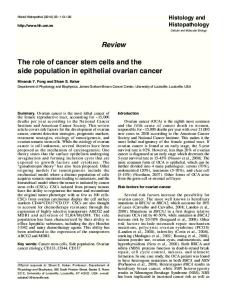
The ovarian cancer tissues were collected from 9 patients with primary high-grade serous carcinomas. As shown in Table 1, the tumor tissues from 6 patients (#2, 3, and 5-8) were successfully engrafted in NOD/ Figure 1. Isolation and enrichment of CD133+ cells from ovarian epitheSCID mice with varying tumor lial cancers. The CD133+ cells were isolated and accumulated with antigrowth or development. Three CD133 coated magnetic beads. The proportions of CD133+ cells were tumor samples (#1, 4, and 9) examined by FACS. A, B. Represent the proportions before and after isolations, respectively. C. Represents the number of CD133+ cells counted in did not grow. Samples 2, 3 and vitro. The amount of CD133+ cells were significantly increased after the 7 grew only 2 passages, while procedure (P