European Journal of Cardio-thoracic Surgery 11 (1997) 1011 – 1016
Surgical treatment of primary malignant chest wall tumours1 Sabaratnam Sabanathan *, Rajesh Shah, Alan J. Mearns Department of Thoracic Surgery, Bradford Royal Infirmary, Duckworth Lane, Bradford BD9 6RJ, UK Received 8 October 1996; received in revised form 27 January 1997; accepted 2 March 1997
Abstract Objective: Primary malignant tumours of the bony chest wall are uncommon and data concerning treatment and results are sparse. Methods: To assess the results of surgical resection and chest wall reconstruction we reviewed our experience with primary malignant chest wall tumours treated since 1958. Results: Of the 49 lesions, 42 were found in the ribs and the remaining 7 in the sternum. These included chondrosarcomas [22], solitary plasmacytoma [18], Ewing’s tumours [7], Askin’s tumour [1] and Desmoid tumour [1]. Skeletal reconstruction was performed in 36 of the 49 patients. Marlex mesh alone was used in 17 patients. Since 1972, a sandwich of two layers of Marlex mesh with a filler of methyl methacrylate was utilised [19] successfully, producing better functional and cosmetic results. Primary soft tissue closure was possible in all but 8 cases in whom latissimus dorsi myocutaneous flaps were used. Bilaterally, partially transposed pectoralis major muscle was used to cover upper sternal defects in 4 cases. All but 1 patient had an uneventful post-operative recovery none requiring ventilatory support. Survival: Overall survival at 5 and 10 years was 68%. The differential figures for 10-year survival were for chondrosarcoma 67%, Ewing’s sarcoma 43%, and solitary plasmacytoma 59%. These were the results of radical en-bloc excisions. The patient with Desmoid tumour is alive at 5 years, following incomplete initial resection and the patient with Askin’s tumour survived for 3 years. Conclusion: Radical en-bloc excision remains the treatment of choice in all primary malignant chest wall neoplasms except large solitary plasmacytomas where incisional biopsy followed by irradiation appears to be the method of preference. In Ewing’s and Askin’s tumours, additional chemotherapy and radiotherapy have to be used. The extent of surgical excision should only be limited by the amount of tissue necessary to remove for adequate malignant tissue clearance, since even large defects can be reconstructed with little functional disturbance. © 1997 Elsevier Science B.V. Keywords: Chondrosarcoma; Solitary plasmacytoma; Askin’s tumour; Ewing’s tumour; Desmoid tumour; Chest wall resection
1. Introduction Primary tumours of the bony chest wall are uncommon [9,14,21], although a wide variety of both benign and malignant tumours arise within the chest wall [21]. To assess the results of therapy, we review our experience of resection and reconstruction of 49 primary malignant chest wall tumours, including 37 patients reported previously in this journal [10]. Since our previ-
* Corresponding author. Tel.: +44 274 364624. 1 Presented at the 10th Annual Meeting of the European Association for Cardio-thoracic Surgery, Prague, Czech Republic, October 6–9, 1996. 1010-7940/97/$17.00 © 1997 Elsevier Science B.V. All rights reserved. PII S 1 0 1 0 - 7 9 4 0 9 7 ) 0 0 0 9 0 - 0
ous report the place of chemotherapy and radiotherapy will be more precise.
2. Patients and methods A retrospective analysis of 49 cases of primary malignant chest wall tumours treated since 1958 is presented. This include 37 cases previously reported [10]. The present study updates their follow-up. The types of tumours encountered and patient characteristics are summarised in Table 1. The commonest tumours were chondrosarcomas (Table 1). Pain and the presence of a mass were the two most common complaints with malignant chest wall tu-
1012
S. Sabanathan et al. / European Journal of Cardio-thoracic Surgery 11 (1997) 1011–1016
Table 1 Primary malignant chest wall tumours Tumours
Number
Average age in years (range)
Sex (M/F)
Tumour location Sternum
Chondrosarcomas Solitary Plasmacytoma Ewing’s tumour Desmoid tumour Askin’s tumour
22 18 7 1 1
21-82 (52.8) 47-78 (68) 14-29 (21) 56 21
mours, and many patients experienced both. Seven patients (14.3%) were asymptomatic, the tumour being discovered during routine radiographic examination (Table 2). All the patients with chondrosarcomas presented with swelling, with 21 of them also complaining of pain. The average duration of symptoms for malignant chest wall tumours was 18 months (range, from 2 weeks to 13 years). We found no difference in the presenting signs and symptoms since our report of 1990 [10]. Radical en-bloc excisions were carried out in all but 9 patients. The latter included 4 patients with chondrosarcoma, 4 patients with Ewing’s tumour treated prior to 1975 and 1 patient with solitary plasmacytoma of the sternum. Those patients who had incomplete resection of Ewing’s tumour as well as the patient whose solitary plasmacytoma of the sternum was only biopsied were treated with radiotherapy. Patients with excision of a chondrosarcoma received no further treatment. Those 3 patients (treated since 1975) with complete excision of Ewing’s tumours underwent chemotherapy following surgical excision and radiotherapy to the tumour bed. Radical resection was done in all but 1 patient with solitary plasmacytoma. They were followed up regularly and chemotherapy only commenced when there was evidence of systemic disease. The single patient with Desmoid tumour had resection alone, while the patient with Askin’s tumour had radical resection, radiotherapy to tumour bed and chemotherapy. Wide resection of a rib tumour included the ribs immediately above and below, the adjacent muscles (at least with 4 cm clear margin) and the underlying pleura. As much of the diseased rib as feasible was excised because of possible intramedullary and periosteal extension. Other tissues adherent to the tumours was also excised. For sternal tumours, the whole or part of the sternum was excised, depending on the size and location of each tumour. Lesions in or near the upper third of the sternum necessitated resection of the medial ends of the clavicles and the adjacent costal cartilages along with surrounding soft tissues to prevent the local recur-
15/7 11/7 2/5 1/0 0/1
4 3 0 0 0
Ribs Anterior
Posterior
7 9 7 1 1
11 6 0 0 0
rence which is especially common in chondrosarcomas. Any involved structures such as lung, thymus, pericardium or chest wall muscles were all included in the resection. Table 3 shows the methods used to achieve skeletal closure. When large defects (\ 7.5 cms) are situated on the posterior chest wall caudal to the fourth rib, skeletal reconstruction should be performed to prevent the scapula from being caught underneath the ribs inside the chest cavity. On the anterior chest wall, defects less than 6 cms can be often closed quite safely with a Marlex patch followed by a primary muscle and cutaneous closure. Larger defects including those of the sternum were closed with Marlex mesh alone until 1972. Since then, reconstructions were performed using a composite prosthesis of Marlex mesh and methyl methacrylate. Such a ‘sandwich’ can be custom made at the time of surgery. A template is made by placing a clean gauze swab on the skeletal defect to outline the shape and size of the defect. Two identical sheets of Marlex mesh are then cut using this template. The filler of methyl methacrylate is prepared and applied between the two layers of Marlex mesh and moulded to fit the contour of the chest wall. The ‘sandwich’ is thus made. This composite prosthesis is then sutured using interrupted 0 polypropylene sutures before the methyl methacrylate is solidified. Primary soft tissue closure was possible in all but 8 cases in whom latissimus dorsi myocutaneous flaps were used. In addition, bilateral, partial transposition of pectoralis major muscle was created to cover upper sternal defects in 4 cases.
3. Results Follow-up was complete in all the patients with a range of 122 years. There was one postoperative death in a patient with an advanced chondrosarcoma. None of the patients required postoperative ventilatory support. All the myocutaneous flaps were viable at the time of discharge. No patient suffered with parodoxical res-
S. Sabanathan et al. / European Journal of Cardio-thoracic Surgery 11 (1997) 1011–1016
1013
Table 2 Presenting symptoms Symptoms and signs
Tumour histology Chondro- sarcoma
Pain Swelling Brachial plexus compression Spinal cord compression Pleural effusion Asymptomatic Duration of symptoms
Total Solitary plasmacytoma Ewing’s tumour
21 12 22 4 5 0 2 0 4 2 0 6 6 weeks-13 years 3 months-4 years
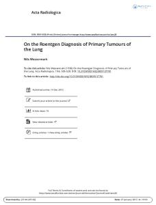
piratory movements from chest wall instability and all patients expressed satisfaction with the cosmetic results. Overall survival of 68% at 5 and 10 years was obtained. The differential figures for chondrosarcoma were 67%, Ewing’s sarcoma 43% and solitary plasmacytoma 59% and Fig. 1). These were the results of radical en-bloc excisions. The patient with the Desmoid is alive at 5 years after resection of a local recurrence. The single patient with Askin’s tumour survived for only 3 years. The 4 patients with chondrosarcoma who did not have radical en-bloc excisions had extensive disease involving the vertebral column (2), the thoracic inlet (1), and the surrounding tissues (1). Of these only 1 patient survived 10 years. Local recurrence did not, however, preclude further excision as shown by 3 patients in this series who have undergone two, two and three excisions, respectively, for chondrosarcoma and are still alive at 10, 15 and 22 years after their initial radical operations. Of the 17 solitary plasmacytomas treated by radical excision, 7 progressed to multiple myeloma with only 1 5-year survivor after chemotherapy. Multiple myeloma developed within 2 years of surgery in all 7 patients. The remaining patient who underwent incisional biopsy and radiotherapy for sternal solitary plasmacytoma survived 2 years. Before 1975, subtotal excision and radiotherapy was performed in 4 patients with Ewing’s tumours with no 5-year survivors. Since then, combined therapy with total excision, radiotherapy and chemotherapy has produced 3-, 5- and 10-year survivors. The use of chemotherapy and radiotherapy did not lead to any local complications in the prosthesis.
4. Discussion Primary chest wall tumours are rare [21]. Limited experience accumulated by each unit may prove to be a considerable challenge in their diagnosis and management. This is compounded by the difficulty in distin-
6 4 0 0 3 1 2 weeks-6 months
Desmoid tumour
Askin’s tumour
1 1 1 0 0 0 6 months
1 1 0 0 0 0 3 months
42 32 6 2 9 7
guishing benign from malignant cartilagenous lesions of the chest wall even by skilled pathologists [20]. Radiological features are not always diagnostic of malignancy except in the presence of cortical destruction and soft tissue swelling [21] Preoperative evaluation of these patients is aimed at diagnosis and assessment of the extent of the tumour. Plain chest X-ray as well as CT scanning is essential. The role of magnetic resonance imaging remains to be evaluated [1]. The clinical features that suggest that a tumour is or has become malignant are recent rapid increase in size, invasion of adjacent structures, and the presence of metastases, particularly to the lungs. However, the majority of malignant neoplasms in this series lacked these features. When visualised together, cortical destruction and soft tissue involvement are the only specific radiological features of malignancy [21]. The fact that a tumour involves only one rib does not necessarily imply that it is benign, since a malignant tumour may be monostotic early in its natural history. The initial biopsy technique to be used remains controversial [1,21]. In general, nonexcisional biopsy of a tumour of the chest wall should be discouraged because of the risk of implantation of tumour tissue along the needle track after aspiration biopsy and in the soft tissues after open biopsy [1,21]. Excisional biopsy remains the preferred mode of treatment of small primary chest wall tumours. For larger resectable chest wall neoplasms (\ 4 cms), incisional biopsy is appropriate provided en-bloc resection of the malignant tumour includes the biopsy site, surrounding skin and the underlying subcutaneous tissue and muscle [1,21]. Advances in pathological techniques, including the use of special stains and immunocytochemistry may help in the eventual diagnosis [20]. It is widely accepted that all tumours of the sternum should be regarded as malignant until proved otherwise [14,21]. Preoperative pulmonary function testing is essential to assess the patient’s fitness for major Radical en-bloc surgical excision with immediate reconstruction is the key to success in the management of
1014
S. Sabanathan et al. / European Journal of Cardio-thoracic Surgery 11 (1997) 1011–1016
Table 3 Skeletal reconstruction Reconstruction
No reconstruction Marlex Mesh Marlex aesh + Methyl methacrylate
Chondrosarcoma
00 12 10
Tumour histology
Total
Solitary plasma- cy- Ewing’s tumour toma
Desmoid tumour
Askin’s tumour
9 4 5
00 00 1
00 1 0
malignant chest wall tumors [1,4,5,9,18 – 21]. In all patient’s, full thickness excision including the skin (if involved), subcutaneous tissue and involved portion of the chest wall should be performed. In this series this was possible in all but nine cases. Recent advances in the techniques of skeletal and musculocutaneous reconstructions have facilitated the treatment of these tumours so that tumour size is not a contraindication to radical excision. The extent of the resection in anterior and lateral chest wall tumours is determined only by what needs to be done to effect local control of the disease. For small defects less than 5 cm in diameter or those located posteriorly under scapula above the fourth rib, the skeletal component can be ignored and the defect closed with only soft tissue. Aggressive wide radical resection must be balanced by durable reconstruction, and this can be achieved with a number of methods. At present this is achieved by the use of synthetic mesh such as Marlex, Prolene or polytetrafluroethylene either alone or reinforced with methyl methacrylate [10,14,16,18]. Bony Reconstruction can be tailored to the extent of resection. Stabilisation of the chest wall obviates the need for prolonged ventilation as patients are able to maintain their pulmonary function postoperatively [12]. Morbidity and mortality rates after chest wall resection and reconstruction in our series and in those recently reported series have been consistently low [10–14]. Familiarity with the techniques of chest wall reconstruction allows satisfactory management not only of primary chest wall tumours but also of breast carcinoma, carcinoma of the lung, radionecrosis and infective and inflammatory conditions. In many cases primary soft tissue closure is possible, as was achieved in 37 of our cases, and this remains the treatment of choice. Muscle is the tissue of choice for soft tissue coverage of full thickness chest wall defects. It can be transposed as muscle alone [18] or as a musculocutaneous flap [8,18]. Today, the common myocutaneous flaps used are lattissimus dorsi, pectoralis major, serratus anterior, rectus abdominis, external oblique and trapezius [11]. Their axial blood supply permit elevation and rotation for a significant distance [8,18]. Omental flaps are usually reserved for infected
4 00 3
13 17 19
and ulcerating wounds or as a second line procedure should muscle transposition fail [2]. For chondrosarcomas, radical en-bloc excision produces better long term survival rates compared to limited local excision [15,22] since chondrosarcomas are not responsive to chemotherapy or radiotherapy [1,4,14,15]. Inadequate excision will lead to local recurrence. This may have serious consequences for the patients as each successive recurrence tends to be more malignant in its behaviour [20]. Even so, repeated excision of recurrences may be still worthwhile, if localised, as illustrated by the 3 patients in this series who are still alive at 10, 15 and 22 years, respectively. Our 5 and 10 years survival of chondrosarcomas of 67% concurs with that of other reported studies [4,14,15]. The most important prognostic factors influencing survival are completeness of the relation and the grade of the tumour [5,14]. The location of the tumour, tumour size, age and sex are not prognostic factors [5]. Solitary plasmacytomas are rare tumours of plasma cell origin. Despite much attention by many investigators, their nature and relation to multiple myeloma remains unclear, since they occasionally remain solitary throughout the life of the patient [21]. In our series, in 38.9% of patients multiple myeloma eventually developed. This incidence is lower than in most reported series [5]. However, the 5 and 10 year survival of 59% is similar to those reported in the literature [4,7,21]. The results reported here suggest that the long term survival in patients with solitary plasmacytoma depends on the subsequent development of multiple myeloma rather than on the method of local control. The surgical resection may prevent systemic dissemination, thus yielding long term survival [4,14,19,21]. However, the role of surgery is controversial. Periodic laboratory screening tests and a careful clinical surveillance are used in the follow-up to achieve early detection of local or systemic recurrences. The role of chemotherapy is not well-defined in an adjuvant setting. In patient’s with Ewing’s tumour, increasing evidence indicates that an integral therapeutic approach consisting of wide surgical excision, irradiation to tumour bed and adjuvant chemotherapy results in a high rate of local tumour control and improved survival
S. Sabanathan et al. / European Journal of Cardio-thoracic Surgery 11 (1997) 1011–1016
1015
Fig. 1. Survival curve (Kaplan-Meter) for primary malignant chest wall tumour patients following radical resection: chondrosarcoma, Solitary plasmacytoma and Ewing’s tumour.
[5,17,21,23]. This is reflected in our own series. Total excision combined with chemotherapy and radiotherapy have produced 3 long term survivors. The 5- and 10-year survival rate of 43% in this series is comparable to that of other reports [5,17,21,23]. Askin’s tumour is a rare disease in the paediatric age group, and even more rare in adults [3,6]. An indistinguishable [t(11;22)] translocation has been demonstrated in Askin’s and Ewing’s tumour [24], perhaps indicating a common origin for these tumours. The treatment for Askin’s tumour is aggressive chemotherapy following radical excision and radiotherapy [6]. The prognosis is often poor as in our case. The Desmoid tumours are of uncertain aetiology. Although these lesions are considered to be benign, they have a tendency to recur locally as in our single case. Therefore, desmoids should be treated by wide surgical excision similar to primary malignant chest wall tumours [18]. Radical en-bloc excision remains the treatment of choice in all primary malignant chest wall neoplasms except large solitary plasmacytomas where incisional biopsy followed by irradiation appears to be the method of preference. In Ewing’s tumours, additional chemotherapy and radiotherapy have to be used. Residual tumour precludes long-term survival and degrades the quality of life by early recurrence. Tumour
size is not a contraindication to radical excision. The extent of surgical excision should only be determined by the amount of tissue needed to be removed to affect adequate tissue clearance, since even large defects can be reconstructed with little functional disturbance. The operation can be performed safely. Prolonged survival in this and other series justifies this aggressive approach.
References [1] Anderson BO, Burt ME. Chest wall neoplasms and their management. Ann. Thorac. Surg. 1994;58:1774 – 81. [2] Arnold PG, Witzke DJ, Irons GB, Woods JE. Use of omental transposition flaps for soft tissue reconstruction. Ann Plastic Surg 1983;11:508 – 12. [3] Askin FB, Rosai J, Sibley RK, Dehner LP, McAlister WH. Malignant small-cell tumour of the thoracopulmonary region in childhood. Cancer 1979;43:2438 – 51. [4] Burt M, Fulton M, Wessner-Dunlap S, Karpeh M, Huvos AG, Bains MS, Martini N, McCormack PM, Rusch VW, Ginsberg RJ. Primary bony and cartilaginous sarcomas of chest wall: results of therapy. Ann Thorac Surg 1992;54:226 – 32. [5] Burt M, Karpeh M, Ukoha O, Bains MS, McCormack PM, Rusch VW, Ginsberg RJ, Martini N. Medical tumors of the chest wall: Solitary plasmacytoma and Ewing’s sarcoma. J Thorac Cardiovasc Surg 1993;105:89 – 96. [6] Cabezali R, Lozano R, Bustamante E, Castiella T, Guemes A, Ramirez Moncada E, Sousa R, Gil I. Askin’s tumour of the
S. Sabanathan et al. / European Journal of Cardio-thoracic Surgery 11 (1997) 1011–1016
1016
[7]
[8] [9] [10]
[11]
[12]
[13] [14]
[15]
[16]
[17]
[18]
[19]
[20] [21] [22]
[23]
[24]
chest wall: a case report in an adult. J Thorac Cardiovasc Surg 1994;107:960 – 2. Corvin J, Lindberg RD. Solitary plasmacytoma of bone vs extramedullary plasmacytoma and their relationship to multiple myeloma. Cancer 1979;43:1007–13. Dingman RO, Argenta LC. Reconstruction of the chest wall. Ann Thorac Surg 1981;32:202–8. Eng J, Sabanathan S, Pradhan GN. Primary sternal tumours. Scand J Cardiovasc Surg 1989;23:289–92. Eng J, Sabanathan S, Mearns AJ. Chest wall reconstruction after resection of primary malignant chest wall tumours. Eur J Cardio-thorac Surg 1990;4:101–4. Eschapasse H, Gaillard J, Henry E, Vassallo, B, Lacheheb, M. Chest wall tumours: Surgical management. In: Grillo H and Eschapasse H, editors, International trends in General thoracic surgery. Volume 2. London:WB Saunders, 1987:292-309. Kroll SS, Walsh G, Ryan B, King RC. Risks and benefits of using Marlex mesh in chest wall reconstruction. Ann Plast Surg 1993;31:303 – 6. Mansour KA, Anderson TM, Hester TR. Sternal resection and reconstruction. Ann Thorac Surg 1993;55:838–43. Martini N, Huvos AG, Burt ME, Hellan RT, Bains MS, McCormack PM, Rusch VW, Weber M, Downey RJ, Ginsberg RJ. Predictors of survival in malignant tumours of the sternum. J Thorac Cardiovasc Surg 1996;111:96–106. McAfee MK, Pairolero PC, Bergstralh EJ, Piehler JL, Unni KK, McLeod RA, Bertnatz PE, Payne WS. Chondrosarcoma of the chest wall. Factors affecting survival.. Ann Thorac Surg 1985;40:535 – 41. McCormack P, Bains MS, Beattie EJ, Martini N. New trends in skeletal reconstruction after resection of chest wall. Ann Thorac Surg 1981;31:45 – 52. Nesbit ME, Gehan EA, Burgert EO, et al. Multimodal therapy for the management of primary, nonmetastatic Ewing’s sarcoma of bone: a long term follow-up of the first intergroup study. J Clin Oncol 1990;8:1664–74. Pairolero PC, Arnold PG. Chest wall tumors: Experience with 100 consecutive patients. J Thorac Cardiovasc Surg 1985;90:367 – 72. Pezzella AT, Fall SM, Pauling FW, Sadler TR. Solitary plasmacytoma of the sternum: Surgical resection with long-term followup. Ann Thorac Surg 1989;48:859–62. Pringle JAS. Pathology of bone tumours. Balliere’s Clin Oncol 1987;1:21 – 63. Sabanathan S, Salama FD, Morgan WE, Harvey JA. Primary chest wall tumours. Ann Thorac Surg 1985;39:4–15. Soyeal O, Walsh GL, Nesbitt JC, McMurtrey MJ, Roth JA, Putnam JB. Resection of sternal tumours: extent, reconstruction and survival. Ann Thorac Surg 1995;60:1353–9. Thomas PRM, Foulkes MA, Gilula LA, Burgert ED, Evans RG, Kissane J, Nesbit ME, Pritchard DJ, Tefft M, Vieetti TJ. Primary Ewing’s sarcoma of the ribs: a report from the intergroup Ewing’s sarcoma study. Cancer 1983;51:1021–7. Whang Peng J, Triche TJ, Knutsen T, Miser J, Kao-Shan S, Tsai S, Israel MA. Cytogenetic characterization of selected small round cell tumours of childhood. Cancer Genet Cytogenet 1986;21:185 – 208.
Appendix A. Conference discussion Mr Moghissi (Hull, England): Thank you. That was a very good presentation and a very interesting series. Two questions. Firstly, how many patients had sternal tumors? The second one concerns the sandwich method for Marlex mesh. Presumably these were the thin Marlex mesh, not heavy Marlex mesh. I think that if you use heavy Marlex mesh, if need be, supported by some sort of strut underneath it, the combination provides a good stable chest wall. My worry about the sandwich method is that the tissue, as you say, will grow beneath the mesh, and above the mesh, but actually you will not have a complete coverage of the mesh. Can you comment on that? Mr Shah: The answer to the first question was we had 7 sternal tumors in the entire series out of 49. And the answer to the second question is, we are very comfortable using double-layered Marlex with methyl methacrylate. And we did not use the thick Marlex; we used two layers of thin Marlex mesh. I think it is a matter of personal choice as to what one should use in the chest wall reconstruction, and we find this technique is very good as compared to using a bar in addition to a thick Marlex mesh. Dr Dosios (Athens, Greece): I would like to express my congratulations on your excellent results. You presented your experience on the surgical treatment of the primary malignant chest wall tumours. My question is: did you have any patient with primary malignant soft tissue tumour of the chest wall? Mr Shah: Only the bony chest wall tumors were included in the series. Dr Dosios: My second question regards the so called infected chest wall tumours. In my series these tumours are encountered mainly in old ladies who had had mastectomy a long time ago and have now developed either a local recurrence with ulceration and infection or actinonecrosis. There are also some patients with primary malignant ulcerated chest wall tumours. After radical resection of these infected tumours we avoid to use Marlex mesh to cover the defect of the chest wall. Instead of Marlex mesh we use myocutoneous flaps with satisfactory results. I would like to hear of your experience and your comments on this kind of management. Mr Shah: We have excluded in this series all the secondary tumors of the chest wall, which means cancer of the breast or cancer of the lung which is invading the chest wall. So those tumors have been completely excluded. We have just included primary malignant chest wall tumors. But yes, there is anxiety to avoid using foreign material in the cases of infected chest wall tumors, and we have done a few cases of this. What we do in these cases is we still use the sandwich prosthesis but we cover with omentum. When we have to resect the muscle, we never leave the prosthesis just underneath the skin. In the literature, there is quite a bit of argument about this. And if the foreign material, the reconstruction, is covered with well-vascularized tissue, then the chances of infection are much, much less, because omentum is a very good vascular pedicle. It will take care of the infection, it will cover the mesh properly, and we shouldn’t have too many problems. The cosmetic result will be poor if you don’t use the mesh, but in the elderly age group, if you are perhaps not worried about the cosmesis in a patient with recurrent carcinoma, then you can avoid the mesh and just reconstruct using soft tissues.
.
.