Rheumatology 2015;54:827835 doi:10.1093/rheumatology/keu409 Advance Access publication 13 October 2014
RHEUMATOLOGY
Original article Prevalence of knee pain and knee OA in southern Sweden and the proportion that seeks medical care Aleksandra Turkiewicz1,2, Maria Gerhardsson de Verdier3, Gunnar Engstro¨m4, Peter M. Nilsson5, Carl Mellstro¨m3, L. Stefan Lohmander1,6,7 and Martin Englund1,2,8 Abstract Objective. The aim of this study was to estimate the prevalence of frequent knee pain in radiographic, symptomatic and clinically defined knee OA in middle-aged and elderly patients and the proportion that seeks medical care. Methods. In 2007 a random sample of 10 000 56- to 84-year-old residents of Malmo¨, Sweden, were questioned about knee pain. We classified subjects reporting knee pain with a duration of at least 4 weeks as having frequent knee pain. A random sample of 1300 individuals with frequent knee pain and 650 without were invited for assessment by the ACR clinical knee OA criteria and for bilateral weight-bearing knee radiography. We considered a KellgrenLawrence grade 52 as radiographic knee OA and that in combination with frequent knee pain as symptomatic knee OA. By linkage with the Ska˚ne Healthcare Register, we determined the proportion of subjects that had consulted for knee OA or pain.
Conclusion. Fifteen per cent of middle-aged or elderly individuals have knee OA and symptoms. About one in three of those do not consult a physician. Inefficient care of OA and self-coping may be an explanation. Key words: knee osteoarthritis, knee pain, prevalence, radiography.
Introduction OA is one of the most common causes of pain and functional impairment among the elderly and adults of working
1 Department of Orthopaedics, Clinical Sciences Lund, Lund University, 2Epidemiology and Register Centre South, Ska˚ne University Hospital, Lund, 3Astra Zeneca R&D Mo¨lndal, Mo¨lndal, 4 Cardiovascular Epidemiology, Clinical Sciences Malmo¨, Lund University, 5Department of Clinical Sciences, Ska˚ne University Hospital, Malmo¨, 6Research Unit for Musculoskeletal Function and Physiotherapy, 7Department of Orthopedics and Traumatology, University of Southern Denmark, Odense, Denmark and 8Clinical Epidemiology Research and Training Unit, Boston University School of Medicine, Boston, MA, USA
Submitted 5 February 2014; revised version accepted 20 August 2014 Correspondence to: Aleksandra Turkiewicz, Ska˚nes Universitetssjukhus, Klinikgatan 22, 22185 Lund, Sweden. E-mail:
[email protected]
age [18]. OA is mainly a clinical diagnosis, but findings on radiographs, including joint space narrowing, subchondral sclerosis and osteophyte formation, are commonly used for epidemiological study purposes, even though there is often a discrepancy between radiographic changes and symptoms [9]. Asymptomatic radiographic OA is common and so is knee pain due to knee OA not yet detectable on plain radiographs, as these changes may appear relatively late in the natural course of the disease. Hence, to obtain estimates of the occurrence of OA in society is challenging, as the definitions are plentiful and often ambiguous [10, 11]. The knee is one of the most common sites affected. The first presenting symptom of knee OA is often pain in the joint, and in patients >55 years of age, knee pain is often attributable to OA [12]. The disease is expected to
! The Author 2014. Published by Oxford University Press on behalf of the British Society for Rheumatology. All rights reserved. For Permissions, please email:
[email protected]
CLINICAL SCIENCE
Results. The 10 000 subjects had a mean age of 70 years (S.D. 7.6), a mean BMI of 27.1 kg/m2 and 62% were women. The prevalence of frequent knee pain was 25.1% (95% CI 24.1, 26.1), higher in women and similar across age groups. The prevalence of radiographic knee OA was 25.4% while 15.4% had either symptomatic or clinically defined knee OA. Of these, 68.9% consulted a physician for knee OA or pain during 200411.
Aleksandra Turkiewicz et al.
become increasingly common due to ageing and the increasingly obese populations in many countries [13]. Thus updated estimates reflecting the beginning of the 21st century are needed. Evidence from morbidity surveys in primary care from England and Wales confirms that OA and joint pain contribute substantially to the workload of general practice [14]. The proportion of older adults with knee pain that consult a physician varies from 15 to 50% depending on the knee pain definition and time period studied [12, 1517]. However, there is also a substantial lack of information on what proportion of subjects with symptomatic or clinically defined knee OA consults a physician. We used a combination of questionnaire, clinical examination, interview by trained personnel and radiography in a random sample of residents of the Malmo¨ region in southern Sweden to determine the current prevalence of knee pain and radiographic and symptomatic knee OA. In addition, using health care register data covering all levels of health care in the entire region, we assessed the proportion of subjects with symptomatic or clinically defined knee OA who consulted a physician for their knee symptoms.
Methods Study sample The Malmo¨ OA (MOA) study was carried out between 2007 and 2008 and originated from the Malmo¨ Diet and Cancer Study (MDCS) cohort established between 1991 and 1996 [1822]. The first part of the MOA study consisted of a knee pain questionnaire sent to a random sample of 10 000 subjects from the MDCS cohort who were still alive and resident in the Malmo¨ area at the beginning of 2007. Respondents answered a question about whether they had knee pain during the previous 12 months and its duration (3 months). We classified subjects with pain in one or both knees in the past 12 months and duration at least 4 weeks as having frequent knee pain. In the second part of the study a random sample of 1300 subjects with frequent knee pain and 650 subjects without were invited to a clinical visit and radiographic examination (Fig. 1). At the clinical visit the trained study nurse performed a physical examination where weight and height were measured, subjects were asked if they had had previous knee arthroplasty and a questionnaire was completed assessing, among other things, pain in the whole body and its location. Data on education and BMI from the MDCS examination in 199196 were available for the whole study sample. The study was approved by the Regional Ethics Committee in Lund and informed consent was obtained from all participants in accordance with the Declaration of Helsinki.
Knee pain As a main knee pain outcome measure, we used the question from the first part of the study about frequent knee pain. In addition, we used the knee pain question
828
from the ACR clinical criteria at the clinical assessment, i.e. have you had pain in the knee on most days of the previous month? This question was used to determine clinically defined knee OA.
Knee OA Both knees were radiographed in a weight-bearing and semi-flexed position (knees in 1015 degrees of flexion) using a posterioranterior beam direction (film focus distance 110 cm, 60 kV and 10 mA) with the aid of fluoroscopy to optimally align the tibia plateau. We also obtained patella axial images with knees in 3040 degrees of flexion. An independent senior radiologist specialized in musculoskeletal conditions who was blinded to the clinical data assessed joint space narrowing and osteophytes according to the atlas from the Osteoarthritis Research Society International [23]. We classified a knee as having radiographic knee OA if one or more of the following criteria were fulfilled in either the medial, lateral or patellofemoral compartment: joint space narrowing grade 2 or worse, the sum of marginal osteophyte grades in the same compartment 2 or worse, joint space narrowing grade 1 and osteophyte grade 1 in the same compartment [approximating a KellgrenLawrence (KL) grade 52] [24]. We considered those with radiographic knee OA and frequent knee pain as having symptomatic knee OA. Clinically defined knee OA status was determined by the study nurse blinded to radiographic status using the ACR clinical criteria according to the recursive positioning method [25]. Subjects who had a history of knee replacement or osteotomy were considered as fulfilling all three knee OA definitions.
Health care consultations The Ska˚ne Healthcare Register (SHR) contains information about every health care visit made in the region and includes data on the health care provider, profession (physician, physical therapist, etc.), type of visit (primary/ specialist care, in- or outpatient visit, clinic, etc.) and date of visit. The register contains the publicly practising physicians’ diagnostic codes according to the International Classification of Diseases, 10th revision (ICD-10) system. Using the MOA subjects’ unique personal identification number, we retrieved data on all doctor visits for MOA subjects for 200411. We identified subjects that received a diagnosis of knee OA (ICD-10 code M17) or pain in a joint (joint unspecified, ICD-10 code M25.5) during that time.
Statistical analysis We used weighting to adjust for a possible selection bias that could arise from non-response that arose in the first or second part or in the MOA study [26]. A logistic regression model with sex, age on 1 January 2007 and BMI as well as the highest education level measured at the baseline MDCS examination as covariates was used to estimate the probability of response in the survey, and the reciprocal was used as a weight. Covariates in models for willingness to participate as well as for attendance at
www.rheumatology.oxfordjournals.org
Knee OA prevalence and consultation in Sweden
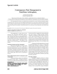
FIG. 1 Study sample
a
Frequent knee pain: knee pain in the last 12 months with a duration of at least 4 weeks. bNo frequent knee pain: no knee pain or knee pain with a duration of 98% of cases, we used only visits to those clinics in the model. Of all visits, 45% were made as private care and thus had no ICD-10 code assigned. In public care, 9% of doctor visits had a missing ICD-10 code. A multivariate normal model with random intercept was used to impute 20 datasets. Variables included in the model were diagnosis, age, sex, clinic, BMI, if having knee pain, if having radiographic knee OA, if having symptomatic knee OA, if having clinically defined knee OA, year of health care visit, if visited a physiotherapist and income. The correlation between visits made by the same person was accounted for by including the random effect in the model [30]. The imputed values were rounded using cutoff values determined by simulation [31]. We used STATA 12.0 (StataCorp, College Station, TX, USA) and R version 2.15.2, package pan (R Project for Statistical Computing, Vienna, Austria), for the analyses.
FIG. 2 The 2008 prevalence of frequent knee pain and knee OA in Swedish adults 5684 years old
Results
Frequent knee pain: knee pain in one or both knees in the last 12 months with a duration of at least 4 weeks; radiographic knee OA: changes on radiographs approximating KellgrenLawrence grade 52; clinically defined knee OA: knee OA according to the ACR clinical criteria, recursive positioning method; symptomatic knee OA: frequent knee pain as defined above in combination with radiographic knee OA as defined above.
Study sample The 10 000 MOA subjects were 5684 years old [mean 70 years (S.D. 7.6)], 62% were women and mean BMI was 27.2 kg/m2 (S.D. 5). The response rate to the mailed survey was 77.4%, and 72.0% of the responders were willing to participate in the clinical examination. Further, 1527 of 1950 sampled subjects (78.3%) attended the clinical visit. Of those, 42 subjects had missing information from the radiographic examination (41 did not attend and 1 could not participate due to Parkinson’s disease) (Fig. 1).
Prevalence of knee pain The prevalence of frequent knee pain in one or both knees during the last 12 months was 25.1% [(95% CI 24.1, 26.1), 20.8% in men and 27.7% in women]. The prevalence of knee pain on most days of the previous month was 20.3% [(95% CI 18.2, 22.6) 17.9% in men and 21.7% in women]. The prevalence remained stable across age groups (Fig. 2).
Prevalence of knee OA The prevalence of radiographic knee OA was 25.6% [(95% CI 22.7, 28.6) 24.3% in men and 26.4% in women] using the definition approximating a KL grade of 52 in the medial, lateral or patellofemoral compartment (Table 1). The prevalence increased with increasing age and was 21.0% when the patellofemoral compartment was excluded (Fig. 2). The prevalence of symptomatic knee OA was 10.5% [(95% CI 9.8, 11.3) 9.7% in men and 11.0% in women]. The prevalence of clinically defined knee OA was 9.0% [(95% CI 7.9, 10.3) 8.0% in men and 9.6% in women] (Table 1). The prevalence of those fulfilling either the clinically defined knee OA criteria or symptomatic knee OA definition was 15.4% [(95% CI 14.2, 16.7) 13.6% in men and 16.5% in women].
830
Relationship between knee pain and radiographic knee OA The prevalence of frequent knee pain in subjects with radiographic knee OA was 41.5% (95% CI 36.5, 46.7), similar in men and women (Fig. 3). The prevalence ratio of frequent knee pain in subjects with and without radiographic knee OA, adjusted for age, sex, current BMI and having pain other than knee pain, was 2.3 (95% CI 1.9, 2.7). The prevalence of radiographic knee OA in the population with frequent knee pain was 43.2% [(95% CI 40.1, 46.4) 47.5% in men and 41.3% in women]. In the study sample, 11.8% of subjects reported frequent knee pain but did not fulfil OA criteria, for either clinical ACR or radiographic knee OA.
Health care consultations Between 2004 and 2011, 74.7% (95% CI 70.0, 79.3) of the subjects classified as having symptomatic knee OA consulted a physician and received a diagnosis of either knee OA or pain in a joint, while 63.0% (95% CI 57.8, 68.2) were diagnosed with knee OA specifically. The corresponding percentages for the subjects fulfilling the clinical knee OA criteria were 66.8% (95% CI 59.1, 74.6) and 49.9% (95% CI 41.8, 58.1) (Table 2). Of those with either symptomatic or clinically defined knee OA, 53.3% (95% CI 47.6, 58.9) consulted a physician for knee OA during an 8-year time period, while 68.9% (95% CI 63.8, 74.0) consulted for either knee OA or pain in a joint. Generally the proportion that consulted was similar in men and women, irrespective
www.rheumatology.oxfordjournals.org
Knee OA prevalence and consultation in Sweden
TABLE 1 The 2008 prevalence (%) of knee pain and knee OA in a population aged 5486 in southern Sweden Age group and sex 5664 years
Number Frequent knee paina Radiographic knee OAb Symptomatic knee OAc Clinically defined knee OAd Symptomatic or clinically defined knee OAe
6574 years
7584 years
Men
Women
All
Men
Women
All
Men
Women
All
130 21.1 18.8 9.6 10.4 14.6
381 28.2 16.7 8.1 9.8 14.4
511 26.2 17.3 8.5 10.0 14.5
256 21.2 19.8 10.6 8.0 14.1
346 26.1 20.3 9.7 9.7 15.7
602 24.0 20.1 10.1 9.0 15.1
164 20.3 31.7 8.9 6.7 12.5
250 29.0 42.5 15.5 9.3 19.6
414 25.4 40.0 12.7 8.3 16.6
a Frequent knee pain: knee pain in one or both knees in the last 12 months with duration at least 4 weeks. bRadiographic knee OA: changes on radiographs approximating KellgrenLawrence grade 52. cSymptomatic knee OA: frequent knee pain as defined above in combination with radiographic knee OA as defined above. dClinically defined knee OA: knee OA according to the ACR clinical criteria, recursive positioning method. eSyptomatic or clinically defined knee OA: symptomatic knee OA as defined above and/or clinically defined knee OA as defined above.
FIG. 3 The 2008 prevalence and overlap of frequent knee pain and knee OA in Swedish adults 5684 years old
Numbers are percentages describing the prevalence of knee OA or knee pain and their combinations. Radiographic knee OA: changes on radiographs approximating KellgrenLawrence grade 52; clinically defined knee OA: knee OA according to the ACR clinical criteria; frequent knee pain: knee pain in one or both knees in the last 12 months with duration at least 4 weeks. of the OA definition used (Table 2). In 98% of consultations the diagnosis of knee OA or pain in a joint was made by a primary care provider or by a specialist (or physician under specialty training) in orthopaedics or emergency medicine.
Discussion We found that in the Swedish population aged 5684 years, one in four reported frequent knee pain and the
www.rheumatology.oxfordjournals.org
same fraction had radiographic knee OA approximating KL grade 52. The criteria for symptomatic OA or clinically defined (according to ACR criteria) OA were fulfilled by 15.4%, of which 69% had consulted a physician for knee complaints during an 8-year time frame. In 1982, some 25 years prior to our study, a populationbased study from the city of Gothenburg, Sweden, reported a prevalence of knee complaints (pain, stiffness or swelling) in 79-year-olds (25% in women and 11% in men) that was lower than the prevalence of frequent knee pain in those 7880 years of age in our study (30% in women and 14% in men) [32]. Methodological differences, non-response and sampling bias and different knee pain questions make comparisons challenging. While the prevalence of radiographic knee OA in our study is in line with numbers from a Danish study in a population aged 5579 years, it is much lower than the prevalence in the USA [3336]. The higher prevalence of obesity in the USA than in Sweden is a probable explanation, as well as the different age and ethnic structure of the population. For instance, in our sample the mean BMI was 27, compared with 31 reported from Johnston County, NC, USA, study participants of a similar age [37]. In spite of the differences in the prevalence of radiographic knee OA, the prevalence of symptomatic knee OA is similar in our study and in the studies from the USA. In the age group 6079 years the US prevalence ranges from 9.3% to 11.8%, with our estimate in the middle of this range (10.9%) [35, 36]. This holds even for sex-specific estimates, with women having a slightly higher prevalence than men. The prevalence of frequent knee pain was higher in our study than in studies from the USA, which may be one explanation of the similar prevalence of symptomatic knee OA. Our estimate of the prevalence of frequent knee pain, 25%, is in line with estimates from England for a population aged 555, while studies from the USA and the Netherlands reported lower numbers [12, 3840]. However, slightly different knee pain questions, composition of study groups and methods of data collection may explain the variation in the prevalence of knee pain.
831
Aleksandra Turkiewicz et al.
TABLE 2 The percentage of 2008 prevalent subjects with knee OA who received a diagnosis of knee OA or pain in a joint from a physician during 200411
OA definition All
Men
Women
Radiographic knee OAa Clinically defined knee OAb Symptomatic knee OAc Symptomatic or clinically defined knee OAd Radiographic knee OAa Clinically defined knee OAb Symptomatic knee OAc Symptomatic or clinically defined knee OAd Radiographic knee OAa Clinically defined knee OAb Symptomatic knee OAc Symptomatic or clinically defined knee OAd
Diagnosis of knee OA or pain in a joint, % (95% CI) 56.8 66.8 74.7 68.9 60.8 67.2 76.1 71.3 54.5 66.7 73.9 67.7
(49.1, (59.1, (70.0, (63.8, (48.6, (56.4, (68.4, (63.8, (44.7, (56.5, (67.9, (61.0,
64.5) 74.6) 79.3) 74.0) 72.9) 78.0) 83.8) 78.8) 64.4) 76.8) 79.9) 74.4)
Diagnosis of knee OA, % (95% CI) 43.4 49.9 63.0 53.3 50.8 51.7 66.3 58.5 39.4 49.1 61.2 50.7
(36.5, (41.8, (57.8, (47.6, (39.2, (38.8, (57.5, (49.5, (30.5, (38.6, (54.6, (43.4,
50.4) 58.1) 68.2) 58.9) 62.4) 64.6) 75.2) 67.6) 48.2) 59.6) 67.8) 58.1)
Diagnosis of pain in a joint, % (95% CI) 31.3 39.7 40.8 40.1 30.9 41.4 39.3 39.4 31.6 38.9 41.6 40.5
(23.7, (30.9, (35.3, (34.3, (20.0, (27.6, (30.1, (30.2, (21.6, (27.4, (34.6, (32.8,
38.9) 48.4) 46.4) 45.9) 41.8) 55.3) 48.6) 48.6) 41.5) 50.3) 48.6) 48.1)
a
Radiographic knee OA: changes on radiographs approximating KellgrenLawrence grade 52. bClinically defined knee OA: OA according to the ACR clinical criteria, recursive positioning method. cSymptomatic knee OA: knee pain of duration at least 4 weeks in the last 12 months in combination with radiographic OA as defined above. dSypomtomatic or clinically defined knee OA: symptomatic knee OA as defined above and/or clinically defined knee OA as defined above.
The estimated prevalence of symptomatic knee OA and clinically defined knee OA according to ACR criteria were similar, but the overlap between subjects fulfilling those two definitions was relatively low. The low overlap was also found by Peat et al. [41] in patients 550 years of age and with knee pain during the previous 12 months. In this group the prevalence of symptomatic knee OA was 32.9% and the prevalence of knee OA according to the ACR clinical criteria was 30.2%. Our estimates in subjects with knee pain in the last 12 months were lower, 20.4% and 18.5%, respectively. It is somewhat counterintuitive that the prevalence of ACR-defined OA did not increase with age, and this may be explained by the fact that crepitus and stiffness was less frequently found and reported by the elderly in our cohort, perhaps part of adaption (i.e. these symptoms are increasingly considered as normal and are not reported even if they are present). During an 8-year period (200411) only two of three patients with clinically defined knee OA or symptomatic knee OA consulted health care and received a diagnosis of knee OA or pain in a joint. In a study of the performance of ACR clinical criteria in the general population, Peat et al. [41] found that among subjects with knee pain and fulfilling the ACR clinical criteria for knee OA, 29.8% consulted for knee OA or knee pain during the 18 months preceding the study examination, while 37.1% of those with symptomatic knee OA consulted during the same time period. In the corresponding MOA study sample subgroup (knee pain during the past year, consultations within 18 months before the first MOA survey), 27.8% of subjects fulfilling the clinical ACR knee OA criteria and 33.5% of those with symptomatic knee OA consulted for knee OA or pain in a joint. Both results suggest that only a minority of knee OA patients with knee pain actually consult health care [3]. Self-management or coping strategies as well as
832
over-the-counter pain treatments may partly explain this. Another explanation may be the perception of knee pain in the general population or among physicians. Older people may often view chronic joint pain and other symptoms of OA as a part of normal ageing and are more likely to consult when symptoms come on suddenly and severely or disturb sleep or when they have mobility problems [6, 42]. Both international and Swedish national guidelines for treatment of OA of the knee first recommend non-drug treatments. However, only a minority of knee OA patients are referred to a physical therapist [43]. In one questionnaire, only about half of UK-based physical therapists agreed that knee problems are improved by exercise, and adherence was seen as the patient’s responsibility [44]. On the other hand, patients delay seeking medical care for musculoskeletal pain and many do not take the treatment and/or lack information about their disease [45]. Physical activity guidelines and recommended daily steps are followed by less than half of the people with knee OA [46]. Fewer than half of obese patients with knee OA in a study from the USA have been advised by a health care professional to lose weight, and people with knee pain continue to have persistent problems regardless of whether they consult a physician or not [4, 47]. Better management of patients with OA in primary care and improved awareness of non-drug treatments in society could result in more symptomatic subjects seeking health care [48, 49]. Non-response is common in surveys and may result in selection bias if participants are systematically different from non-participants [50]. The non-response rate in the MOA postal questionnaire was relatively low, at 22.6%, as was the dropout rate from the MOA clinical examination (21.7% of all invited). The baseline variables available for the whole study sample were age, sex, BMI and education level. All of these were associated with non-response and thus were
www.rheumatology.oxfordjournals.org
Knee OA prevalence and consultation in Sweden
used in the calculation of weights which, together with the sampling weights, were used in the analyses to account for possible selection bias. However, the results might still be affected by selection bias due to factors we could not account for (such as knee pain status in non-responders in part I of the study). Using multiple imputation we accounted for the missing diagnostic codes in the SHR, but we cannot rule out that the missing data in the MOA study or in the register depended on unobserved factors that would introduce bias into our results. However, the majority of the missing diagnostic codes in the SHR were due to the administrative routines, as the codes from private health care providers are not forwarded to the register. In Sweden, both types of health care are equally accessible and financed through the same taxbased system, thus missing data from private providers can be considered as missing and random and will not introduce bias into our estimates. The 10 000 subjects in the MOA study were a random sample from the MDCS cohort. This cohort has been shown to have slightly lower mortality rates than the background population, suggesting a healthy selection bias in persons willing to participate [18]. Between 2004 and 2006 (3 years preceding the MOA examination), 7.3% of the 10 000 MOA subjects consulted health care and received a diagnosis of knee OA, while the corresponding number for the whole Ska˚ne population aged 5684 years was 6.8%, which suggests that with respect to knee OA, the MOA study sample is fairly representative of the background population. For the definition of symptomatic knee OA we required knee pain for at least 4 weeks to exclude persons with milder symptoms. However, for 37 persons we did not have information of whether the pain was in the knee with radiographic changes. In conclusion, we found that in the first decade of the 21st century the prevalence of symptomatic or clinically defined knee OA in the Swedish population aged 5684 years is 15.4%. Of these, two of three saw a physician during an 8-year time period and were diagnosed with knee pain or knee OA. The prevalence of radiographic knee OA, irrespective of symptoms, was 25.4%. Our findings show that there is a large group of people with symptomatic knee OA not seeking health care. This group could potentially benefit from OA education and training. Rheumatology key messages Knee OA prevalence in the middle-aged and elderly in Sweden ranges from 9% to 25%. . One in three with knee OA and symptoms do not consult a physician. . Inefficient care of OA and self-coping strategies may be an explanation for not consulting a physician. .
Acknowledgements The Malmo¨ Osteoarthritis Study was funded by AstraZeneca. We thank the staff at the Clinical Research Department for clinical examinations in Malmo¨ and Charlotte Bergknut for extracting data from the Ska˚ne Healthcare Register. This work was also supported by
www.rheumatology.oxfordjournals.org
grants to M.E. from the Swedish Research Council, the King Gustaf V 80-year Birthday Fund, the Kock Foundations, the Faculty of Medicine Lund University and Region Ska˚ne. Funding: AstraZeneca was the sponsor of this study. AstraZeneca was involved in the overall MOA study design and approved the manuscript for publication, but was not involved in the collection, analysis and interpretation of data, in the writing of the report or in the decision to submit the article for publication. The other funders had no role in the study design, in the collection, analysis and interpretation of data, in the writing of the article or in the decision to submit the article for publication. Disclosure statement: M.G.d.V. has been employed AstraZeneca and is an owner of AstraZeneca shares part of the bonus system. C.M. is an employee AstraZeneca. G.E. was formerly employed AstraZeneca R&D. All other authors have declared conflicts of interest.
by as of by no
References 1 Vos T, Flaxman AD, Naghavi M et al. Years lived with disability (YLDs) for 1160 sequelae of 289 diseases and injuries 19902010: a systematic analysis for the Global Burden of Disease Study 2010. Lancet 2012;380: 216396. 2 Lawrence RC, Felson DT, Helmick CG et al. Estimates of the prevalence of arthritis and other rheumatic conditions in the United States. Part II. Arthritis Rheum 2008;58: 2635. 3 Wilson MG, Michet CJ Jr, Ilstrup DM, Melton LJ III. Idiopathic symptomatic osteoarthritis of the hip and knee: a population-based incidence study. Mayo Clin Proc 1990;65:121421. 4 Lohmander S. [Osteoarthritis is frequent, very frequent. What can we do?]. Lakartidningen 2002;99:43424. 5 Carman WJ, Sowers M, Hawthorne VM, Weissfeld LA. Obesity as a risk factor for osteoarthritis of the hand and wrist: a prospective study. Am J Epidemiol 1994;139: 11929. 6 Dieppe P, Basler HD, Chard J et al. Knee replacement surgery for osteoarthritis: effectiveness, practice variations, indications and possible determinants of utilization. Rheumatology 1999;38:7383. 7 Hochberg MC. Mortality in osteoarthritis. Clin Exp Rheumatol 2008;26(Suppl 51):S1204. 8 Thomas E, Peat G, Croft P. Defining and mapping the person with osteoarthritis for population studies and public health. Rheumatology 2014;53:33845. 9 Schellevis FG, van der Velden J, van de Lisdonk E, van Eijk JT, van Weel C. Comorbidity of chronic diseases in general practice. J Clin Epidemiol 1993;46:46973. 10 Pereira D, Peleteiro B, Araujo J et al. The effect of osteoarthritis definition on prevalence and incidence estimates: a systematic review. Osteoarthritis Cartilage 2011;19: 127085.
833
Aleksandra Turkiewicz et al.
11 Felson DT, McAlindon TE, Anderson JJ et al. Defining radiographic osteoarthritis for the whole knee. Osteoarthritis Cartilage 1997;5:24150. 12 Peat G, McCarney R, Croft P. Knee pain and osteoarthritis in older adults: a review of community burden and current use of primary health care. Ann Rheum Dis 2001;60:917. 13 Turkiewicz A, Petersson IF, Bjork J, Dahlberg LE, Englund M. Prognosis for the year 2030: the consultation prevalence of osteoarthritis in Sweden may increase by 50 [abstract]. Arthritis Rheum 2012;64(Suppl 10):914. 14 McCormick A, Fleming D, Charlton J. Morbidity statistics from general practice: fourth national study 19911992. In: McCormick A, Fleming D, Charlton J, eds. Surveys OoPCa. London: HMSO, 1995. 15 Thorstensson CA, Gooberman-Hill R, Adamson J, Williams S, Dieppe P. Help-seeking behaviour among people living with chronic hip or knee pain in the community. BMC Musculoskel Disord 2009;10:153. 16 Jordan K, Jinks C, Croft P. A prospective study of the consulting behaviour of older people with knee pain. Br J Gen Pract 2006;56:26976. 17 Bedson J, Mottram S, Thomas E, Peat G. Knee pain and osteoarthritis in the general population: what influences patients to consult? Fam Pract 2007;24:44353. 18 Manjer J, Carlsson S, Elmstahl S et al. The Malmo Diet and Cancer Study: representativity, cancer incidence and mortality in participants and non-participants. Eur J Cancer Prev 2001;10:48999. 19 Rosdahl L, Lamm C, Engstro¨m G et al. Generic and disease-specific health-related quality of life—a Swedish population-based study on chronic knee pain and knee osteoarthritis. Osteoarthritis Cartilage 2010;18(Suppl 2): 163S4S. 20 Lamm C, Rosdahl L, Rollof J et al. Comparison of instruments for measuring health-related quality of life—a population-based study of chronic knee pain and knee osteoarthritis. Osteoarthritis Cartilage 2010;18(Suppl 2): 162S3S. 21 Mellstro¨m C, Rosdahl L, Engstro¨m G et al. The costs associated with chronic knee pain and knee osteoarthritis—a population-based study from Sweden. Osteoarthritis Cartilage 2010;18(Suppl 2):S160.
26 Little RJ, Vartivarian S. On weighting the rates in nonresponse weights. Stat Med 2003;22:158999. 27 Sellam J, Berenbaum F. The role of synovitis in pathophysiology and clinical symptoms of osteoarthritis. Nat Rev Rheumatol 2010;6:62535. 28 Rubin DB. Introduction. Multiple Imputation for Nonresponse in Surveys. New York, NY, USA: John Wiley & Sons, 1987:157. 29 Turkiewicz A, Petersson I, Bjo¨rk J, Dahlberg LE, Englund M. Consultation prevalence of osteoarthritis in southern Sweden [abstract]. Arthritis Rheum 2012; 64(Suppl):S3967. 30 Schafer JL, Yucel RM. Computational strategies for multivariate linear mixed-effects models with missing values. J Comput Graph Stat 2002;11:43757. 31 Yucel RM, He Y, Zaslavsky AM. Using calibration to improve rounding in imputation. Am Stat 2008;62:1259. 32 Bagge E, Bjelle A, Eden S, Svanborg A. A longitudinal study of the occurrence of joint complaints in elderly people. Age Ageing 1992;21:1607. 33 van Saase JL, van Romunde LK, Cats A, Vandenbroucke JP, Valkenburg HA. Epidemiology of osteoarthritis: Zoetermeer survey. Comparison of radiological osteoarthritis in a Dutch population with that in 10 other populations. Ann Rheum Dis 1989;48: 27180. 34 Jordan JM, Helmick CG, Renner JB et al. Prevalence of knee symptoms and radiographic and symptomatic knee osteoarthritis in African Americans and Caucasians: the Johnston County Osteoarthritis Project. J Rheumatol 2007;34:17280. 35 Dillon CF, Rasch EK, Gu Q, Hirsch R. Prevalence of knee osteoarthritis in the United States: arthritis data from the Third National Health and Nutrition Examination Survey 199194. J Rheumatol 2006;33:22719. 36 Felson DT, Naimark A, Anderson J et al. The prevalence of knee osteoarthritis in the elderly. The Framingham Osteoarthritis Study. Arthritis Rheum 1987;30:9148. 37 Nelson AE, Golightly YM, Renner JB et al. Brief report: differences in multijoint symptomatic osteoarthritis phenotypes by race and sex: the Johnston County Osteoarthritis Project. Arthritis Rheum 2013;65: 3737.
22 Lohmander LS, Gerhardsson de Verdier M, Rollof J, Nilsson PM, Engstrom G. Incidence of severe knee and hip osteoarthritis in relation to different measures of body mass: a population-based prospective cohort study. Ann Rheum Dis 2009;68:4906.
38 Odding E, Valkenburg HA, Algra D et al. Associations of radiological osteoarthritis of the hip and knee with locomotor disability in the Rotterdam Study. Ann Rheum Dis 1998;57:2038.
23 Altman RD, Hochberg M, Murphy WA Jr, Wolfe F, Lequesne M. Atlas of individual radiographic features in osteoarthritis. Osteoarthritis Cartilage 1995;3(Suppl A): 370.
39 Claessens AA, Schouten JS, van den Ouweland FA, Valkenburg HA. Do clinical findings associate with radiographic osteoarthritis of the knee? Ann Rheum Dis 1990; 49:7714.
24 Englund M, Roos EM, Lohmander LS. Impact of type of meniscal tear on radiographic and symptomatic knee osteoarthritis: a sixteen-year followup of meniscectomy with matched controls. Arthritis Rheum 2003;48: 217887.
40 Anderson JJ, Felson DT. Factors associated with osteoarthritis of the knee in the first national Health and Nutrition Examination Survey (HANES I). Evidence for an association with overweight, race, and physical demands of work. Am J Epidemiol 1988;128:17989.
25 Altman R, Asch E, Bloch D et al. Development of criteria for the classification and reporting of osteoarthritis: classification of osteoarthritis of the knee. Arthritis Rheum 1986;29:103949.
41 Peat G, Thomas E, Duncan R et al. Clinical classification criteria for knee osteoarthritis: performance in the general population and primary care. Ann Rheum Dis 2006;65: 13637.
834
www.rheumatology.oxfordjournals.org
Knee OA prevalence and consultation in Sweden
42 Menz HB, Jordan KP, Roddy E, Croft PR. Musculoskeletal foot problems in primary care: what influences older people to consult? Rheumatology 2010;49:210916. 43 Cottrell E, Roddy E, Foster NE. The attitudes, beliefs and behaviours of GPs regarding exercise for chronic knee pain: a systematic review. BMC Fam Pract 2010;11:4. 44 Holden MA, Nicholls EE, Young J, Hay EM, Foster NE. UKbased physical therapists’ attitudes and beliefs regarding exercise and knee osteoarthritis: findings from a mixedmethods study. Arthritis Rheum 2009;61:151121.
osteoarthritis: population based cohort study. BMJ 2011; 342:d1165. 47 Felson DT, Zhang Y. An update on the epidemiology of knee and hip osteoarthritis with a view to prevention. Arthritis Rheum 1998;41:134355. 48 Hunter DJ, Lo GH. The management of osteoarthritis: an overview and call to appropriate conservative treatment. Med Clin North Am 2009;93:12743, xi.
45 Woolf AD, Zeidler H, Haglund U et al. Musculoskeletal pain in Europe: its impact and a comparison of population and medical perceptions of treatment in eight European countries. Ann Rheum Dis 2004;63:3427.
49 Smink AJ, van den Ende CH, Vliet Vlieland TP et al. ‘‘Beating osteoARThritis’’: development of a stepped care strategy to optimize utilization and timing of non-surgical treatment modalities for patients with hip or knee osteoarthritis. Clin Rheumatol 2011;30: 16239.
46 Nuesch E, Dieppe P, Reichenbach S et al. All cause and disease specific mortality in patients with knee or hip
50 Etter J-F, Perneger TV. Analysis of non-response bias in a mailed health survey. J Clin Epidemiol 1997;50:11238.
www.rheumatology.oxfordjournals.org
835