PAAVO-ILARI KUIKKA
Epidemiology and Magnetic Resonance Imaging-Based Diagnostics of Knee Injuries and Anterior Knee Pain in Young Adults
ACADEMIC DISSERTATION To be presented, with the permission of the board of the School of Medicine of the University of Tampere, for public discussion in the Auditorium of Finn-Medi 5, Biokatu 12, Tampere, on August 23rd, 2012, at 12 o’clock.
UNIVERSITY OF TAMPERE
ACADEMIC DISSERTATION University of Tampere, School of Medicine Centre for Military Medicine Central Military Hospital, Department of Orthopedic Surgery Finland
Supervised by Docent Harri Pihlajamäki University of Helsinki Finland Docent Ville Mattila University of Tampere Finland
Reviewed by Docent Arsi Harilainen University of Helsinki Finland Docent Juhana Leppilahti University of Oulu Finland
Copyright ©2012 Tampere University Press and the author Distribution Bookshop TAJU P.O. Box 617 33014 University of Tampere Finland
Tel. +358 40 190 9800 Fax +358 3 3551 7685
[email protected] www.uta.fi/taju http://granum.uta.fi
Cover design by Mikko Reinikka Taitto Sirpa Randell Acta Universitatis Tamperensis 1753 ISBN 978-951-44-8876-4 (print) ISSN-L 1455-1616 ISSN 1455-1616
Tampereen Yliopistopaino Oy – Juvenes Print Tampere 2012
Acta Electronica Universitatis Tamperensis 1226 ISBN 978-951-44-8877-1 (pdf ) ISSN 1456-954X http://acta.uta.fi
Table of Contents
1 List of Original Publications......................................................................................... 7 2 Abbreviations.................................................................................................................. 8 3 Tiivistelmä....................................................................................................................... 9 4 Abstract...........................................................................................................................11 5 Introduction.................................................................................................................. 13 6 Review of the Literature.............................................................................................. 16 6.1 Health Problems in Young Adults in Finland............................................... 16 6.2 Epidemiology of Knee Injuries in Young Adults.......................................... 17 6.2.1 Overall Incidence of Knee Injuries...................................................... 17 6.2.2 Incidence and Relative Frequency of Common Specified Knee Injuries and Anterior Knee Pain .............................................. 18 6.2.3 Risk Factors for Musculoskeletal Injuries in General....................... 26 6.2.4 Risk Factors for Knee Injuries and Anterior Knee Pain.................. 27 6.3 Diagnostics of Meniscal Tears, Chondral Lesions and Anterior Knee pain........................................................................................................... 32 6.3.1 History and Physical Examination...................................................... 32 6.3.2 Imaging of Knee Injuries and Anterior Knee Pain........................... 35 6.3.2.1 Plain radiographs and Computed Tomography............... 35 6.3.2.2 Magnetic Resonance Imaging............................................. 36 6.4 General Treatment Principles of Meniscal Tears, Chondral Lesions and Anterior Knee Pain .................................................................... 41 6.4.1 Conservative Management................................................................... 41 6.4.2 Arthroscopy and Arthrotomy............................................................. 42 7 Aims of the Study......................................................................................................... 44
8 Materials and Methods................................................................................................ 45 8.1 Study Population............................................................................................... 45 8.1.1 Diagnostic Samples (I–III)................................................................... 46 8.1.2 Epidemiological Sample (IV)............................................................... 48 8.2 Physical Examination Methods (I–III).......................................................... 48 8.3 Imaging Methods (I–III).................................................................................. 48 8.4 Arthroscopies (I–III)........................................................................................ 49 8.5 Epidemiological Methods (IV)........................................................................ 50 8.5.1 Outcomes................................................................................................ 50 8.5.2 Data of Inpatient Care Admissions..................................................... 50 8.5.3 Disability Data........................................................................................ 51 8.6 Statistical Methods of the Diagnostic Section (I–III).................................. 51 8.6.1 Data of Fresh Chondral Lesions (I)..................................................... 51 8.6.2 Data of Meniscal Tears (II)................................................................... 52 8.6.3 Data of Anterior Knee Pain and Patellar Chondral Lesions (III)............................................................ 53 8.7 Statistical Methods of the Epidemiological Section (IV)............................ 54 9 Results............................................................................................................................ 56 9.1 Clinical and Arthroscopic Diagnoses (I–III)................................................ 56 9.1.1 Fresh Traumatic Chondral Lesions (I)................................................ 56 9.1.2 Meniscal Tears (II)................................................................................. 56 9.1.3 Anterior Knee Pain and Patellar Chondral Lesions (III)................. 56 9.2 Radiographs in Anterior Knee Pain (III)....................................................... 57 9.3 Diagnostic Validity of MRI (I–III)................................................................. 58 9.3.1 Fresh Chondral Lesions (I)................................................................... 58 9.3.2 Fresh and Old Meniscal Tears (II)....................................................... 60 9.3.3 Patellar Chondral Lesions Associated with Anterior Knee Pain (III)....................................................................... 64 9.4 Incidence of Knee Injuries Needing Inpatient Care (IV)............................ 66 9.5 Readmissions, Disability, and Need for Surgery (IV).................................. 67 9.6 Risk Factors for Knee Injuries Needing Inpatient Care (IV)...................... 68 10 Discussion...................................................................................................................... 71 10.1 Diagnostics (I–III)............................................................................................. 71 10.1.1 Fresh Chondral Lesions (I)................................................................... 71 10.1.2 Fresh and Old Meniscal Tears (II)........................................................74 10.1.3 Patellar Chondral Lesions Associated with Anterior Knee Pain (III)....................................................................... 77
10.2 Epidemiology of Knee Injuries (IV)............................................................... 81 10.2.1 Incidence of Knee Injuries Needing Inpatient Care......................... 81 10.2.2 Readmissions, Disability, and Need for Surgery............................... 85 10.2.3 Risk Factors for Knee Injuries Needing Inpatient Care................... 85 11 Conclusions................................................................................................................... 88 12 Acknowledgements...................................................................................................... 90 13 References...................................................................................................................... 92 14 Original Publications................................................................................................. 105
7
1
List of Original Publications
This thesis is based on the following original publications, which are referred to in the text by the Roman numerals I–IV: I
Kuikka, P. I., Kiuru, M. J., Niva, M. H., Kröger, H., Pihlajamäki, H. K. (2006). Sensitivity of routine 1.0-Tesla magnetic resonance imaging versus arthroscopy as gold standard in fresh traumatic chondral lesions of the knee in young adults. Arthroscopy, 22(10), 1033–1039.
II
Kuikka, P. I., Sillanpää, P., Mattila, V. M., Niva, M. H., Pihlajamäki, H. K. (2009). Magnetic resonance imaging in acute traumatic and chronic meniscal tears of the knee: A diagnostic accuracy study in young adults. Am J Sports Med, 37(5), 1003– 1008.
III
Pihlajamäki, H. K., Kuikka, P. I., Leppänen, V. V., Kiuru, M. J., Mattila, V. M. (2010). Reliability of clinical findings and magnetic resonance imaging for the diagnosis of chondromalacia patellae. J Bone Joint Surg Am, 92(4), 927–934.
IV
Kuikka, P. I., Pihlajamäki, H. K., Mattila, V.M. (2011). Knee injuries related to sports in young adult males during military service – Incidence and risk factors. Scand J Med Sci Sports. Epub 2011 Oct 19.
Articles I–IV have been reprinted in this thesis with the permission from Elsevier (I), SAGE Publications (II), J Bone Joint Surg Am. (III) and Blackwell Munksgaard (IV).
8
2
Abbreviations
ACL = Anterior cruciate ligament AKP= Anterior knee pain BMI = Body mass index CI= Confidence Interval CMP = Chondromalacia patellae CT = Computed tomography ICRS = International Cartilage Repair Society LCL = Lateral collateral ligament MCL = Medial collateral ligament MRI = Magnetic Resonance Imaging NHDR = National hospital discharge register OR = Odds Ratio PCL = Posterior cruciate ligament PFM = Patellofemoral malalignment PFPS = Patellofemoral pain syndrome SPGR = Spoiled gradient echo T = Tesla US = United States of America
9
3
Tiivistelmä
Tämän väitöskirjan tarkoituksena on selvittää nuorten aikuisten miesten polvivammojen ilmaantuvuutta ja riskitekijöitä sekä magneettikuvauspainotteista diagnostiikkaa tuoreissa polven rustovaurioissa, nivelkierukan repeämissä ja polven etuosan kivussa. Epidemiologisena aineistona tutkimuksessa olivat kaikki tutkimusajanjakson aikana varusmiespalveluksensa suorittaneet 18–30-vuotiaat miespuoliset varusmiehet (n=128 584). Altistumisaika oli 97 503 henkilövuotta. Pakollisen asevelvollisuuden vuoksi varusmiehet edustavat erittäin hyvin perustervettä nuorta aikuista miesväestöä Suomessa. Kaikki sairaalahoitoon johtaneet polvivammat tässä aineistossa selvitettiin kansallisen hoitoilmoitusrekisterin avulla. Polvivammoja käsiteltiin kokonaisuutena ja lisäksi jaoteltuina erillisiin diagnooseihin, joita olivat ristiside- ja sivusidevammat, nivelkierukkarepeämät, traumaattiset rustovauriot ja polvilumpion sijoiltaanmenot. Tiedot palveluskelpoisuusluokan muutoksista ja mahdollisesti polvivammoihin yhteydessä olevista riskitekijöistä saatiin puolustusvoimien sisäisistä rekistereistä. Magneettikuvauksen diagnostista validiteettia tuoreissa rustovaurioissa sekä tuoreissa ja vanhoissa nivelkierukkarepeämissä tutkittiin retrospektiivisesti. Aineistojen kaikille potilaille oli tehty magneettikuvaus ja artroskopia keskussotilassairaalassa normaalin kliinisen käytännön mukaisesti. Tuoreiden rustovaurioiden magneettikuvausta käsittelevään aineistoon otettiin mukaan 32 potilasta, joilla oli todettu artroskopiassa tuore traumaattinen rustovaurio. Potilaiden mediaani-ikä oli 19 vuotta (vaihteluväli 19–21). Tuoreiden ja vanhojen nivelkierukkarepeämien magneettikuvausdiagnostiikkaa vertailevien aineistojen mukaanottokriteerit täytti 82 potilaista, joilla oli tuore polvivamma, ja 40 potilasta, joilla oli pidempikestoinen polviongelma. Potilaiden mediaani-ikä oli 20 vuotta (vaihteluväli 18–25). Polven etuosan kipuun liittyvien oireiden ja statuslöydösten yhteyttä artroskooppisiin löydöksiin tutkittiin prospektiivisesti 56 varusmiehen aineistossa. Kaikilla oli pidempiaikainen, vammaan liittymätön polven etuosan kiputila. Potilaiden mediaani-ikä oli 19.5 vuotta (vaihteluväli 18–25). Kaikille tutkimukseen valituille tehtiin magneettikuvaus ja artroskopia keskussotilassairaalassa. Sairaalahoitoon johtaneiden polvivammojen ilmaantuvuus kokonaisuutena oli 11 potilasta 1000 henkilövuotta kohden [95 % luottamusväli (CI): 10.4–11.7]. Polvivammojen riskitekijöitä analysoitiin logistisella regressiolla. Merkittävimmät riskitekijät polvi-
10
vammoille olivat korkeampi ikä (OR 1.7; 95 % CI: 1.3–2.2) ja ylipaino (OR 1.6; 95 % CI: 1.03–2.5). Kirurginen toimenpide tehtiin kahdelle kolmasosalle kaikista sairaalassa hoidetuista potilaista ja pidempiaikainen haitta (palveluskelpoisuusluokan muutos) jäi yhdelle kolmasosalle potilaista. Tuoreiden traumaattisten rustovaurioiden todentamisessa 1.0 Teslan magneetti kuvauksen sensitiivisyys oli alhainen. Sensitiivisyys oli riippuvainen rustovaurion syvyydestä, ollen pinnallisissa vaurioissa vain 17 %. Koko ruston syvyyden kattaville vaurioille sensitiivisyys oli hieman parempi, 57 %. Tulos tarkoittaa, että lähes puolet syvistä rustovaurioista jäi diagnosoimatta magneettikuvauksessa. Huolimatta normaalista magneettikuvauslöydöksestä saattaa artroskopia paljastaa korjaustoimenpiteisiin soveltuvia syviä rustovaurioita. Tuoreiden ja vanhojen nivelkierukkarepeämien magneettikuvauksen diagnostisessa validiteetissa ei todettu merkittävää eroa. Tutkimustulokset antoivat myös viitteitä siitä, että polvinivelen turvotus tai veripolvi ei heikennä diagnostista validiteettia. Polven etuosan kipua käsittelevässä 56 potilaan aineistossa todettiin polvilumpion rustovaurio 25 tapauksessa (45 %). Nivelkalvon poimu todettiin myös 25 polvessa, nivelkierukan repeämä 4 polvessa, femorotibiaalinen rustovaurio neljässä polvessa ja normaali anatomia kuudessa polvessa. Polvilumpion takainen krepitaatio tai polvilumpion painamisesta provosoituva kipu ei lisännyt rustovaurioiden todennäköisyyttä potilailla, joilla oli tyypilliset polven etuosan kiputilaan liittyvät oireet. Oireiden, löydösten ja polvilumpion rustovaurion asteen välillä ei todettu selkeää yhteyttä (p = 0.83). Tulokset tukevat aiempia havaintoja siitä, että polvilumpion rustovaurioita ei voida luotettavasti diagnosoida oireiden ja statuslöydösten perusteella. 1.0 Teslan magneettikuvauksen sensitiivisyys oli alhainen luokan I rustovaurioille (13 %), mutta selkeästi parempi syvemmille rustovaurioille (83 %). Magneettikuvausta voidaankin käyttää diagnostisena apuvälineenä polvilumpion syvempien, mahdollisesti operatiivista hoitoa tarvitsevien rustovaurioiden diagnostiikassa.
11
4
Abstract
Knee injuries and anterior knee pain are frequently encountered and treated by orthopaedic surgeons and general practitioners in daily clinical practice. Knee injuries are most common in those under 30 years of age and especially in males. Accurate incidence rates of knee injuries requiring hospitalisation (i.e. inpatient care admission) in this high-risk subgroup, however, are not known. Also unclear are the roles of intrinsic modifiable factors, such as body mass index (BMI), weight, aerobic fitness, and muscular strength, as risk factors for knee injuries. The epidemiologic section of this dissertation is based on population-based data among Finnish young adult male conscripts. Our aim was to determine the incidence and possible risk factors for knee injuries requiring inpatient care. Moreover, knee injuries were analysed by specified diagnosis (cruciate and collateral ligament tears, meniscal tears, traumatic chondral lesions, and patellar dislocations). The total number of Finnish male conscripts performing their compulsory military service during the study period was 128,584 and total exposure time was 97,503 person-years. Risk factor analyses were performed by logistic regression. The person-based incidence of inpatient care admissions for knee injury in general was 11 cases per 1000 person-years (95% confidence interval [CI]: 10.4–11.7). The most important risk factors were higher age (odds ratio [OR] 1.7; 95% CI: 1.3–2.2) and obesity (OR 1.6; 95% CI: 1.03–2.5). Two-thirds of all subjects admitted to inpatient care for knee injuries had surgery, and one-third had long-term notable disability. The diagnostic section of this dissertation addresses three diagnostic challenges: fresh traumatic chondral lesions, fresh meniscal tears, and anterior knee pain (AKP). Study populations were based on conscripts treated at the Central Military Hospital in Helsinki, Finland. Arthroscopic results served as the gold standard for calculating the sensitivity, specificity, and accuracy of magnetic resonance imaging (MRI) findings. The validity of MRI for fresh traumatic chondral lesions and for fresh vs. old meniscal tears was studied retrospectively. Study populations comprised young adult conscripts in whom both knee MRI and arthroscopy were performed at the Central Military Hospital. In the first sample, 32 patients, ranging in age from 19 to 21 years (median, 19 years), with arthroscopically proven fresh traumatic chondral lesions of the knee met the inclusion criteria.
12
In the samples used for comparing MRI validity in fresh traumatic and old meniscal tears, 82 patients, ranging in age from 18 to 25 (median, 20 years) met the inclusion criteria with acute knee trauma (MRI within 30 days from trauma) and 40 patients with chronic knee symptoms (symptoms lasting over 6 months before MRI). Diagnostic studies revealed that routine clinical use of 1.0 Tesla (T) MRI has poor sensitivity (36%) for detecting fresh traumatic articular cartilage lesions. Sensitivity was associated with the lesion grade and was only 17% for superficial lesions and moderately better, 57%, for full-thickness lesions. Thus, almost half of the full-thickness cartilage lesions remained undiagnosed following preoperative MRI. Despite negative MRI findings, arthroscopy may reveal lesions amenable to cartilage repair procedures. The diagnostic validity of MRI for meniscal tears in acute knee trauma and in knee symptoms lasting over 6 months in young adults was similar. This study also suggests that effusion and haemarthrosis are not associated with the diagnostic validity of MRI for meniscal tears. The association between the clinical symptoms and arthroscopic findings, and the role of MRI in AKP were studied prospectively. Fifty-six young adult conscripts (median age, 19.5 years) with AKP were prospectively selected for the study and MRI of the knee followed by arthroscopy was performed at the Central Military Hospital in Finland. Arthroscopy confirmed the presence of patellar chondral lesions in 25 (45%) of 56 knees of patients with AKP. Synovial plicae were as common a finding as patellar chondral lesions and was present in 25 knees. Normal anatomy was observed in only six knees. The presence of retropatellar crepitus or pain on manipulation of the patella was not associated with a higher proportion of patellar chondral lesions in patients with typical clinical AKP symptoms. The severity of patellar chondral lesions observed at arthroscopy was not associated with clinical symptoms of AKP syndrome (p = 0.83). This data supports earlier reports that patellar chondral lesions cannot be distinguished from other causes of AKP based on clinical symptoms and physical examination signs. The routine MRI protocol used for patients with AKP showed a sensitivity of only 13% for superficial patellar chondral lesions. For more severe lesions, the sensitivity was substantially higher, 83%, and 1.0T MRI may be considered a sensitive diagnostic tool in these cases.
13
5
Introduction
Injuries are the major cause of morbidity in young adults in Finland (Koskinen, 2005; Haikonen & Lounamaa, 2010). Sports injuries are the most common type of injury and young men in particular are a clear high-risk subgroup (Heiskanen, Sirén, et al., 2004; Parkkari, Kannus, et al., 2004; Haikonen & Lounamaa, 2010). Knee injuries comprise a significant portion of all sport and leisure time injuries (Haikonen & Lounamaa, 2010; Majewski, et al., 2006; Parkkari, et al., 2004). Knee injuries are a major cause of pain and disability in individuals and they are also a public health concern due to the costs associated with health care, work disability, and incapacitation. Population-based studies of the incidence and risk factors of knee injuries in young adult populations, however, are sparse. This information is essential because health differences during young adulthood may predict greater differences in health at an older age. Young adulthood is a critical age to target intervention programs because health habits are usually stabilising during that age period (Koskinen, 2005). Preventive strategies for knee injuries can only be utilised if their risk factors are known. Valid diagnostics of knee injuries are critical for choosing the appropriate treatment methods and avoiding unnecessary treatments. Knee injury diagnostics are based on obtaining an accurate history and physical examination. Imaging modalities such as MRI can be performed when the diagnosis remains unclear after examination by a physician and analysis of plain radiographs. MRI became clinically available in the 1980s (Rappeport, et al., 1996), and is frequently used for diagnosing knee pathologies. Today, MRI of the knee joint is routine practice for the detection of trauma-related lesions, and is considered a sensitive and specific diagnostic method for evaluating meniscus and ligament injuries (Crawford, et al., 2007; Fischer, et al., 1991; Mackenzie, et al., 1996; Oei, et al., 2003; Rappeport, et al., 1996). Lower diagnostic validity is reported, however, when only acute knee injuries are included in the study (Lundberg, et al., 1996). Whether this is due to properties associated with acutely injured knees, such as haemarthrosis or catabolic processes of the meniscal tissue, or to varying methodologies and populations between studies, however, is unclear. There are no studies comparing the diagnostic validity of MRI for fresh and old meniscal lesions in an equivalent population with similar imaging methods. Most previous studies of the diagnostic value of MRI for chondral lesions have focused on older populations in which the prevalence of osteoarthritis is high (Bredella,
14
et al., 1999; Disler, et al., 1996; Felson, 1988; Handelberg, et al., 1990; Hodler, et al., 1992; Potter, et al., 1998; Recht, et al., 1993; Riel, et al., 1999). MRI sensitivity varies widely between these studies (0%–100%) (Handelberg, et al., 1990; Munk, et al., 1998; Spiers, et al., 1993) and continues to be challenging (Figueroa, et al., 2007; von Engelhardt, et al., 2007). It remains unclear whether fresh traumatic chondral lesions of the knee in young adults can be diagnosed preoperatively by the routine MRI protocols used for overall knee examination. AKP is one of the most common knee complaints in young adults. The pathophysiology behind AKP is poorly known and controversial. Patellar chondral lesions (chondromalacia patellae, CMP) was previously thought to be the reason for AKP, but this has been called into question because not all patients with AKP have patellar chondral lesions (Leslie & Bentley, 1978). According to the recently proposed tissue homeostasis theory, increased loading of the patellofemoral joint leads to a loss of tissue homeostasis in the surrounding innervated tissues, which causes the pain. Only in the most severe cases is loss of tissue homeostasis characterized by macrostructural damage such as chondral lesions (Dye, 2005). The widely accepted patellofemoral malalignment (PFM) theory is complementary to the tissue homeostasis theory. Patellofemoral malalignment may cause increased loading to the patellofemoral joint, which leads to a loss of tissue homeostasis (Sanchis-Alfonso, Vicente, 2011). Further, the association between clinical symptoms and arthroscopic findings in AKP is not clear. Whether MRI can confirm possible patellar chondral lesions as an underlying cause of AKP is also uncertain, even if cartilage-specific sequences such as axial T1-weighted three-dimensional (3D) spoiled gradient echo (SPGR) is included in the routine knee evaluation protocol (Disler, et al., 1996; Gagliardi, et al., 1994; von Engelhardt, et al., 2007). A reliable diagnosis of possible articular cartilage lesions as well as many other knee pathologies can be reached by arthroscopy, which allows for a direct view of the patellofemoral joint (Casscells, 1971; Figueroa, et al., 2007). Arthroscopic examination and treatment of the knee, however, also has some potential disadvantages. Because the arthroscopy procedure is invasive, it may lead to work disability, pain, and stress for the patient, and it is also associated with risks related to anaesthesia and surgery. Furthermore, unnecessary arthroscopies consume already-limited health care resources. In cases in which no surgically treatable lesion is found, arthroscopy can be considered an unnecessary diagnostic method and should be avoided. Valid preoperative diagnostics of chondral lesions are especially important in the young adult population. Within the last two decades, new treatment methods for chondral lesions, such as autologous chondrocyte implantation, have evolved and their results are generally moderate to good with a followup of a few years (Brittberg, et al., 1994; Gomoll, et al., 2010; Peterson, et al., 2002). These methods are especially useful for young adults with traumatic chondral lesions (Gomoll, et al., 2010; Kiviranta & Vasara, 2004; Peterson, et al., 2002; Vasara, et al., 2006). These
15
advanced surgical procedures are available at only a few hospitals in Finland (Vasara, et al., 2006). Valid, noninvasive diagnostic methods for deep symptomatic chondral lesions could help to choose the appropriate cartilage repair procedure.
16
6
Review of the Literature
6.1
Health Problems in Young Adults in Finland
The results of the Health 2000 Survey (Koskinen, 2005) conducted to evaluate the health of 18 to 29-year-old Finnish young adults indicated that young adults in Finland are generally rather healthy. For example cardiovascular diseases, diabetes, permanent injuries and severe disabilities are relatively rare in this age group. Injuries are the major cause of morbidity in young adults in Finland as well as internationally (Haikonen & Lounamaa, 2010; Heiskanen, et al., 2004; Jones & Knapik, 1999; Parkkari, et al., 2004). In military populations, injuries can cause 10 times more limited duty days than illnesses (Jones & Knapik, 1999). In the Health 2000 Survey, 9% of the 18 to 29 year-old males and 4% of the females reported some permanent injury (Koskinen, 2005). Several studies demonstrated that sports injuries are the most common injury type in Finland, especially in younger people (Haikonen & Lounamaa, 2010; Heiskanen, et al., 2004; Parkkari, et al., 2004; Peltonen, et al., 2008) In a national victimisation survey, the incidence of sports injuries in the 15 to 24-year age group was 178 cases/1000 persons per year in 2009 (Haikonen & Lounamaa, 2010). Sports injuries were followed by injuries at home (90/1000 persons per year), at work (70/1000 persons per year), traffic-related (50/1000 persons per year), and during other leisure-time activities (30/1000 persons per year). Sports and traffic-related injuries were most common in the younger age groups, and clearly decreased with increasing age. Young men are especially a clear high-risk subgroup. Men aged 15 to 25 had 204 sports-related injuries per 1000 persons in 2009 and women in this age group reported 155 sports-related injuries per 1000 persons. The higher incidence rates in young persons may be due to more intensive sports participation (Parkkari, et al., 2004). In the Health 2000 study, approximately two-thirds of the 18 to 29 year-old responders reported taking part in leisure time sports activities two or more times a week (Koskinen, 2005). In Finnish conscripts, the person-based incidence of hospitalised injuries is close to 100 per 1000 conscripts annually, whereas the event-based incidence of musculoskeletal injuries leading to a military primary care visit is reported to be approximately 10 to 40 times higher (Heir, T. & Glomsaker, 1996; Mattila, et al., 2006; Taanila, et al., 2010). Lower limb injuries comprise a significant
17
portion of all musculoskeletal injuries (Almeida, et al., 1999; Heir, T. & Glomsaker, 1996; Mattila, et al., 2007; Mattila, et al., 2006; Smith & Cashman, 2002).
6.2
Epidemiology of Knee Injuries in Young Adults
6.2.1
Overall Incidence of Knee Injuries
Of the all physical activity-related injuries, knee injuries comprise approximately 15% in Finland (Haapasalo, et al., 2007). A study of sports injuries in the general population in Finland revealed that the knee was the most commonly injured body part, accounting for approximately 12.5% of all sports-related injuries in men and 17% in women (Parkkari, et al., 2004). In the national victimisation survey of 2009 (Haikonen & Lounamaa, 2010), the most commonly injured body part during sports was the ankle (26% of the sports injuries), followed by the knee (17%) and back (9%). In other leisure time activities, the knee was injured most often (25%), followed by the ankle (13%). In the US army and in Norwegian conscripts, knee injuries comprise approximately 25% of all injuries (Almeida, et al., 1999; Heir, T. & Glomsaker, 1996) and similar results are reported in Finnish conscripts for knee and shin injuries combined (Mattila, et al., 2007). At sports injury clinics, knee injuries may account for up to 40% of all injuries (Majewski, et al., 2006). Knee pain is also a common complaint. In the Health 2000 Survey, 8% of males aged 18 to 29 and 6% of females reported knee pain in the preceding month (Koskinen, 2005). The difference between the sexes was not statistically significant. In a UK population, the prevalence of knee pain in males between 16 to 44 years was reported to be 15%, of which about 16% was disabling and 7% intense and disabling (Webb, et al., 2004). The prevalence of knee pain and osteoarthritis, which is a main reason for knee pain, increases with age in both sexes (Kaila-Kangas, 2007). Accurate injury incidence rates of knee injuries in young adults are still unknown, especially in Finland. Knee injuries are most common in those under 30 years of age and especially in males (Gianotti, et al., 2009; Kannus & Järvinen, 1989; Majewski, et al., 2006). Kannus and coworkers studied the incidence of knee injuries in Finnish community health centre patients in the 1980s (Kannus & Järvinen, 1989). In their prospective study of 148 patients, the cumulative incidence of knee injuries was 11 cases per 1000 persons during a 1-year study period. Most of the injuries occurred between the ages 10 to 39, but the incidence rates for different age groups were not reported. Ten percent of the visits led to surgical consultation. The study population was older than the mean age of the general population in Finland. In Denmark, the cumulative incidence of acute knee injuries treated in emergency departments at the primary health care institutions is 13.3 per 1000 inhabitants annually in males and 9.7 in females (Nielsen & Yde, 1991). Not
18
all knee injuries in the area were treated at the same institution and only injuries less than 24 hours old were included, which lowered the reported incidence rate. Also, twothirds of the patients were classified as having a contusion or distortion, which may have caused only minor morbidity. In the US in 2000, a markedly lower cumulative annual incidence rate of less than 3 acute knee injuries requiring a physician’s attention per 1000 persons annually was reported (Yawn, et al., 2000). Men aged 25 to 29 had a peak incidence of approximately 6 injuries per 1000 persons per year. The lower cumulative incidence rate may be due to differences in inclusion criteria, a smaller number of sportsrelated injuries in the US population, or even differences in insurance policies that may decrease the capture of mild injuries in the US study (Yawn, et al., 2000). In Finnish conscripts, the event-based incidence of knee disorders leading to a visit to the garrison clinic is reported to be approximately 730 cases per 1000 person years with a mean of two health care visits per knee disorder (Taanila, et al., 2010). Another report of the same study group showed that 35% of the knee disorders at the garrison clinics are acute injuries (Taanila, et al., 2009). Based on these data, the patient-based incidence of acute knee injuries treated as outpatients in garrison clinics are estimated to be as high as 130 cases per 1000 person-years. Most of the above-mentioned studies reported injuries only according to anatomical location (knee); did not describe specified injury diagnoses, or included nonspecific diagnoses such as knee sprains, strains, or contusions (Kannus & Järvinen, 1989; Taanila, et al., 2010; Yawn, et al., 2000); or the study methodology led to a clear underestimation of the injury incidence rate (Nielsen & Yde, 1991). Also, rather than reporting exact person-time (i.e., first admission due to knee injury, death, or moving out of the area as a censoring event), investigators usually reported the cumulative annual incidence, which assumes that the person is at risk for the whole study period (Kannus & Järvinen, 1989; Nielsen & Yde, 1991; Yawn, et al., 2000) and leads to a minor overestimation of time at risk and thus lowers the incidence rate. To our knowledge, there are no comprehensive studies in the literature reporting the accurate incidence rates of knee injuries needing inpatient care in general and according to specified diagnoses in the young adult population.
6.2.2
Incidence and Relative Frequency of Common Specified Knee Injuries and Anterior Knee Pain
The National Institute for Health and Welfare collects statistics from all outpatient visits to the special health care clinics in Finland and the most common main diagnoses are reported on a yearly basis. In the latest report from the year 2009, 28,035 patients were included in the diagnosis group S80–89 (injury of knee or shin). Of these, diagnosis code S83 (dislocation or distortion of the knee joint or ligaments) was applied to 8127 patients. A main diagnosis of M23, internal derangement of the knee, including old meniscal tears, was applied to 10,964 patients. A main diagnosis of M22, patellar diseases, was applied
19
to 2579 patients (Forsström & Pelanteri, 2011). Majewski and coworkers (Majewski, et al., 2006) studied the epidemiology and relative frequency of knee injuries in sports. Knee sprain without clearly identifiable internal derangement was the most common injury reported. The most commonly identified internal knee derangement was an ACL tear (20.3%), followed by a medial meniscal tear (10.8%), chondral lesion (10.6%), MCL tear (7.9%), contusion injury due to direct trauma (5.5%), lateral meniscal tear (3.7%), and patellar dislocation (3.3%). Also, a few lateral collateral ligament injuries (1.1%) and PCL injuries (0.65%) were reported. In acute knee haemarthrosis patients, the most common arthroscopic finding was ACL injury (45% of patients), followed by patellar dislocation (23%), MCL or meniscal rupture (23% each), chondral lesions (18%), and PCL rupture (3%) (Sarimo, et al., 2002). The most common nontraumatic knee disorder in young adults is AKP (Devereaux & Lachmann, 1984; Sanchis-Alfonso, Vicente, 2011). Most ACL injuries occur during sports (Gianotti, et al., 2009). In the Finnish adolescent and young adult population, the injury incidence rate for hospitalisations due to cruciate ligament injuries is reported to be 1 per 1000 person-years for males and 0.3 per 1000 person years for females (Parkkari, et al., 2008). A slightly higher incidence rate in this age group is reported for surgically-treated ACL injuries in the general population of New Zealand (Gianotti, et al., 2009). For all age groups, the cumulative incidence rate for ACL surgeries was only 0.4 per 1000 persons annually. The injury incidence rate was highest among young adult males in their early thirties, reaching 1.6 cases per 1000 persons annually. In females, the peak incidence occurred in the late thirties: 0.8 cases per 1000 persons annually. The study is limited, however, in that the number of nonsurgically treated ACL injuries was not reported. In a US population of all ages, the incidence rate for ACL injuries treated at the orthopaedic knee injury clinic was 0.4 per 1000 persons annually (Miyasaka, et al., 1991). The incidence rate included only those injuries with pathological anteroposterior motion in physical examination. In a general populationbased UK study, the cumulative person-based incidence of ACL ruptures treated at the orthopaedic trauma unit was markedly lower, 0.08 per 1000 persons annually (Clayton & Court-Brown, 2008). Both inpatient and outpatient care were taken into account and the age of the patient ranged from 12 to 89 years. The cumulative incidence was highest in males aged 20 to 29 years, nearly 0.4/1000 persons per year, and declining rapidly after that. In females, the age-related incidence curve was similar, but peak cumulative incidence was clearly lower, less than 0.1/1000 persons per year. The overall male to female ratio was 76/24. It must be noted that many of the patients were referred to a knee surgeon instead of the clinic involved, leading to an underestimation of the incidence rates. The reported cumulative incidence rates in primary health care emergency department patients in Denmark are only 0.4 for isolated or combined acute cruciate ligament injuries per 1000 inhabitants per year for males and 0.2 for females (Nielsen &
20
Yde, 1991). Only injuries less than 24 hours old were included, which lowers the incidence reported. Posterior cruciate ligament injury typically occurs in car collisions when the shin forcefully strikes the dashboard or during sports when a player falls forcefully on the flexed knee (Moore & Dalley, 2006). Patients are usually young adult males (Schulz, et al., 2003). Posterior cruciate ligament and posterolateral corner injuries are frequently combined with other ligamentous injuries (LaPrade, et al., 2007). Isolated PCL injuries are present in 5% of the knee injuries with acute knee haemarthrosis (LaPrade, et al., 2007). The accurate population-based incidence of PCL injuries is not known. In a study by Mattila and co-workers (Mattila, et al., 2008), PCL injuries constituted less than 10% of all cruciate ligament injuries in Finnish conscripts. Similar results were reported in Denmark (Nielsen & Yde, 1991), suggesting that the cumulative incidence of acute PCL injuries leading to a primary health care visit within 24 hours of injury might be as low as 0.02 cases per 1000 inhabitants per year. The cumulative person-based incidence of PCL injuries with associated pathologic anteroposterior motion was approximately 0.04 per 1000 persons annually in the US (Miyasaka, et al., 1991). Only those PCL injuries that were repaired were reported in the UK study mentioned above (Clayton & Court-Brown, 2008). The clearly underestimated incidence was only 0.005/1000 persons annually. The male to female ratio was 83/17 and the incidence was highest in males in the second or third decade, and declined thereafter (Clayton & Court-Brown, 2008). Meniscal tears may represent as many as 15% of all sports-related knee injuries and 25% of all knee injuries requiring surgery (Majewski, et al., 2006). Slightly more than one-third of meniscal tears include associated ACL injury. This occurs most commonly in males aged 21 to 30, whereas solitary meniscal tears are most common in those 31 to 40 years of age (Poehling, et al., 1990). A cumulative incidence of 0.24 to 0.27 meniscal tears per 1000 persons annually was reported at orthopaedic clinics in the UK and US (Clayton & Court-Brown, 2008; Miyasaka, et al., 1991). The overall male to female ratio in a UK study was 75/25 and the incidence was highest in young adult males, declining after the third decade. The prevalence of asymptomatic degenerative meniscal tears was high, as much as 6%, in older populations, which may, among other factors, lower the reported incidence rate (LaPrade, et al., 1994; Negendank, et al., 1990). Higher incidence rates are reported in primary health care emergency department patients in Denmark where the corresponding incidence for males is 1/1000 persons per year and for females 0.6/1000. This is also considered a possible underestimate. In Finnish primary health care, meniscal tears are reported to account for 20% of all knee injury visits, which can be recalculated to correspond to a cumulative annual person-based incidence of 2 cases per 1000 persons (Kannus & Järvinen, 1989). The highest person-based incidence rates reported are from the garrison clinics of Norwegian military conscripts, approximately 15.6 cases per 1000 person-years (Heir, T. & Glomsaker, 1996).
21
Traumatic chondral lesions of the knee are common in physically active young adults. Detected injuries are not always reported as a separate diagnosis, and the incidence rates and localisations are not well known (Kiviranta & Vasara, 2004). Curl and coworkers examined 31,516 knee arthroscopies, and chondral lesions were found in 63% of the patients with the mean age of 43 years (Curl, et al., 1997). The male to female ratio was 62/38. The prevalence of osteochondritis dissecans in performed arthroscopies was 0.7%, articular fractures 1.3%, grade I chondral lesions 28%, grade II 28%, grade III 41%, and grade IV 19%. Sarimo and coworkers arthroscopically examined 320 consecutive knee haemarthrosis patients in Turku University Hospital and found that 18% of the patients had chondral lesions. The precise incidence rate was not reported, but the mean annual incidence of acute traumatic haemarthrosis was 0.5 per 1000 inhabitants. Based on these statistics, the cumulative annual incidence of chondral lesions in traumatic knee haemarthrosis patients is approximately 0.1 per 1000 inhabitants. This assumption, however, has two severe limitations. First, only acute knee haemarthroses were included in the study and thus only a minor part of the all knee injuries requiring treatment were captured in the study. Second, these knee injuries were treated at the university hospital and not all haemarthrosis patients of the area were treated there. Acute dislocations of the patella typically occur in young adults (Atkin, et al., 2000). Redislocations are common (Mäenpää, et al., 1997). The incidence of primary traumatic patellar dislocations in Finnish young adult male conscripts is 0.77 per 1000 personyears (Sillanpää, et al., 2008). In the US, the cumulative incidence rates of acute first time patellar dislocations (also nontraumatic) for Kaiser Foundation health plan members in the second decade of life are 0.3 cases per 1000 persons annually for females and 0.3 for males. In the third decade of life, the annual incidence rates are reported to be 0.09/1000 for females and 0.14/1000 for males (Atkin, et al., 2000). In the general population of Denmark, the annual incidence of patellar dislocations is 0.2/1000 inhabitants for males and 0.5/1000 for females. It must be taken into account that the US numbers are based on patients visiting a knee injury clinic and the Denmark study did not include all injuries in the study area. Of these, the study of Sillanpää and coworkers is the most generalisable to the general population of Finnish young adult males without known underestimations of incidence rates. The annual cumulative incidence of acute collateral ligament injuries leading to an immediate visit to emergency department is 1 case per 1000 Danish men and 0.5 per 1000 women (Nielsen & Yde, 1991). Of the collateral ligament injuries, MCL injuries are the most common by far. In the US, cumulative person-based incidence of collateral ligament injuries treated at orthopaedic knee injury clinics was reported to be 0.68/1000 persons annually for MCL and 0.04/1000 for LCL injuries (Miyasaka, et al., 1991). The corresponding UK incidence rates reported in 2008 were 0.05 per 1000 persons annually for MCL injuries compared with 0.002/1000 for LCL injuries (Clayton & Court-Brown, 2008). The male to female ratio was 75/25 for MCL injuries. For LCL injuries, the male to
22
female ratio was 100/0, but there were only 5 cases included in the study. The incidence curve for both MCL and LCL injuries was similar to PCL injuries, highest in males in the second or third decade of life, and subsequently declining. Anterior knee pain is suggested to be the most common knee complaint in adolescents and young adults (Sanchis-Alfonso, Vicente, 2011). The multifactorial uncertain aetiology, and confusing and changing terminology makes comprehensive analysis of the condition challenging. The earliest theory was that pain was caused by CMP (i.e., patellar chondral lesions) alone. This theory was brought into question by the finding that only half of the patients with clinical symptoms show patellar chondral lesions in arthroscopy (Leslie & Bentley, 1978). Later, the term CMP was applied only when chondral lesions and degeneration of the articular cartilage of the patella were present. Since the 1970s, PFM has been the most important and widely used theory to explain the genesis of AKP and the term patellofemoral pain syndrome (PFPS) has replaced the term CMP. The PFM theory, however, represents similar problems as CMP. While some people with remarkable PFM are asymptomatic, others have AKP and normal patellofemoral alignment (SanchisAlfonso, Vicente, 2011). Various other knee pathologies can also cause pain to the anterior aspect of the knee and new theories have challenged the traditional views of the origin of the pain. Some of the pathological conditions most commonly linked to AKP other than PFM and patellar chondral lesions are patellar instability and synovial impingement syndrome, including pathological plicae and fat pad impingement, patellar tendinopathy, osteochondroses, iliotibial band syndrome (runner’s knee), and bursitis (Houghton, 2007; Kodali, et al., 2011; Llopis & Padron, 2007). New theories of the origins of the pain includes pathophysiological processes such as neural proliferation in the lateral retinaculum due to periodic short-term ischaemia (Sanchis-Alfonso, V. & Rosello-Sastre, 2003) or a tissue homeostasis imbalance (Dye, 2005). The tissue homeostasis theory has received widespread attention over the last few years. The basic idea behind the theory is that the knee joint is a living, metabolically active unit and normal tissue homeostasis is required for proper maintenance and healing of the tissues. Increased loading of the patellofemoral joint leads to a loss of tissue homeostasis in the surrounding innervated tissues, which causes pain. The normal knee joint can withstand a wide range of loading and maintains its tissue homeostasis. This is called the zone of homeostatic loading. Increased repetitive loading or a single loading event sufficient to cause loss of tissue homeostasis but not macrostructural damage is considered supraphysiological. When loading is increased further, it may cause macrostructural damage such as a chondral lesion or ligament rupture. This is termed the zone of macrostructural failure. According to Dye, the various pathological conditions described above can be considered together as a loss of tissue homeostasis (Dye, 2005). One of the key points of the tissue homeostasis theory is that it can explain pain in knees with normal anatomy. The tissue homeostasis theory can be considered complementary to the PFM theory. PFM may cause increased
23
loading to the patellofemoral joint, thus making it more susceptible to a loss of tissue homeostasis (Sanchis-Alfonso, Vicente, 2011). PFPS can be considered only as a nonsensical replacement of the term CMP (SanchisAlfonso, Vicente, 2011). Abandonment of both terms, CMP and PFPS, has been proposed (Grelsamer, 2005; Sanchis-Alfonso, Vicente, 2011). Instead of CMP, the term chondral or cartilage lesion should be used with a clear description of the detected injury. AKP appears to be the most appropriate term to be used as a main heading to describe pain in the anterior aspect of the knee that may be caused by various pathological knee conditions. The reported incidence rates of AKP are highly varied and there are no accurate statistics taking actual exposure time into account. In sports medicine clinics, AKP may comprise up to 25% of all knee problems (Devereaux & Lachmann, 1984). In athletic young adult students, AKP was reported to occur in 24 of 282 persons during a 2-year study period, but the total exposure time and incidence rates were not reported (Witvrouw, et al., 2000). Calculations based on these numbers indicate a cumulative incidence rate of up to 43 persons per 1000 persons annually. In Norwegian conscripts, the person-based incidence of overuse knee injury, such as PFPS, iliotibial band friction syndrome, and patellar tendinitis, during basic military training is reported to be more than 200 per 1000 persons annually (Heir, T. & Glomsaker, 1996). In Israeli male infantry recruits, AKP occurred in 60 (15%) of a total of 390 study subjects during 14 weeks of basic training (Milgrom, et al., 1991). Again, total exposure time and incidence rate were not reported, but the statistics presented indicate that annual cumulative person-based incidence could be as high as 573 per 1000 persons. These statistics provide general information concerning the importance and relative frequency of knee injuries in Finland, as well as internationally. The main results are represented in Table 1. Accurate injury incidence rates of even the most common specified knee injuries in young adults remain unclear. In Finland, the incidence rates for this high-risk subgroup are available only for cruciate ligaments and primary traumatic patellar dislocations (Parkkari, et al., 2008; Sillanpää, et al., 2008). Also, many other available studies reporting the incidence of knee injuries or disorders are based on patient populations of specialized clinics and do not include all injuries from the study area (Atkin, et al., 2000; Clayton & Court-Brown, 2008), specific patient groups like athletes or military cadets (Uhorchak, et al., 2003; Witvrouw, et al., 2000), or only very specific subgroups of injuries like surgically-treated ACL tears, knee haemarthrosis, or only injuries less than 24 hours old (Gianotti, et al., 2009; Nielsen & Yde, 1991; Sarimo, et al., 2002). This makes generalisation of these results challenging and their application for clinical practice difficult.
UK
US
Finland
Finland
Denmark
Finland
New Zealand General population
Clayton & Court-Brown, 2008
Yawn, et al., 2000
Kannus & Järvinen, 1989
Sarimo, et al., 2002
Nielsen & Yde, 1991
Parkkari, et al., 2008
Gianotti, et al., 2009
Male conscripts
General Population based cohort
General population
General population
General population
General population
General population
Conscripts (99% male)
Finland
Norway
Mattila, et al., 2006
Male Conscripts
Sample
Finland
Country
Heir, T. & Glomsaker, 1996
Taanila, et al., 2010
Authors
NA
Baseline 14–18, followup in 2001 19–33.
NA
NA Patients (16 to 68)
NA (Patients 138˚ was considered to be shallow (Merchant, et al., 1974). A line bisecting the sulcus angle was then compared with a line drawn from the apex of the sulcus angle through the lowest point of the articular ridge of the patella (the congruence angle) to detect possible patellar lateralization.
54
Table 6. Classification of patellar chondral lesions (CMP) according to the system described by Shahriaree (Shahriaree, 1985). Grade I II III IV
Fibrillation Fissure formation Fragmentation Crater formation and eburnation
Slightly reformatted from Table I in original publication III (Pihlajamäki, et al., 2010).
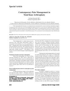
Figure 1. Patellofemoral radiograph of a left knee shows normal patellar anatomy. Lateral patellofemoral angle (white lines) opens laterally approximately 7˚ and sulcus angle is 135˚ (red lines).
8.7
Statistical Methods of the Epidemiological Section (IV)
For the epidemiological section of this study, the follow-up time for each conscript was calculated from entry into military service to the first inpatient care admission due to knee injury or to the end of the service if no hospitalisations occurred. Total exposure time for the population at risk was calculated from the total number of realized service days during the study period, which was obtained from the register. Total exposure time for the population at risk during the study period was 97,503 person-years. The person-time incidence rates for knee injuries leading to inpatient care were calculated by dividing the number of persons with a knee diagnosis by the total exposure time, and the 95% CIs were reported. Because subjects might have been at inpatient care multiple times for the same diagnosis, only first admissions were taken into account for calculating the incidence. The number of possible readmissions, surgical operations, and service class changes by diagnoses were also reported. For the risk factor analysis, the height and weight of the conscripts were divided into quartiles. Age was divided into three groups matched by size. The BMI was calculated as weight in kilograms divided by the square of the height in meters (kg/m2). Conscripts were then categorized as underweight, overweight, or obese based on the international classification according to BMI by the World Health Organization, as follows: BMI