The
n e w e ng l a n d j o u r na l
of
m e dic i n e
clinical practice
Osteoarthritis of the Knee David T. Felson, M.D., M.P.H. This Journal feature begins with a case vignette highlighting a common clinical problem. Evidence supporting various strategies is then presented, followed by a review of formal guidelines, when they exist. The article ends with the author’s clinical recommendations.
A 66-year-old woman who is overweight reports bilateral knee pain of gradual onset during the past several months that increasingly has limited her activities. Last week, when walking down the stairs, she nearly fell when her knee gave way. She does not recall having injured her knee, and she has no morning stiffness and no pain in other joints. She has tried taking up to eight extra-strength (500 mg each) acetaminophen tablets daily without success and has never had ulcers or stomach bleeding. How should the patient be evaluated and treated?
The Cl inic a l Probl e m Approximately 25 percent of persons 55 years of age or older have had knee pain on most days in a month in the past year,1 and about half of them have radiographic osteoarthritis in the knee, a group considered to have symptomatic osteoarthritis. Many without radiographic osteoarthritis of the knee probably have osteoarthritis that is not yet visible on radiography, an imaging procedure insensitive to early disease. Osteoarthritis of the knee increases in prevalence with age and is more common in women than in men. Risk factors include obesity, knee injury, previous knee surgery, and occupational bending and lifting.2 Osteoarthritis of the knee can be part of a generalized diathesis, including osteoarthritis of the hand, which may be inherited. The natural history of osteoarthritis of the knee is highly variable, with the disease improving in some patients, remaining stable in others, and gradually worsening in others. Osteoarthritis is a leading cause of impaired mobility in the elderly.3 Many persons with knee pain have limitations in function that prevent them from engaging in their usual activities. Osteoarthritis affects all structures within a joint. Not only is hyaline articular cartilage lost, but bony remodeling occurs, with capsular stretching and weakness of periarticular muscles. In some patients, synovitis is present, laxity of the ligaments occurs, and lesions in the bone marrow develop that may represent trauma to bone.4 Osteoarthritis involves the joint in a nonuniform and focal manner. Localized areas of loss of cartilage can increase focal stress across the joint, leading to further cartilage loss. With a large enough area of cartilage loss or with bony remodeling, the joint becomes tilted, and malalignment develops. Malalignment is the most potent risk factor for structural deterioration of the joint,5 since it increases further the degree of focal loading, creating a vicious cycle of joint damage that ultimately can lead to joint failure. Local inflammation in the synovium and the cartilage may contribute to pain and joint damage.6 The following three joint compartments combine to form the knee: the lateral tibiofemoral compartment, the medial tibiofemoral compartment, and the patellofemoral compartment. Although any of these three compartments may be a source
n engl j med 354;8
www.nejm.org
From the Boston University School of Medicine, Boston. Address reprint requests to Dr. Felson at A203, 80 E. Concord St., Boston University School of Medicine, Boston, MA 02118, or at
[email protected]. N Engl J Med 2006;354:841-8. Copyright © 2006 Massachusetts Medical Society.
february 23, 2006
Downloaded from www.nejm.org at UC SHARED JOURNAL COLLECTION on April 5, 2006 . Copyright © 2006 Massachusetts Medical Society. All rights reserved.
841
The
n e w e ng l a n d j o u r na l
of the pain associated with osteoarthritis, pain emanates most often from the patellofemoral joint.7 Bone,8 synovial inf lammation, and a stretched joint capsule filled with f luid9 are likely to be sources of pain; bursitis can also cause symptoms.10 Hyaline articular cartilage is unlikely to be a source of pain, since it contains no nociceptive fibers.
S t r ategie s a nd E v idence Diagnosis
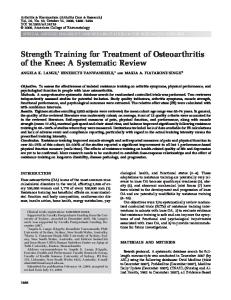
The pain of osteoarthritis is usually related to activity. For osteoarthritis of the knee (Fig. 1), activities such as climbing stairs, getting out of a chair, and walking long distances bring on pain. Morning stiffness usually lasts less than 30 minutes.11 Patients often note that their knees “give way,” a so-called instability symptom.
Femur
Worn articular cartilage Narrowed joint space Osteophytes
Damaged medial meniscus
Tibia
Bony sclerosis and cysts
Fibula
Figure 1. Osteoarthritis of the Medial Side of the Knee.
842
n engl j med 354;8
of
m e dic i n e
Since the knee does not bend much during walking on level ground, the patella does not articulate with the underlying femur, and pain during this activity is not likely to originate in the patellofemoral joint. With more knee bending, such as that which occurs during sitting, stair climbing, or jumping, the patella articulates with the femoral trochlea, and pain during these activities is typical of that originating in the patellofemoral joint. A history of the knee giving way may indicate the presence of an internal derangement such as a meniscal tear or a tear of the anterior cruciate ligament. However, it may also reflect weakness of the muscles that support the joint. Pain in the knee at night reflects either severe symptomatic disease or pain from causes other than osteoarthritis, such as inflammatory arthritis, tumors, infection, or crystal disease (Table 1). Examination of the patient should include testing for various possible causes of knee pain (Table 1). Since arthritis of the hip can cause referred pain to the knee, range of motion of the hip should be assessed to see whether movement at the hip joint induces knee pain or whether there is groin tenderness. Bursitis (either anserine or trochanteric) should also be ruled out. Trochanteric bursitis is part of a syndrome of lateral hip and thigh pain that can extend distally to the tensor fascia lata and even to the iliotibial band, causing lateral knee pain that occurs especially with bending of the knee. Examination of the iliotibial band and more proximal structures in the lateral thigh can identify the source of pain (Table 1). Both anserine and trochanteric bursitis can be treated effectively with a local injection of a corticosteroid. Tenderness at the junction of the femur and tibia (the joint line) should be evaluated, as should the presence of an effusion. Examination of the patient should include an evaluation of whether the legs are varus (bowlegged) or valgus (knockkneed), a physical finding that usually signifies marked malalignment. The knees are farther apart than the feet in the frontal plane when a person with varus malalignment is standing, and the knees are closer together than the feet in a person with valgus malalignment. Varus and valgus malalignment are strong risk factors for worsening radiographic disease4,5 and are probably associated with functional limitations.5 In addition, gait should be observed to determine wheth-
www.nejm.org
february 23, 2006
Downloaded from www.nejm.org at UC SHARED JOURNAL COLLECTION on April 5, 2006 . Copyright © 2006 Massachusetts Medical Society. All rights reserved.
clinical pr actice
Table 1. Features That Distinguish Various Causes of Chronic Knee Pain from Osteoarthritis.* Condition
Features According to History
Features of Physical Examination
Laboratory Features
Chronic inflammatory arthritis, including rheumatoid arthritis
Prominent morning stiffness Other joints affected
Other joints swollen or tender
Increased erythrocyte sedimentation rate Inflammatory synovial fluid
Gout or pseudogout
Other joints affected (especially in cases of gout)
Other joints swollen or tender
Inflammatory synovial fluid containing crystals
Hip arthritis
Pain with hip rotation Groin tenderness
Chondromalacia patellae
Relatively young age of the patient Predominance of patellofemoral symptoms
Anserine bursitis Trochanteric bursitis
Tenderness only over the patellofemoral joint Tenderness distal to the knee over the medial tibia
Lateral hip pain
Tenderness in the region of the lateral hip
Iliotibial band syndrome
Tenderness of the iliotibial band†
Joint tumors
Nocturnal or continuous pain
Bloody synovial fluid Possibility of an abnormal radiograph
Meniscal tear
Prominent mechanical symptoms (e.g., buckling or locking)
Tenderness over the joint line Positive McMurray test‡
Meniscal tear on MRI
Anterior cruciate ligament tear
Prominent mechanical symptoms
Positive Lachman test§
Anterior cruciate ligament tear on MRI
* Knee pain is defined as chronic if it is present for at least six weeks. MRI denotes magnetic resonance imaging. † Tenderness of the iliotibial band is usually lateral to the knee over the insertion site of the iliotibial band in the fibular head or superior to that, where it courses over the lateral femoral condyle. ‡ No physical examination maneuver for meniscal tears has both high sensitivity and specificity.12 Tenderness at the joint line has a sensitivity of 79 percent and a specificity of 15 percent, whereas a McMurray test has a sensitivity of 53 percent and a specificity of 59 percent. A McMurray test is positive if a click is palpable over the medial or lateral tibiofemoral joint line during flexion and extension of the knee during varus (medial tear) or valgus (lateral tear) stress. These data are derived from studies of acute tears,12 and diagnostic data are not available for chronic tears. § A Lachman test is positive if there is excessive anterior translation of the tibia at 30 degress of knee flexion.
er there is antalgia (a limp secondary to pain) and whether gait has slowed because of knee pain. If the patient uses a cane, appropriate use of the cane should be assessed during gait. The location of tenderness in the knee is sometimes helpful in diagnosis, although its reproducibility is limited.13 Tenderness over the medial or lateral joint lines often signals disease there but is also common with meniscal tears.12 Patellofemoral tenderness provides evidence of involvement of the patellofemoral compartment with either osteoarthritis, inflammatory arthritis, or other conditions (Table 1). Tears of the anterior cruciate ligament, if acute, may cause pain. The anterior cruciate ligament prevents translation of the tibia anteriorly during flexion of the knee, and when there is anterior cruciate ligament insufficiency, a Lachman test is more often positive than is an anterior drawer test (Table 1).12 In patients with advanced osteoarthritis, meniscal tears are nearly universal14 and anterior cruciate
n engl j med 354;8
ligament tears are common15; diagnosing them is not likely to change treatment. Repairing meniscal tears in patients with osteoarthritis is unlikely to improve the disease course or ameliorate pain; meniscal tears are not associated with pain in osteoarthritis.14,16 Laboratory Tests
No blood tests are routinely indicated in the workup of a patient with chronic knee pain unless symptoms and signs suggest rheumatoid arthritis or other forms of inflammatory arthritis (Table 1). Examination of synovial fluid is indicated if inflammatory arthritis or gout or pseudogout is suspected or if joint infection is a concern; a white-cell count below 1000 per cubic millimeter in the synovial fluid is consistent with osteoarthritis, whereas higher white-cell counts suggest inflammatory arthritis. The presence of crystals is diagnostic of either gout or pseudogout. Radiography is indicated in the workup of a
www.nejm.org
february 23, 2006
Downloaded from www.nejm.org at UC SHARED JOURNAL COLLECTION on April 5, 2006 . Copyright © 2006 Massachusetts Medical Society. All rights reserved.
843
The
n e w e ng l a n d j o u r na l
of
m e dic i n e
patient if knee pain is nocturnal or is not activity-related. If knee pain persists after effective therapy for osteoarthritis, a radiograph may reveal clues to a missed diagnosis. In patients with osteoarthritis, the radiographic findings correlate poorly with the severity of pain (Fig. 2), and radiographs may be normal in persons with disease.17 Although chondrocalcinosis may be seen on the radiograph, it is an age-related finding that is inconsistently associated with knee pain.18 Avascular necrosis can be diagnosed with radiography, although if it is seen, it is often too late Figure 2. Radiograph Showing Osteoarthritis of the Medial Side of the Knee. to treat it. Magnetic resonance imaging (MRI) is Narrowing of the medial joint space (arrow) and osteolikely to reveal changes that indicate the presence phytes (arrowhead) are shown. of osteoarthritis, but it is not suggested in the workup of older persons with chronic knee pain. MRI findings of osteoarthritis, including menis- ity of conventional NSAIDs has been the use of cal tears, are common in middle-aged and older COX-2 inhibitors,23 although the results of recent adults14 with and without knee pain. trials showing increased cardiovascular risk with these agents has limited their use.24 Alternatively, Treatment the combination of NSAIDs and misoprostol or Treatment of osteoarthritis involves alleviating proton-pump inhibitors has been shown in ranpain, attempting to rectify mechanical malalign- domized trials to reduce the number of endoscopment, and identifying and addressing manifesta- ically confirmed ulcers associated with NSAIDs tions of joint instability. (Table 2). Nonsteroidal Antiinflammatory Drugs, Cyclooxygenase-2 Inhibitors, and Acetaminophen
For treating the pain of osteoarthritis of the knee, head-to-head randomized trials showed that nonsteroidal antiinflammatory drugs (NSAIDs) and cyclooxygenase-2 (COX-2) inhibitors are more efficacious than acetaminophen.19,20 However, the superiority of NSAIDs over acetaminophen (at doses of 4 g per day) is modest.20 In one large crossover trial,19 the average reduction in pain during the first treatment period, on a scale of 0 to 100, was 21 in patients treated with NSAIDs and 13 in those given acetaminophen (P