Hindawi Publishing Corporation Journal of Oncology Volume 2011, Article ID 620523, 12 pages doi:10.1155/2011/620523
Clinical Study Prevalence of Epithelial Ovarian Cancer Stem Cells Correlates with Recurrence in Early-Stage Ovarian Cancer Karina Dahl Steffensen,1, 2 Ayesha B. Alvero,3 Yang Yang,3 Marianne Waldstrøm,4 Pei Hui,5 Jennie C. Holmberg,3 Dan-Arin Silasi,3 Anders Jakobsen,1, 2 Thomas Rutherford,3 and Gil Mor3, 6 1
Department of Oncology, Vejle Hospital, Kabbeltoft 25, 7100 Vejle, Denmark of Regional Health Services Research, University of Southern Denmark, 5230 Odense, Denmark 3 Department of Obstetrics, Gynecology and Reproductive Sciences, Yale University, School of Medicine, New Haven, CT 06510, USA 4 Department of Pathology, Vejle Hospital, 7100 Vejle, Denmark 5 Department of Pathology, Yale University School of Medicine, New Haven, CT 06510, USA 6 Reproductive Immunology Unit, Department of Obstetrics, Gynecology and Reproductive Sciences, Yale University, School of Medicine, 333 Cedar Street, LSOG 305A, New Haven, CT 06520, USA 2 Institute
Correspondence should be addressed to Karina Dahl Steffensen,
[email protected] and Gil Mor,
[email protected] Received 16 March 2011; Revised 3 June 2011; Accepted 13 June 2011 Academic Editor: Peter E. Schwartz Copyright © 2011 Karina Dahl Steffensen et al. This is an open access article distributed under the Creative Commons Attribution License, which permits unrestricted use, distribution, and reproduction in any medium, provided the original work is properly cited. Epithelial ovarian cancer stem cells (EOC stem cells) have been associated with recurrence and chemoresistance. CD44 and CK18 are highly expressed in cancer stem cells and function as tools for their identification and characterization. We investigated the association between the number of CD44+ EOC stem cells in ovarian cancer tumors and progression-free survival. EOC stem cells exist as clusters located close to the stroma forming the cancer stem cell “niche”. 17.1% of the samples reveled high number of CD44+ EOC stem cells (>20% positive cells). In addition, the number of CD44+ EOC stem cells was significantly higher in patients with early-stage ovarian cancer (FIGO I/II), and it was associated with shorter progression-free survival (P = 0.026). This study suggests that quantification of the number of EOC stem cells in the tumor can be used as a predictor of disease and could be applied for treatment selection in early-stage ovarian cancer.
1. Introduction Epithelial ovarian cancer (EOC) is the fourth leading cause of cancer-related deaths in women in the United States and the leading cause of gynecologic cancer deaths with a 5-year survival of only 30–40% [1–5]. Most patients are diagnosed with advanced-stage disease and the majority recurs despite optimal surgical debulking and initial response to chemotherapy. Recurrence is almost always accompanied by the development of chemoresistance and carcinomatosis, which may not be amenable to surgery [2]. Thus, patients with recurrent ovarian cancer usually succumb to the disease.
Current studies suggest that the tumor is initiated and maintained by a unique population of cells with stem-like properties [6]. The cancer stem cell (CSC) hypothesis implies that the inherently chemoresistant CSC can persist after chemotherapy and repopulate the tumor leading to recurrence [7–10]. Contrary to the stochastic model of cancer (clonal expansion), the Cancer Stem Cell model holds that tumors are hierarchically organized and only some cells have the capacity to indefinitely self-renew and sustain tumor growth [11, 12]. It is thought that CSCs are able to survive conventional chemotherapies, which usually target fast dividing cells, and give rise to recurrent tumors that are more
2 resistant and more aggressive [13]. Thus, detection of the CSC population has implications for the diagnosis and treatment of most cancers. One of the major problems in elucidating the cellular origin and pathogenesis of ovarian cancer is that it is a heterogeneous disease. Indeed, ovarian cancer can be classified into multiple types (serous, endometrioid, clear cell, and mucinous), with each type having widely different clinicopathologic properties. It is therefore possible that each of these types of ovarian cancer has different cellular origin. Consequently, the CSC population for each type may also be variable. It is therefore not surprising that stem cell properties have been reported in ovarian cancer cells isolated using different cell surface markers, including CD44, CD133, or CD24 [14–21]. Each of these ovarian cancer cell types may represent either a hierarchy of CSC or an entirely different population of CSC for that particular ovarian histotype. Recently we, and others, demonstrated the presence of epithelial ovarian cancer stem cells (EOC stem cells) in tissue samples and cell lines [16–19, 22]. Several markers have been used for the identification of EOC stem cells, which reflect the heterogeneity of ovarian cancer. These markers include CD44, CD133, CD24, ALDH1, MyD88, and CD117. Of these markers, the cell surface protein CD44 has been most extensively described to potently enrich the EOC stem cells. CD44+ EOC stem cells express pluripotency markers such as β-catenin, Oct-4, and SSEA-4 [14] and have been demonstrated to be the chemoresistant progenitors in vivo and are able to differentiate into the heterogenous cell types comprising the tumor [14, 22]. The objectives of the present study were twofold: (i) to characterize the location of CD44+ EOC stem cells in tissue samples and (ii) to determine whether the CD44+ EOC stem cell “load” correlates with clinical parameters in ovarian cancer patients. Using ovarian cancer tissue sections from 117 patients with primary disease, we investigated the relationship between the number of CD44+ EOC stem cells and various clinical parameters, which include chemoresponse and progression-free survival.
2. Materials and Methods 2.1. Ovarian Cancer Cells. The experiments described here were performed using five EOC stem cells (CD44+) and five mature ovarian cancer cells (mEOC cells, CD44−) that our laboratory isolated and established from either ascites or ovarian tumors [14]. mEOC cells correspond to the CD44− component of the tumor or from cells derived from CD44+ EOC cells following in vitro and in vivo differentiation. We found the same characteristics in CD44− cells isolated from the original tumor or CD44− cells originated from CD44+ EOC cells following in vitro and in vivo differentiation [15]. We generated fluorescence-labeled EOC stem cell clones by stable transfection with lentiviral constructs expressing the red fluorescence protein Tomato under the ubiquitin promoter-driven L2G (pFU-L2T) as described elsewhere [23]. This construct led to the most efficient or stable labeling and brightest bioluminescent signal [24].
Journal of Oncology 2.2. Protein Preparation. Protein extraction was done as previously described [25]. Briefly, cell pellets were lysed on ice in 1× phosphate-buffered saline with 1% NP40, 0.1% SDS and freshly added 20 mL/mL protease inhibitor cocktail (Sigma Chemical, St Louis, MO, USA) and 2 mM phenylmethylsulfonyl fluoride (Sigma Chemical). Protein concentration was determined by BCA Protein Assay (Pierce Biotechnology, Rockford, IL, USA), and proteins were stored at −80◦ C until further use. 2.3. SDS–PAGE and Western Blots. A quantity of 20 μg of each protein sample was denatured in sample buffer and subjected to 12% SDS-polyacrylamide gel electrophoresis (PAGE) as previously described [25]. The following antibody dilutions were used: CD44 antibody (1 : 2000) MEM-263 (Novus Biologicals, Littleton, CO, USA), monoclonal Ck18 antibody DC10 (1 : 1000) (Cell Signaling, Danvers, MA, USA), and rabbit anti-human β-actin (1 : 10,000). Specific protein bands were visualized using enhanced chemiluminescence (Pierce Biotechnology). 2.4. Flow Cytometry. Flow cytometry analysis was performed as previously described [14]. Briefly, cells were trypsinized and pelleted cells were incubated with either PE-anti CK18 or APC-anti CD44 antibodies (eBioscience, San Diego, CA). Data was acquired using BD FACS Calibur and analyzed using Cell Quest Pro (BD Bioscience). 2.5. Study Population. Tumor tissue and patients’ clinical data were collected from a prospective translational research protocol. The patients were all newly diagnosed with ovarian cancer and referred for first-line platinum-based chemotherapy at the departments of clinical oncology at Vejle, Aalborg, Odense, and Herning Hospitals. Collected data were entered into case report forms and all tumor specimens underwent central pathology evaluation. Patients received both oral and written study-related information before they signed a consent form prior to collection of biological material. The Danish Biomedical Research Ethics Committee and the Danish Data Protection Agency approved the study. A majority of the patients underwent primary debulking surgery, while a minor portion (N = 3, 2.6%) were treated with neoadjuvant chemotherapy. All the patients in this cohort received first-line combination chemotherapy with carboplatin (AUC5) and paclitaxel (175 mg/m2 ). Treatment was administered every 3 weeks for at least four cycles. Response to chemotherapy was assessed according to GCIG CA125 criteria [26, 27] and/or RECIST criteria by CT or MRI scans. 2.6. CD44 and Ck18 Immunohistochemical Staining. Formalin-fixed, paraffin-embedded tissue blocks obtained during primary tumor debulking and prior to first-line chemotherapy were used for immunohistochemical staining for CD44 or Ck18 (Cell Signaling) 1 : 100 dilution. The slides from the primary debulking operations were collected from nine regional Danish Departments of Pathology and underwent central pathology revision (MW). The tumors were classified
Journal of Oncology according to the WHO histological classification and graded according to Shimizu et al. [28]. One representative paraffinembedded formalin-fixed tumor block from each patient was selected and 4 μm sections were cut and stored at −80◦ C until further analysis. One section from each patient was used for IHC with monoclonal CD44 antibody (1 : 2000) MEM263 (Novus Biologicals, Littleton, CO, USA) and monoclonal Ck18 antibody DC10 (1 : 1000) (Cell Signaling, Danvers, MA, USA). In brief, the sections were deparaffinized in Tissue clear (Tissue Tec, Sakura Finetek, Zoeterwoude, Netherlands) followed by washes in a graded series of ethanol for rehydration in Tissue Tec Prism (Sakura, Prohosp, Vaerloese, Denmark). The sections were then treated with 3% H2 O2 to block endogenous peroxidase activity. Heat-induced epitope retrieval was done in a microwave oven using TEG pH 9.0 with 15 minutes boiling and 15 minutes for cooling down. The sections were incubated with the primary antibody in 1% bovine serum albumin/tris-buffered saline for 30 minutes at room temperature. Immunohistochemical staining was performed by the Autostainer Plus Link (AS 10030 DAKO, Glostrup, Denmark) according to manufacturer’s instruction. DAKO Envision+ (DAKO, Glostrup, Denmark) was used for antibody detection and was followed by visualization with DAB+ (DAKO, Glostrup, Denmark). After washing, the reaction was enhanced by 0.5% copper sulphate in TBS for 10 minutes, and the slides were counterstained with Mayers sour hematoxyline before dehydration and mounting. To validate the immunohistochemical procedure, negative and positive controls were included in each run. A small tissue microarray containing ovarian tumors was used together with tissue from appendix and tonsil for positive controls. The same tissue was incubated in 1% bovine serum albumin/tris-buffered saline but without the primary antibody for negative control. 2.7. Evaluation of CD44 Immunohistochemical Staining. The study pathologist (MW) scored all the samples: the whole tumor slide was evaluated, and the percentage of CD44 positive stained cells was divided into 0%, >0–5%, >5%– 10%, >10%–20%, >20%–50%, and >50%. For classification, we divided the patients into those with 20% CD44+ cells. 2.8. Statistical Analyses. The correlation between CD44 expression and clinicopathological parameters was assessed by χ 2 statistics and the same applied to the association between CD44 expression and response to chemotherapy. Progression-free survival was defined as the elapsed time from date of diagnosis (date of primary surgery) until progression or death attributable to any cause. Univariate progression-free survival analysis was performed using the Kaplan-Meier estimates and log-rank statistics for comparison of survival plots. Multivariate progression-free survival analysis was determined by the Cox regression model. The parameters entered in the Cox analysis were CD44 status (Low expression: 20%
3 positive cells), FIGO stage, grade, and residual tumor as categorical variables, and age at diagnosis as a continuous variable. Statistical analyses were performed with the NCSS software (version 2007, Kaysville, Utah, http://www.ncss .com/). A value of P < 0.05 was considered statistically significant.
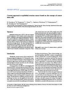
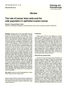
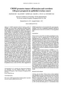
3. Results 3.1. Characteristics of Ovarian Cancer Stem Cells. We previously demonstrated that the ovarian cancer stem cells are CD44+, represent the chemoresistant population, and are able to differentiate in vitro and in vivo to CD44− cells [14]. Recent studies have shown that ovarian cancer stem cells are also ALDH1+. Therefore, we evaluated ALDH1 expression on CD44+ and CD44− EOC cells. As shown in Figure 1, CD44+, but not CD44−, EOC cells express high levels of ALDH1, further confirming that the CD44+ EOC stem cells express the majority of identified markers for tumor initiating cells (Figure 1) [15, 21]. To closely monitor the process of differentiation, we labeled pure clones of CD44+ EOC cells with a fluorescent reporter, which allows flow cytometry analysis and in vivo imaging. Thus, CD44+ EOC stem cell clones were stably transfected with a viral vector expressing the red fluorescence protein “Tomato” (pFU-L2T) [23]. CD44+/Tomato+ EOC cells were injected into nude mice, and the established tumor (60 days later) was evaluated for CD44 and Tomato. As shown in Figure 2, prior to injection the EOC stem cells are 99.5% CD44+/Tomato+. The xenograft established, however, is only 4.5 % CD44+/Tomato+ and 95.5% CD44−/ Tomato+. These results demonstrate that the CD44− cells originated from the CD44+/Tomato+ EOC cells (Figure 2). 3.2. Cytokeratin 18 (Ck18) Is Preferentially Expressed by the EOC Stem Cells. We previously showed, using gene expression microarray, that Ck18 expression is 7-fold higher (P = 0.0007) in CD44+/MyD88+ EOC stem cells compared to the CD44−/MyD88− mature ovarian cancer stem cells (mOCCs) [14]. To validate this finding, we determined the levels of Ck18 in five EOC stem cell clones, three mOCC clones, and in the EOC cell line A2780 using western blot analysis. As shown in Figure 3, Ck18 expression is limited to the EOC stem cells and not the mOCCs. Correlation between CD44 and Ck18 expression was also observed by flow cytometry and western blot (Figures 3(a), 3(b)). Evaluation of the location of CD44+ and Ck18+ cells in tumor tissues obtained from ovarian cancer patients showed that CD44+ (Figures 3(c), 3(d)) and Ck18+ cells (Figures 3(e), 3(f)) are surrounded by CD44-/Ck18− mOCCS. Within tumor nests, single (Figure 4(a)) and clusters (Figures 4(b)–4(d)) of Ck18+ cancer cells were observed. These cells morphologically appear less differentiated with larger size, higher nuclear to cytoplasm (N/C) ratio, more prominent nucleoli, and a vesicular chromatin pattern (Figure 5). In cells with a more differentiated phenotype (smaller size and lower N/C ratio), Ck18 staining was weak to absent (Figures 3 and 4). Some of the Ck18+ clusters were observed in close
4
Journal of Oncology Phase
ALDH1
CD44+
CD44−
Figure 1: Correlation of CD44 and ALDH1 Expression in epithelial ovarian cancer stem cells. A panel of ovarian cancer cells was evaluated for the expression of CD44 and ALDH1 by immunofluorescence. Only CD44+ EOC stem cells are also positive for ALDH1 expression. representative figure of five independent experiments using five clones of CD44+ cells and their derived CD44− cells.
Pre-differentiation
Post-differentiation
104
104 99.5%
95.5%
4.5%
103 FL2-H
FL2-H
Tomato red
103
102
101
102
101
100
100 100
101
102 FL1-H
103
104
100
101
102 FL1-H
103
104
CD44 (a)
(b)
Figure 2: CD44+ EOC cells undergo in vivo differentiation into CD44− cells. Flow cytometry analysis of CD44+ cells stable transfected with a lentivirus expressing the fluorescent protein Tomato (red). (a) Cells prior to injection into the mice are 99.5% double positive for CD44 and Tomato. (b) 95.5% of the cells isolated from the tumor remain positive for the fluorescence protein Tomato but are negative to CD44. Only 4.5% of the injected cells remained double positive.
proximity to the stroma and showed a clear and defined basal membrane (Figure 5). The observed distribution of the Ck18+ cancer cells follows the description of the niche associated with CSC [9, 29]. A similar pattern of localization was observed with CD44 staining [14].
3.3. Variable Expression of CD44+ EOC Stem Cells in Ovarian Cancer Tissues . Our next objective was to determine whether the prevalence of EOC stem cells has a prognostic value. For this study, we focused using a single marker and selected CD44 as a widely accepted marker for the identification of
Journal of Oncology
5 OCSC1 CD44
CK18
200
200 97.15%
120
99.77%
160 Counts
Counts
160
M1
80 40
120 80
M1
40
0
0 100
101
102 FL1-H
103
104
100
101
102 FL2-H
103
104
(a)
OCSC1 OCSC3 OCSC4 OCSC5 OCSC6 T182
T127
01-19 A2780
Ck18
CD44
β-actin (b)
(d)
(c)
(e)
(f)
Figure 3: Expression of CD44 and CK18 in multiple clones of ovarian cancer cells. (a)-(b). CD44+ cells also express CK18 as determined by flow cytometry (a) and western blot analysis (b). Flow cytometer is representative of the eight evaluated clones. EOC stem cells, determined by either CD44 (c and d) or Ck18 (e and f) expression, are found in clusters surrounded by CD44− or Ck18− negative cancer cells.
ovarian cancer stem cells. Thus, we analyzed CD44 staining in ovarian cancer tissue sections obtained from 117 patients. The clinical-pathological data of the study cohort is presented in Table 1. The majority of the patients were older than 50 years with histopathologic diagnosis of serous ovarian cancer. In addition, most of the patients were classified FIGO stage II and higher, with moderate or poorly differentiated tumors (grade > 1) (Table 1). We detected CD44+ cancer cells in all but one tissue section tested. However, we observed variability in the number, distribution, and location of CD44+ cancer cells amongst patients. Of all patients tested, 39 patients had between 1–5% CD44+ cancer
cells, 38 patients had >5–10% CD44+ cells, 19 patients had >10–20% CD44+ cells, 9 patients had >20–50% CD44+ cells, and 11 patients had >50% CD44+ cells. Only one sample was negative for CD44 staining. Due to this high variation, we classified the samples as low expression of EOC stem cells if they had less than 20% positive cells (20% EOC stem cells). Thus, of the 117 patients, 20 patients were considered high expression and 97 patients were considered low expression (Table 1). We then determined if the percentage of EOC stem cells has clinical correlation.
6
Journal of Oncology
(a)
(b)
(c)
(d)
Figure 4: EOC stem cells present unique morphological characteristics. CD44+ EOC stem cells are characterized by high nuclear to cytoplasm (N/C) ratio, contain vesicular chromatin pattern, and have prominent nucleoli. The cells can be found as single cells (a) or clusters of two or more cells (b–d).
3.4. CD44 Levels Inversely Correlate with FIGO Stage and Tumor Grade . Patients with FIGO stage I tumors had a higher number of CD44+ EOC stem cells (>20% CD44+ cells) with 57.1% of the stage I patients expressing >20% CD44+ cells. For FIGO stages II, III, and IV, 18.2%, 12.9%, and 4.5% expressed >20% CD44+ cells (Table 1). Thus, a significant percent had FIGO stage I (P = 0.00025; x2 = 19.2). Similarly, the majority of patients with grade I tumors showed high expression of EOC stem cells (>20% CD44+ cells) (P = 0.021, x2 = 7.7, Table 1). High expression of CD44+ EOC cells was seen in fifty percent, 14.3%, and 12.1% of grade 1, 2, and 3 disease, respectively. This indicates that in patients with primary disease, tumors tend to have a lower number of CD44+ EOC stem cells as the disease progresses. 3.5. Correlation between Number of CD44+ EOC Stem Cells and Chemoresponse. We then evaluated whether a correlation exists between percentage of CD44+ EOC stem cells and response to treatment. All the patients in this cohort received treatment. The 14 patients with FIGO stage I cancer comprised 2 patients with stage IA cancer (one clear cell cancer and one grade 2 serous = patients with adverse histological
features/high risk patients that routinely receive chemotherapy) and 12 patients with stage IC tumors that according to guidelines are treated with adjuvant chemotherapy. Although it was only marginally statistically significant, we observed an obvious trend (P = 0.06) for poorer response rates among patients with >20% CD44+ EOC stem cells. Only 73% of these patients had complete or partial response compared to 90% in patients with low number of EOC stem cells (20% positive cells for CD44 had stable or progressive disease during or by the end of first line carboplatin and paclitaxel treatment compared to only 10% in the patients with a low number of CD44+ cells (20%) had significantly shorter progression-free survival compared to patients with a low number of CD44+ cells EOC stem cells (20% CD44+ EOC stem cells had a shorter progression-free survival compared to patients with tumors having 20%) or more than 20% CD44 positive cells (1 cm (Unknown: 8 ) CD44 immunostaining Less than 20% CD44+ cells More than 20% CD44+ cells
No. of patients
%
20% CD44 Cells N (%)
10 54 53
8.6 46.2 45.3
6 (60.0) 40 (74.1) 51 (96.2)
4 (40.9) 14 (25.9) 2 (3.8)
0.0013
0.00025 14 11 70 22
12.0 9.4 59.8 18.8
6 (42.9) 9 (81.8) 61 (87.1) 21 (95.5)
8 (57.1) 2 (18.2) 9 (12.9) 1 (4.5)
8 42 58
6.8 35.9 49.6
4 (50.0) 36 (85.7) 51 (87.9)
4 (50.0) 6 (14.3) 7 (12.1)
9
7.7
100 5 8 1
85.5 4.3 6.8 0.9
86 (86.0) 4 (80.0) 5 (62.5) 0 (0.0)
14 (14.0) 1 (20.0) 3 (37.5) 1 (100)
3
2.6
2 (66.7)
1 (33.3)
56
51.4
42 (75.0)
14 (25.0)
53
48.6
49 (92.5)
4 (7.5)
97 20
82.9 17.1
NA
NA
0.0209
0.0760
0.014
Table 2: Correlation between percentage of CD44+ EOC stem cells and response to first-line carboplatin/paclitaxel treatment. Groups are classified based on their CD44 expression as tumors containing less than 20% CD44 positive (>20%) or more than 20% CD44 positive cells ( cm % E O C stem cells 20% CD44 Cells ∗
Cox regression model.
Survival plot
Progression-free survival probability
1
0.8
0.5
0.3
0 0
6.7
13.3 20 26.7 33.3 Progression-free survival (months)
40
CD44 low CD44 high FIGO stage I/II. P = 0.026
Figure 6: Progression-free survival in ovarian cancer patients with stage I/II. In early-stage ovarian cancer (FIGO stage I/II), patients with a high percentage of CD44+ positive EOC stem cells (>20%) (CD44 High) had significantly shorter progression-free survival compared to patients with a low number of CD44+ EOC stem cells (CD 44 low) (P = 0.026).
However, it may also be a result of the heterogeneous nature of ovarian cancer and its multiple sources of origin [34, 35]. An ovarian cancer stem cell originated in the fallopian tube might present different surface markers than a CSC originated from the endometrium or the surface epithelium of the ovaries. Our group previously identified at least two types of EOC cells based on their response to chemotherapy: Type
I-chemoresistant and Type II-chemosensitive EOC cells [36, 37]. Further characterization showed that these cells have additional differences in terms of growth, cytokine production, and intracellular markers [38]. While Type II EOC cells represent the “classical” ovarian cancer cells characterized by fast growth and lack of cell to cell contact inhibition, Type I EOC cells are characterized by slower growth, which is inhibited upon cell to cell contact. In addition, Type I, but not Type II, EOC cells have constitutive NF-kB activity and constitutively secrete IL6, IL8, MCP-1, and GROα [14]. Gene expression microarray analysis comparing these two types of cells further showed that Type I EOC cells expressed significantly higher levels of the stem cell markers, CD44 and SSEA-4, the TLR adapter protein MyD88, Cytokeratin 18, Trop-1, and others [14]. In contrast, Type II EOC cells were negative for all these markers. These findings suggest that Type I EOC cells may represent the population that has stem-like properties. Indeed, we demonstrated that Type I EOC cells, as selected by CD44, are able to form xenografts in mice and resulted in tumors containing both CD44+ and CD44− cells. In this study, we evaluated additional markers present in our recently isolated CD44+ EOC cells [14]. We observed that these cells are also CK18+, a marker associated with epithelium of the fallopian tubes. Aldehyde dehydrogenase (ALDH1) has been proven useful for the identification of cancer stem cells, including ovarian cancer [19]; therefore, we evaluated the expression of ALDH1 in the identified CD44+ EOC stem cell clones. We found high levels of ALDH on EOC stem cells by immunofluorescence, suggesting that ALDH1 could be used also as a marker to monitor the presence of cancer stem cells. We described additional evidence in support of previous findings showing that Type I EOC cells (CD44+) are the source of Type II cells or CD44−. To closely monitor EOC stem cell fate and function in mice, we labeled CD44+ cells
10 with dual-function reporter genes encoding the sequence of the florescence protein Tomato (red color). Using a xenograft tumor model, we demonstrated that following injection of double positive CD44+/Tomato+ cells, the newly formed tumor originating from these double positive cells is characterized by CD44− cells, which maintain the expression of the fluorescent protein Tomato. This demonstrates that CD44+ EOC stem cells can both self-renew and differentiate [39]. Moreover, microscopic analysis of the xenografts showed that CD44+ EOC cells were able to recapitulate the morphology of the original tumor [14]. Finally, in vitro differentiation of the chemoresistant CD44+ EOC stem cells resulted in chemosensitive cultures that have lost CD44. In this study, we showed that the presence of CD44+ EOC stem cells correlates with poor prognosis. Since the CD44+ cells are in general more chemoresistant, they can persist after chemotherapy and may initiate recurrence upon the completion of treatment. We found EOC stem cells localized in clusters surrounded by differentiated ovarian cancer cells and in close proximity with the stroma. Emerging evidence indicates that a specialized environment, the stem cell niche, is one of the factors regulating stem cell maintenance and self-renewal [9, 40, 41]. Alterations to the stroma may affect the control of self-renewal [30]. This is illustrated by the studies of Yauch et al. who showed that inhibition of Hh pathway in pancreatic associated stroma cells resulted in suppression of tumor growth [42]. In contrast, inhibition of the same pathway in the cancer cells did not have any effect on tumor growth. This suggests that the variation on the number of cancer stem cells observed in our study may be the result of alteration in the interaction between the stroma and the cancer stem cells. A functional stroma might maintain a small pool of cancer stem cells while promoting differentiation. However, disruption of the stroma-cancer stem cells interaction might lead to uncontrolled self-renewal and significant increase in the pool of chemoresistant EOC stem cells and consequent poor prognosis. CD44 is a cell surface glycoprotein receptor with several isoforms [43]. All isoforms are encoded by a single gene and result from alternative splicing. CD44 is expressed by most cells, including hematopoietic cells and tumors. Several studies have evaluated CD44 expression in ovarian cancer tumors and correlated with survival outcome. CD44 expression has been reported to correlate with a significantly shorter disease survival than for patients with CD44 negative tumors [44, 45]. However, studies investigating CD44 expression in terms of IHC and survival are contradictory [46, 47]. Differences between these studies that could account for differences in their findings could be attributed to technical factors, including the use of different monoclonal or polyclonal antibodies that exhibit variable efficacy in paraffin-embedded tissues and to different methods used for assessment of immunostaining. In this study, we focused on CD44 expression as a marker of the cancer stem cells and its evaluation is based on the percentage of ovarian cancer stem cells present in the tumor. CD44 is more than a marker; this transmembrane receptor has been shown to be important in various cellular
Journal of Oncology processes such as growth, differentiation, and motility [43]. The most studied function of CD44 is its role as the receptor for hyaluronan (HA) [48]. Binding of HA to CD44 controls cell-cell interactions, as well as interactions of the cell with the extra-cellular matrix. Furthermore, it can function as detector of tissue damage and promote tissue repair. Therefore, it is possible that CD44 expression in EOC stem cells might play a central role in self-renewal and the response to tissue damage.
5. Conclusion We describe the intratumoral localization of EOC stem cells in ovarian tumor samples. We show their existence as clusters located close to the stroma forming what has been described as the CSC “niche”. Furthermore, we demonstrate a correlation between the percentage of CD44+ EOC stem cells and survival in early-stage ovarian cancer. Although it is a small cohort, especially the early stage, the findings from this study are important since they suggest that quantification of the number of EOC stem cells present in the tumor can be used as a predictor of disease and could be applied for treatment selection in early-stage ovarian cancer.
Conflict of Interests The authors declare that there is no conflict of interests.
Acknowledgments The authors thank laboratory technologist Tinna Herløv Jensen for her work with the CD44 immunohistochemical staining. This study was supported in part by grants from Vejle Hospital, The Cancer Foundation, NCI/NIH RO1CA127913, RO1CA118678, The Janet Burros Memorial Foundation, The Sands Family Foundation, and the Discovery to Cure Research Program.
References [1] A. Jemal, R. Siegel, E. Ward, Y. Hao, J. Xu, and M. J. Thun, “Cancer statistics, 2009,” CA Cancer Journal for Clinicians, vol. 59, no. 4, pp. 225–249, 2009. [2] P. E. Schwartz, “Current diagnosis and treatment modalities for ovarian cancer,” Cancer Treatment and Research, vol. 107, pp. 99–118, 2002. [3] M. S. Piver, F. M. Muggia, M. F. Brady, and R. Alvarez, “Maintenance chemotherapy in advanced ovarian cancer,” Journal of Clinical Oncology, vol. 18, no. 8, pp. 1803–1805, 2000. [4] P. G. Rose, “Chemotherapy for newly diagnosed and relapsed advanced ovarian cancer,” Seminars in Oncology Nursing, vol. 19, no. 2, pp. 25–35, 2003. [5] M. V. Seiden, “Ovarian cancer,” Oncologist, vol. 6, no. 4, pp. 327–332, 2001. [6] T. Reya, S. J. Morrison, M. F. Clarke, and I. L. Weissman, “Stem cells, cancer, and cancer stem cells,” Nature, vol. 414, no. 6859, pp. 105–111, 2001. [7] E. H. Huang, D. G. Heidt, C. W. Li, and D. M. Simeone, “Cancer stem cells: a new paradigm for understanding tumor
Journal of Oncology
[8]
[9]
[10]
[11]
[12]
[13]
[14]
[15]
[16]
[17]
[18]
[19]
[20]
[21]
[22]
[23]
[24]
progression and therapeutic resistance,” Surgery, vol. 141, no. 4, pp. 415–419, 2007. N. J. Maitland, S. D. Bryce, M. J. Stower, and A. T. Collins, “Prostate cancer stem cells: a target for new therapies,” Ernst Schering Foundation Symposium Proceedings, vol. 5, pp. 155– 179, 2006. N. J. Maitland and A. Collins, “A tumour stem cell hypothesis for the origins of prostate cancer,” BJU International, vol. 96, no. 9, pp. 1219–1223, 2005. M. Dean, T. Fojo, and S. Bates, “Tumour stem cells and drug resistance,” Nature Reviews Cancer, vol. 5, no. 4, pp. 275–284, 2005. T. Lapidot, C. Sirard, J. Vormoor et al., “A cell initiating human acute myeloid leukaemia after transplantation into SCID mice,” Nature, vol. 367, no. 6464, pp. 645–648, 1994. J. Vormoor, T. Lapidot, F. Pflumio et al., “SCID mice as an in vivo model of human cord blood hematopoiesis,” Blood Cells, vol. 20, no. 2-3, pp. 316–320, 1994. R. Morrison, S. M. Schleicher, Y. Sun et al., “Targeting the mechanisms of resistance to chemotherapy and radiotherapy with the cancer stem cell hypothesis,” Journal of Oncology, vol. 2011, Article ID 941876, 13 pages, 2011. A. B. Alvero, R. Chen, H. H. Fu et al., “Molecular phenotyping of human ovarian cancer stem cells unravel the mechanisms for repair and chemo-resistance,” Cell Cycle, vol. 8, no. 1, pp. 158–166, 2009. G. Mor, G. Yin, I. Chefetz, Y. Yang, and A. Alvero, “Ovarian cancer stem cells and inflammation,” Cancer Biology and Therapy, vol. 11, no. 8, pp. 708–713, 2011. S. A. Bapat, A. M. Mali, C. B. Koppikar, and N. K. Kurrey, “Stem and progenitor-like cells contribute to the aggressive behavior of human epithelial ovarian cancer,” Cancer Research, vol. 65, no. 8, pp. 3025–3029, 2005. N. K. Kurrey, K. Amit, and S. A. Bapat, “Snail and Slug are major determinants of ovarian cancer invasiveness at the transcription level,” Gynecologic Oncology, vol. 97, no. 1, pp. 155– 165, 2005. S. Zhang, C. Balch, M. W. Chan et al., “Identification and characterization of ovarian cancer-initiating cells from primary human tumors,” Cancer Research, vol. 68, no. 11, pp. 4311– 4320, 2008. S. Deng, X. Yang, H. Lassus et al., “Distinct expression levels and patterns of stem cell marker, aldehyde dehydrogenase isoform 1 (ALDH1), in human epithelial cancers,” PLoS ONE, vol. 5, no. 4, Article ID e10277, 2010. I. A. Silva, S. Bai, K. McLean et al., “Aldehyde dehydrogenase and CD133 define angiogenic ovarian cancer stem cells that portend poor patient survival,” Cancer Research, vol. 71, no. 11, pp. 3991–4001, 2011. S. Dyall, S. A. Gayther, and D. Dafou, “Cancer stem cells and epithelial ovarian cancer,” Journal of Oncology, vol. 2010, Article ID 105269, 9 pages, 2010. A. B. Alvero, H. H. Fu, J. Holmberg et al., “Stem-like ovarian cancer cells can serve as tumor vascular progenitors,” Stem Cells, vol. 27, no. 10, pp. 2405–2413, 2009. P. Ray, R. Tsien, and S. S. Gambhir, “Construction and validation of improved triple fusion reporter gene vectors for molecular imaging of living subjects,” Cancer Research, vol. 67, no. 7, pp. 3085–3093, 2007. T. F. Massoud, R. Paulmurugan, A. De, P. Ray, and S. S. Gambhir, “Reporter gene imaging of protein-protein interactions in living subjects,” Current Opinion in Biotechnology, vol. 18, no. 1, pp. 31–37, 2007.
11 [25] M. Kamsteeg, T. Rutherford, E. Sapi et al., “Phenoxodiol— an isoflavone analog—induces apoptosis in chemoresistant ovarian cancer cells,” Oncogene, vol. 22, no. 17, pp. 2611–2620, 2003. [26] G. J. Rustin, “Use of CA-125 to assess response to new agents in ovarian cancer trials,” Journal of Clinical Oncology, vol. 21, no. 10, pp. 187–193, 2003. [27] G. J. Rustin, A. E. Nelstrop, P. McClean et al., “Defining response of ovarian carcinoma to initial chemotherapy according to serum CA 125,” Journal of Clinical Oncology, vol. 14, no. 5, pp. 1545–1551, 1996. [28] Y. Shimizu, S. Kamoi, S. Amada, K. Hasumi, F. Akiyama, and S. G. Silverberg, “Toward the development of a universal grading system for ovarian epithelial carcinoma I. Prognostic significance of histopathologic features-problems involved in the architectural grading system,” Gynecologic Oncology, vol. 70, no. 1, pp. 2–12, 1998. [29] S. J. Leedham, M. Brittan, S. A. McDonald, and N. A. Wright, “Intestinal stem cells,” Journal of Cellular and Molecular Medicine, vol. 9, no. 1, pp. 11–24, 2005. [30] M. F. Clarke and M. Fuller, “Stem cells and cancer: two faces of eve,” Cell, vol. 124, no. 6, pp. 1111–1115, 2006. [31] S. K. Singh, I. D. Clarke, M. Terasaki et al., “Identification of a cancer stem cell in human brain tumors,” Cancer Research, vol. 63, no. 18, pp. 5821–5828, 2003. [32] R. W. Cho, X. Wang, M. Diehn et al., “Isolation and molecular characterization of cancer stem cells in MMTV-Wnt-1 murine breast tumors,” Stem Cells, vol. 26, no. 2, pp. 364–371, 2008. [33] R. Galli, E. Binda, U. Orfanelli et al., “Isolation and characterization of tumorigenic, stem-like neural precursors from human glioblastoma,” Cancer Research, vol. 64, no. 19, pp. 7011–7021, 2004. [34] J. W. Carlson, A. Miron, E. A. Jarboe et al., “Serous tubal intraepithelial carcinoma: its potential role in primary peritoneal serous carcinoma and serous cancer prevention,” Journal of Clinical Oncology, vol. 26, no. 25, pp. 4160–4165, 2008. [35] E. A. Jarboe, A. K. Folkins, R. Drapkin, T. A. Ince, E. S. Agoston, and C. P. Crum, “Tubal and ovarian pathways to pelvic epithelial cancer: a pathological perspective,” Histopathology, vol. 53, no. 2, pp. 127–138, 2008. [36] R. Chen, A. B. Alvero, D. A. Silasi et al., “Regulation of IKKβ by miR-199a affects NF-κB activity in ovarian cancer cells,” Oncogene, vol. 27, no. 34, pp. 4712–4723, 2008. [37] M. G. Kelly, A. B. Alvero, R. Chen et al., “TLR-4 signaling promotes tumor growth and paclitaxel chemoresistance in ovarian cancer,” Cancer Research, vol. 66, no. 7, pp. 3859–3868, 2006. [38] R. Chen, A. B. Alvero, D. A. Silasi, K. D. Steffensen, and G. Mor, “Cancers take their Toll—the function and regulation of Toll-like receptors in cancer cells,” Oncogene, vol. 27, no. 2, pp. 225–233, 2008. [39] G. Yin, R. Chen, A. B. Alvero et al., “TWISTing stemness, inflammation and proliferation of epithelial ovarian cancer cells through MIR199A2/214,” Oncogene, vol. 29, no. 24, pp. 3545–3553, 2010. [40] S. J. Leedham, A. T. Thliveris, R. B. Halberg, M. A. Newton, and N. A. Wright, “Gastrointestinal stem cells and cancer: bridging the molecular gap,” Stem Cell Reviews, vol. 1, no. 3, pp. 233–242, 2005. [41] S. J. Leedham and N. A. Wright, “Expansion of a mutated clone: from stem cell to tumour,” Journal of Clinical Pathology, vol. 61, no. 2, pp. 164–171, 2008.
12 [42] R. L. Yauch, S. E. Gould, S. J. Scales et al., “A paracrine requirement for hedgehog signalling in cancer,” Nature, vol. 455, no. 7211, pp. 406–410, 2008. [43] D. Naor, R. V. Sionov, and D. Ish-Shalom, “CD44: structure, function, and association with the malignant process,” Advances in Cancer Research, vol. 71, pp. 241–319, 1997. [44] M. Uhi-Steidl, E. Muller-Holzner, A. G. Zeimet et al., “Prognostic value of CD44 splice variant expression in ovarian cancer,” Oncology, vol. 52, no. 5, pp. 400–406, 1995. [45] S. Kayastha, A. N. Freedman, M. S. Piver, J. Mukkamalla, M. Romero-Guittierez, and B. A. Werness, “Expression of the hyaluronan receptor, CD44s, in epithelial ovarian cancer is an independent predictor of survival,” Clinical Cancer Research, vol. 5, no. 5, pp. 1073–1076, 1999. [46] L. Rodr´ıguez-Rodr´ıguez, I. Sancho-Torres, C. Mesonero, D. G. Gibbon, W. J. Shih, and G. Zotalis, “The CD44 receptor is a molecular predictor of survival in ovarian cancer,” Medical Oncology, vol. 20, no. 3, pp. 255–263, 2003. [47] C. Ricciardelli and R. J. Rodgers, “Extracellular matrix of ovarian tumors,” Seminars in Reproductive Medicine, vol. 24, no. 4, pp. 270–282, 2006. [48] R. V. Sionov and D. Naor, “Hyaluronan-independent lodgment of CD44+ lymphoma cells in lymphoid organs,” International Journal of Cancer, vol. 71, no. 3, pp. 462–469, 1997.
Journal of Oncology
MEDIATORS of
INFLAMMATION
The Scientific World Journal Hindawi Publishing Corporation http://www.hindawi.com
Volume 2014
Gastroenterology Research and Practice Hindawi Publishing Corporation http://www.hindawi.com
Volume 2014
Journal of
Hindawi Publishing Corporation http://www.hindawi.com
Diabetes Research Volume 2014
Hindawi Publishing Corporation http://www.hindawi.com
Volume 2014
Hindawi Publishing Corporation http://www.hindawi.com
Volume 2014
International Journal of
Journal of
Endocrinology
Immunology Research Hindawi Publishing Corporation http://www.hindawi.com
Disease Markers
Hindawi Publishing Corporation http://www.hindawi.com
Volume 2014
Volume 2014
Submit your manuscripts at http://www.hindawi.com BioMed Research International
PPAR Research Hindawi Publishing Corporation http://www.hindawi.com
Hindawi Publishing Corporation http://www.hindawi.com
Volume 2014
Volume 2014
Journal of
Obesity
Journal of
Ophthalmology Hindawi Publishing Corporation http://www.hindawi.com
Volume 2014
Evidence-Based Complementary and Alternative Medicine
Stem Cells International Hindawi Publishing Corporation http://www.hindawi.com
Volume 2014
Hindawi Publishing Corporation http://www.hindawi.com
Volume 2014
Journal of
Oncology Hindawi Publishing Corporation http://www.hindawi.com
Volume 2014
Hindawi Publishing Corporation http://www.hindawi.com
Volume 2014
Parkinson’s Disease
Computational and Mathematical Methods in Medicine Hindawi Publishing Corporation http://www.hindawi.com
Volume 2014
AIDS
Behavioural Neurology Hindawi Publishing Corporation http://www.hindawi.com
Research and Treatment Volume 2014
Hindawi Publishing Corporation http://www.hindawi.com
Volume 2014
Hindawi Publishing Corporation http://www.hindawi.com
Volume 2014
Oxidative Medicine and Cellular Longevity Hindawi Publishing Corporation http://www.hindawi.com
Volume 2014