Original Article Effects of malaria in pregnancy on newborn anthropometry Catherine O. Falade1, Olukemi O. Tongo2, Oluwatoyin O. Ogunkunle 2, Adebola E. Orimadegun3 1
Clinical Pharmacology Department, College of Medicine, University of Ibadan, Ibadan, Nigeria Department of Paediatrics, College of Medicine, University of Ibadan, Ibadan, Nigeria 3 Institute of Child Health, College of Medicine, University of Ibadan 2
Abstract Background: Malaria in pregnancy remains a major cause of infant mortality through its contribution to preterm delivery, low birth weight and intrauterine death. Methodology: During a cross-sectional study of 983 mothers delivering in a secondary health care facility in Ibadan, southwestern Nigeria, an area of high malaria transmission, the effect of maternal and placental malaria parasitaemia on newborn anthropometry was evaluated. Malaria parasitemia was detected by microscopy of Giemsa stained thick blood smears. Results: Placental, maternal and combined placental and maternal malaria parasitaemia rates at the time of delivery were 13.1%, 12.7% and 11.1% respectively. The geometric mean parasite densities in maternal and placental smears were significantly higher in primigravid mothers than others (p = 0.004 and 0.002 respectively). Low birth weight rate was higher among babies born to mothers with maternal parasitaemia compared to those without (8.0 % versus 6.3%, p < 0.05). The mean birth weight was lower in neonates of mothers with peripheral and placental parasitaemia by 138 g and 122 g (p = 0.01 and 0.02) respectively, while the respective difference was up to 168 g and 151 g among primigravidae (p = 0.03 and 0.04). Neonates of mothers with maternal and placental parasitaemia had a lower mean length than those without parasitaemia (48.2 vs 49.2cm, p = < 0.0001 and 48.5 vs 49.2cm p = 0.02 respectively). Occiptofrontal circumference and ponderal indices were not significantly affected by maternal malaria parasitaemia. Conclusion: Malaria in pregnancy results in symmetric foetal growth restriction and the effect is more marked among primigravid mothers. Keywords: malaria, pregnancy, newborn, anthropometry J Infect Dev Ctries 2010; 4(7):448-453. (Received 16 July 2009 – Accepted 1 May 2010) Copyright © 2010 Falade et al. This is an open-access article distributed under the Creative Commons Attribution License, which permits unrestricted use, distribution, and reproduction in any medium, provided the original work is properly cited.
Introduction Malaria in pregnancy is of public health concern due to its adverse effects on the mother as well as the foetus. In sub-Saharan Africa, where malaria transmission is high and perennial, approximately 25 million pregnant women are at risk of Plasmodium falciparum infection every year, and at least 27.8% of pregnant women are reported to have evidence of peripheral/placental infection at the time of delivery [1]. The consequences of malaria in pregnancy are grave for both mother and foetus. In endemic countries, up to 26% of severe anemia in pregnancy is attributable to malaria, with malaria-related maternal deaths reaching up to 0.5% - 23% [2]. Malaria during pregnancy may result in preterm delivery, intrauterine growth restriction (IUGR), reduced length and low birth weight (LBW) or stillbirth. Up to 20% of LBW in sub-Saharan Africa has been attributed to malaria in pregnancy and this figure represents 35% of preventable low birth
weight in the region [1,3,4]. Malaria-induced LBW is reported to account for 62,000 to 363,000 infant deaths annually [1,5]. It is also an important determinant of perinatal mortality. Studies have demonstrated a 27% reduction in perinatal mortality with successful implementation of malaria prevention strategies in pregnancy [1]. Pregnancy and malaria are known to be mutually aggravating in endemic areas, especially among primigravidae including in those who were previously immune. Though previous reports have emphasized that the greatest burden of malaria in pregnancy in terms of incidence and outcome is among the paucigravida, it has been increasingly recognized that women of higher gravidities, especially in areas of low transmission, are also at risk [1]. The present study was undertaken to determine the association between malaria parasitaemia in pregnancy and the prevalence of low birth weight, IUGR, and preterm delivery among mothers who
Falade et al. - Malaria in pregnancy and newborn anthropometry
J Infect Dev Ctries 2010; 4(7):448-453.
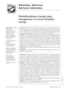
Figure 1. Map of Ibadan metropolis
delivered in Ibadan, Nigeria. In addition, the effects of maternal and placental malaria on anthropometric measurements (length, occipitofrontal circumference and ponderal index) among various gravidity groups in the study population were evaluated. Materials and methods This study, conducted at Saint Mary’s Catholic Hospital, Eleta, Ibadan (Figure 1), was part of a larger analytical cross-sectional evaluation of the epidemiology of congenital malaria in Ibadan, southwestern Nigeria, between May 2003 and October 2004. The participants were 983 consecutive parturient mothers who provided written informed consent. The hospital is a secondary mission health facility which is highly subsidized by the church organization and accessible to the entire community in Ibadan metropolis. It is well patronized because of affordability. Ethical approval was obtained from the University of Ibadan/University College Hospital ethical review committee. Demographic details, blood smears from maternal finger prick blood samples, placental aspirate, cord blood, and neonatal heel pricks were obtained. The demographic details, pregnancy history including vector control measures, and malarial
chemoprophylaxis were obtained directly from the mothers using an interviewer-administered form. Infants’ birth weights (to the nearest 0.1 gram on an electronic baby weighing scale), length in centimeters (using an infantometer), and occipitofrontal circumference (OFC) (in centimeters using a non stretchable tape) were measured. The Rohrer’s ponderal index (PI) was calculated using weight and length of babies (weight (g)/ height (cm3). A PI of < 2.32 is regarded as indicative of disproportionate foetal growth, while 2.32-2.85 is normal and greater than 2.85 is obese [6]. The gestational age was determined using the Ballard scoring system. Babies born at < 37 weeks were classified as pre-term, those between 37 and 42 weeks as term, and over 42 weeks as post-term. Babies weighing less than 2,500 gm at birth were regarded as having low birth weight. The rectal temperature of the babies was taken using a digital thermometer. Stratification into five social classes was according to Oyedeji’s classification using fathers’ and mothers’ levels of education and current employment status [7]. By this stratification, those in classes 1 and 2 represent the upper class, while 3 to 5 represent the lower socioeconomic classes. 449
Falade et al. - Malaria in pregnancy and newborn anthropometry
J Infect Dev Ctries 2010; 4(7):448-453.
Table 1. Prevalence of maternal and placental parasitaemia in relation to gravidity of parturient mothers in southwestern Nigeria +ve Placental +ve Peripheral +ve Placenta & maternal Gravidity Frequency parasitaemia parasitaemia parasitaemia n (%) n (%) n (%) n (%) 1 230 (23.4) 47 (18.7) 41 (17.8) 37 (16.1) 2 212 (21.6) 26 (12.3) 23 (10.8) 19 (9.0) 3 205 (20.9) 26 (12.7) 23 (11.2) 22 (10.7) 4 149 (15.2) 17 (11.4) 19 (12.8) 16 (10.7) ≥5 187 (18.9) 16 (8.6) 19 (10.2) 15 (8.1) +ve = positive parasitaemia
Blood sampling Thick and thin blood smears were prepared from placental aspirates and the cord blood was obtained within one hour of delivery. Thick and thin peripheral blood smears of the mothers and babies were also prepared within four hours of delivery by finger and heel pricks respectively collected directly on glass slides (2 slides for each site). Blood smears were air dried and stained with 10% freshly prepared Giemsa stain maintained at a PH of 7.2. Thin blood smears were fixed with 100% methanol prior to staining. The stained blood smears were viewed under a light microscope (Olympus, Japan) at x1000 magnification. The diagnosis of malaria was based on the identification of asexual stages of Plasmodium on the thick blood smears, while thin blood smears were used to identify species of Plasmodium. Plasmodium parasite density was determined by counting the number of asexual parasites against 200 leucocytes on the thick blood film and converted to parasites per µl using an assumed total white blood cell (WBC) count of 8,000⁄µl () [8]. Blood films were declared negative if no parasite was seen after viewing 500 WBC. Data was analyzed using the SPSS for Windows 11.0 (SPSS Inc. USA). Results The mothers were aged between 17 and 48 years with a mean (SD) of 29.6 years (5.17). The majority (82.1%) of the participants were from the lower socioeconomic classes 3 to 5 and 17.9% were from the upper classes 1 and 2. Two hundred and thirty (23.4%) were primigravid, 21.6% gravida 2; 20.9% gravida 3; 15.2% gravid 4; 10.1% gravid 5; and the rest were gravida 6 – 11 with a median of gravida 3.
Over 98% (956/983) of the study participants admitted to using at least one form of anti-vector (anti mosquito) measure. The various anti-vector measures used included mosquito windows screens (77.9%), insecticide sprays (69.1%), mosquito coils (25%), herbs (6.6%), untreated bed nets (6.6%) and mosquito repellants (0.5%). Insecticide-treated bed nets (ITNs) were used by only 1.1% of the subjects. Most (85.5%) of the mothers received antimalarial chemoprophylaxis in pregnancy comprising intermittent presumptive therapy with sulfadoxinepyrimethamine (IPT-SP) 60.8% (598/983) and pyrimethamine monotherapy 21.8% (214/983). Other agents used by mothers were chloroquine (2.3%), herbs (0.4%), and proguanil (0.1%). A total of 128 (13.1%) mothers had patent placental malaria parasitaemia, while 125 (12.7%) had peripheral parasitaemia, and 109 (11.1%) had both placental and peripheral parasitaemia at the time of delivery. The mean (SD) maternal ages of the mothers with and without patent parasitemia were 28.3 years (5.4) and 29.8 years (5.1) respectively. The social class distribution was similar for motherwith and without parasitaemia, with 85.2% of mothers with parasitaemia being in the lower socioeconomic classes while the corresponding figure in those without parasitaemia was 81.6%. The prevalence of maternal parasitemia among the various gravidity groups is shown in Table 1. The mean parasite density in peripheral and placenta smears is shown in Table 2. The geometric mean peripheral parasite density was significantly higher among primigravid mothers than in the others (2641/μl vs 874/μl; ρ = 0.004), so also was the placental parasite density (2640/μl vs 1223/μl; ρ = 0.002).
Table 2. Mean malaria parasite density in peripheral and placental smears by gravidity No with positive Peripheral GMPD per No with positive Gravidity (n) smear μl smear Primigravid Others
41 84
2641.7 19176
43 85
Placental GMPD per μl 2640.1 1223
GMPD - Geometric Mean Parasite density
450
Falade et al. - Malaria in pregnancy and newborn anthropometry
J Infect Dev Ctries 2010; 4(7):448-453.
Table 3. Low birth weight rate and preterm delivery rates in various parasitaemia groups Parasitaemia group Rates% (n) OR 95% CI
Low birth rate Maternal peripheral Placental Maternal and placental Cord Neonatal Preterm delivery Maternal peripheral Placental Maternal and placental Cord Neonatal +ve = positive parasitaemia
Parasitaemia +ve
Parasitaemia ve
8.0(10/125) 6.3(8/128) 6.4(7/109) 5.9(2/34) 12.5(1/8)
6.3(54/858) 6.5(56/855) 6.5(57/874) 6.5(62/949) 6.5(63/975)
1.30 0.95 1.10 0.89 2.07
0.64–2.61 0.44–2.04 0.44 – 2.22 0.21 – 3.82 0.25–17.1
22.4(28/125) 20.3(26/128) 22.0(24/109) 11.8(4/34) 50.0(4/8)
13.9(119/857) 14.2(121/855) 14.1(123/874) 15.1(143/949) 14.7(143/975)
1.79 1.54 1.72 0.75 5.8
1.12 – 2.84 0.96 – 2.48 1.05 – 2.81 0.26 – 2.16 1.43 – 23.5
-ve = negative parasitaemia
The gestational ages of the neonates at delivery was between 30 and 44 weeks with a mean (SD) of 38.4 weeks (1.64) while the birth weight of the babies was 1,300 g to 4,700 g with a mean (SD) of 3,158.5 g (326.4). Sixty-four (6.5%) neonates weighed less than 2,500 g and were classified as low birth weight. Low birth weight rates in the various groups by parasitaemia are shown in Table 3. Malaria parasitaemia (peripheral or placental) at parturition was not significantly associated with increased risk of delivering a low birth weight infant, whereas maternal peripheral but not placenta parasitaemia was significantly associated with the risk of preterm delivery (Table 3). This risk was increased three-fold when the baby also had patent parasitaemia. Details of anthropometric measurements of the babies with peripheral and placental parasitaemia are shown in table 4. The mean birth weight was 138 g lower and mean length 1 cm less among babies born to mothers with peripheral parasitemia. These reductions were more marked in primigravid mothers (168 g and 1.6 cm, respectively). The occipitofrontal circumference and ponderal index were not significantly different among babies whose mothers and placentae had patent parasitemia. The mean birth weight was also lower by 122 g and mean length lower by 0.7 cm among mothers with placenta parasitaemia, with more marked reduction in mean birth weight among the primigravidae (151g). Other anthropometric measurements were not affected by gravidity as shown in Table 4. As shown in Table 5, the mean birth weight of babies born to primigravid mothers was significantly lower and the reduction was more marked among mothers with peripheral parasitaemia (138 g vs 168 g; p= 0.03).
Using a ponderal index cut-off value of < 2.32g/cm3, 22.5% (221/983) of all the babies had disproportionate foetal growth. The difference in the frequency of disproportionate foetal growth in the presence of maternal parasitaemia (19.7%), placental parasitaemia (23.4%), cord parasitaemia (25.8%), and neonatal parasitaemia (42.9%) was not statistically significant (p > 0.05 in each case). Using the spearman correlation coefficient, there was no significant correlation between parasite density at any of the sampling sites and any of the anthropometric measurements (p > 0.05). Discussion Studies from various geographical areas, especially areas of high transmission, have reported malaria in pregnancy to be a cause of varying degrees of foetal growth restriction as well as other unfavorable pregnancy outcomes [3,4,9,10]. In this study, the prevalence of placenta parasitaemia of 13.0% is within the range of 9.5 – 37.1% reported for sub-Saharan Africa [8] though much lower than what was reported from a centre in the same part of Nigeria by Adebami et al. [11] probably because of the widespread use of malaria preventive measures. Low birth weight rate in this study was not significantly increased in association with placenta or peripheral parasitaemia as widely reported in most previous studies [1,9,10]. Low birthweight rates of 8.0% or 6.3% among mothers with patent peripheral and placental parasitaemia respectively is lower than 8 – 25% reported from other parts of sub-Saharan Africa [1]. The reason for this is not clear but may be due to the widespread use of chemoprophylaxis 451
Falade et al. - Malaria in pregnancy and newborn anthropometry
J Infect Dev Ctries 2010; 4(7):448-453.
Table 4. Comparison of means of anthropometric measurements of babies born to mothers with and without peripheral and placental parasitaemia among different gravidity groups All (n = 983) Primigravidae Other gravidities Anthropometric (n = 230) (n = 753) parameters mean mean mean +ve Maternal parasitemia Weight (g) Length (cm) OFC (cm) P. index Placental parasitemia Weight (g) Length (cm) OFC (cm) P. index
-ve
p value
+ve
-ve
p -value
+ve
-ve
p value
3037 48.2 34.1 2.75
3175 49.2 34.1 2.69
0.01