NEURAL REGENERATION RESEARCH Volume 8, Issue 25, September 2013
www.nrronline.org
doi:10.3969/j.issn.1673-5374.2013.25.006 [http://www.nrronline.org; http://www.sjzsyj.org] Ju ZY, Cui HS, Guo XH, Yang HY, He JS, Wang K. Molecular mechanisms underlying the effects of acupuncture on neuropathic pain. Neural Regen Res. 2013;8(25):2350-2359.
Molecular mechanisms underlying the effects of acupuncture on neuropathic pain** Ziyong Ju1, Huashun Cui2, Xiaohui Guo1, Huayuan Yang1, Jinsen He1, Ke Wang3 1 College of Acumox and Tuina, Shanghai University of Traditional Chinese Medicine, Shanghai 201203, China 2 Department of Acupuncture, Shuguang Hospital Affiliated to Shanghai University of Traditional Chinese Medicine, Shanghai 201203, China 3 Department of Cardiothoracic Surgery, Shuguang Hospital Affiliated to Shanghai University of Traditional Chinese Medicine, Shanghai 201203, China
Research Highlights (1) Previous studies of the mechanisms underlying acupuncture analgesia mainly focused on stimulating the release of endogenous opioid peptides, on adjusting the levels of pain-related neurotransmitter, and on neuropeptide levels. Chronic neuropathic pain not only refers to allodynia, but also the recovery of damaged nerves. (2) Ephrin-B/EphB signaling participates in the modulation of neural development, synaptic plasticity or pain. However, few studies have addressed whether ephrin-Bs/EphBs are involved in the effects of acupuncture on neuropathic pain. (3) This study investigated the expression of ephrin-Bs/EphBs in the spinal dorsal horn using rat models of neuropathic pain, and first verified that acupuncture for neuropathic pain and the recovery of neurological function are associated with the activation of ephrin-B/EphB signaling, indicating a potential mechanism of action for acupuncture in improving pain and repairing injured nerves.
Abstract Acupuncture has been used to treat neuropathic pain for a long time, but its mechanisms of action remain unknown. In this study, we observed the effects of electroacupuncture and manual acupuncture on neuropathic pain and on ephrin-B/EphB signaling in rats models of chronic constriction injury-induced neuropathic pain. The results showed that manual acupuncture and electroacupuncture significantly reduced mechanical hypersensitivity following chronic constriction injury, especially electroacupuncture treatment. Real-time PCR results revealed that ephrin-B1/B3 and EphB1/B2 mRNA expression levels were significantly increased in the spinal dorsal horns of chronic constriction injury rats. Electroacupuncture and manual acupuncture suppressed the high expression of ephrin-B1 mRNA, and elevated EphB3/B4 mRNA expression. Electroacupuncture significantly enhanced the mRNA expression of ephrin-B3 and EphB3/B6 in the dorsal horns of neuropathic pain rats. Western blot results revealed that electroacupuncture in particular, and manual acupuncture, significantly up-regulated ephrin-B3 protein levels in rat spinal dorsal horns. The results of this study suggest that acupuncture could activate ephrin-B/EphB signaling in neuropathic pain rats and improve neurological function.
Key Words neural regeneration; acupuncture; neuropathic pain; chronic constriction injury; electroacupuncture; spinal dorsal horn; ephrin-B/EphB signaling; nerve repair; grants-supported paper; neuroregeneration
2350
Ziyong Ju, M.D. Ziyong Ju and Huashun Cui contributed equally to this work. Corresponding authors: Jinsen He, M.D., Professor, College of Acumox and Tuina, Shanghai University of Traditional Chinese Medicine, Shanghai 201203, China,
[email protected]. Ke Wang, M.D., Department of Cardiothoracic Surgery,, Shuguang Hospital Affiliated to Shanghai University of Traditional Chinese Medicine, Shanghai 201203, China, wangke8430@ 163.com. Received: 2013-06-26 Accepted: 2013-08-25 (N20121022001) Funding: This study was supported by the China Postdoctoral Science Foundation, No. 20100480643; and the Program of Shanghai Municipal Education Commission, No. 2011JW13. Author contributions: Ju ZY, He JS and Wang K conceived and designed the experiments, wrote the manuscript, provided critical revision of the manuscript for intellectual content, and were responsible for statistical expertise. Ju ZY, Cui HS, Guo XH, Yang HY, and Wang K performed the experiments and data analysis. Wang K and Ju ZY obtained funding. All authors approved the final version of the paper.
Ju ZY, et al. / Neural Regeneration Research. 2013;8(25):2350-2359.
Conflicts of interest: None declared. Ethical approval: All experimental protocols received full approval from the Shanghai Committee for Accreditation of Laboratory Animal. Author statements: The manuscript is original, has not been submitted to or is not under consideration by another publication, has not been previously published in any language or any form, including electronic, and contains no disclosure of confidential information or authorship/patent application/funding source disputations.
INTRODUCTION Peripheral nerve injury often leads to neuropathic pain, a chronic condition that can manifest behaviorally as spontaneous pain, hyperalgesia and allodynia[1-3], and which also results in neurological dysfunction[4-6]. Management of neuropathic pain in patients still constitutes a remarkable therapeutic challenge owing to the modest and variable efficacy as well as the side effects of drugs[7-9]. Therefore, investigations into effective therapies for the treatment of neuropathic pain, with fewer adverse effects, are highly relevant. Acupuncture has been widely used to alleviate pain in human subjects and experimental animals with neuropathic pain[10-12], which mainly involves spinal opioid, adrenergic, dopaminergic, serotonergic, and cholinergic receptors[13-17]. In addition, acupuncture has been used to relieve neurological dysfunction in neurodegenerative disorders and induces functional improvement following central nervous system injuries[18-20]. However, the molecular mechanisms underlying the effects of acupuncture treatment on peripheral nerve injury remain unknown. Eph receptor tyrosine kinases and their membrane-bound ligands, ephrins, are involved in a variety of biological processes, such as tissue patterning, angiogenesis, and cardiovascular and skeletal development[21-23]. There are three ephrin-Bs (ephrin-B1–3) and five EphBs (EphB1–4 and EphB6). The interactions of ephrin-Bs/EphBs are well known to lead to bidirectional signals, forward and reverse signaling, and play a critical role in modulating multiple aspects of physiology and pathology in the central nervous system[24-26]. On the one hand, ephrin-B/EphB signaling can modulate synaptic efficacy in the spinal cord, contributing to sensory abnormalities in persistent pain conditions. Recent studies have demonstrated that activation of spinal ephrin-B1–2/ EphB1 signaling plays a critical role in the development and maintenance of chronic pain after peripheral nerve injury, cancer pain[27-29]. On the other hand, ephrin-B/EphB signaling actively participates in various as-
pects of neuronal development, such as neuronal migration, axon guidance and synaptic plasticity[26, 30-31]. Furthermore, ephrin-B/EphB signaling also plays significant roles in key aspects of the nervous system’s response to damage, and is involved in axon regeneration, cellular remodeling and scar formation[32-34]. These studies indicated that ephrin-B/EphB signaling may be involved in pain modulation and functional recovery at the spinal cord level after peripheral nerve injury. We hypothesized that ephrin-B/EphB signaling in the spinal cord involved in acupuncture effectively attenuates pain and ameliorates functional outcomes after peripheral nerve injury. Manual acupuncture and electroacupuncture are frequently used in clinical practice, and both have shown therapeutic effects on neuropathic pain and functional improvement after neural injuries in pre-clinical and clinical studies[13, 35-38]. The acupoints Zusanli (ST36) and Sanyinjiao (SP6) are often used to modulate neural functions and to enhance the immune system, resulting in alleviation of pain and improvement of neurological disorders[39-41]. The present study aimed to explore whether neuropathic pain affects the expression of ephrin-Bs/EphBs in the spinal dorsal horn of chronic constriction injury rat models of neuropathic pain, and then examined whether manual acupuncture and electroacupuncture could interfere with the neuropathic pain by modulating ephrin-B/EphB expression.
RESULTS Quantitative analysis of experimental animals A total of 37 male Sprague-Dawley rats were used in the experiment to establish chronic constriction injury models. Chronic constriction injury-operated rats were divided into three groups: (1) chronic constriction injury (n = 9), (2) chronic constriction injury + electroacupuncture treatment (n = 9), and (3) chronic constriction injury + manual acu2351
Ju ZY, et al. / Neural Regeneration Research. 2013;8(25):2350-2359.
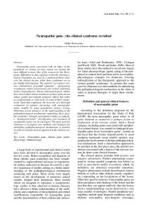
puncture treatment (n = 10). Sham-operated rats were used as controls (n = 9). All rats were involved in the final analysis. nal dorsal horn changed under chronic constriction injury-induced neuropathic injury and after Effects of electroacupuncture and manual electroacupuncture and manual acupuncture treatments. acupuncture on mechanical hypersensitivity of Ephrin-B1, ephrin-B3, EphB1, and EphB2 mRNA levels chronic constriction injury rats in the spinal dorsal horn were significantly enhanced As shown in Figure 1A, the basal paw withdrawal after chronic constriction injury-induced neuropathic inthreshold was similar for all groups before surgery. jury (P < 0.05 to P < 0.01) (Figure 2). Furthermore, this Compared with control rats, chronic constriction injury upward trend of these genes was not inhibited by rats displayed a profound decrease in ipsilateral paw electroacupuncture and manual acupuncture treatments withdrawal threshold in a time-dependent manner, except for ephrin-B1, whose mRNA levels were not difreaching the lowest level at day 9 after chronic conferent between the electroacupuncture and control striction injury of the sciatic nerve. No significant change groups. In contrast, the mRNA levels of EhpB3 and was observed among chronic constriction injury, EphB4 were significantly up-regulated after electroacupuncture and manual acupuncture groups at electroacupuncture and manual acupuncture treatments day 7 post- injury. The paw withdrawal threshold in the (P < 0.01; Figure 2). Specifically, the mRNA levels for contralateral hind paw of all the rats was not significantly EphB3, EphB6 and ephrin-B3 were dramatically indifferent from the pre-lesion baseline value at all time creased in the spinal dorsal horn after points (data not shown). The paw withdrawal threshold in electroacupuncture stimulation compared with the conthe ipsilateral hind paw of chronic constriction injury trol, chronic constriction injury and manual acupuncture group without treatments remained significantly lower groups. than that in the control group at various time points measured after chronic constriction injury of the sciatic A nerve (P < 0.01). Electroacupuncture and manual acupuncture stimulations were performed every other day from day 8. One day after the first treatments (day 9), hypersensitivity remained unchanged. However, the paw withdrawal threshold in the ipsilateral hind paw increased gradually following the electroacupuncture and manual acupuncture treatments. At day 17, mechanical allodynia was significantly alleviated in the electroacupuncture and manual acupuncture groups compared with the chronic constriction injury group (P < 0.01), and this trend lasted until the end of the experiment (Figure 1A). On the other hand, the electroacupuncture stimulation showed a better tendency to improve the paw withdrawal threshold than manual acupuncture stimulation (Figure 1A). The area under the curve for the paw withdrawal threshold changes over time (Figure 1B) also indicated that electroacupuncture treatment had a better overall effect than manual acupuncture treatment (P < 0.05). Effects of electroacupuncture and manual acupuncture on mRNA expression of ephrin-Bs and EphBs in the spinal dorsal horns of chronic constriction injury rats Real-time PCR analyses were used to investigate whether the mRNA expression levels of ephrin-Bs (ephrin-B1, ephrin-B2, and ephrin-B3) and EphBs (EphB1, EphB2, EphB3, EphB4, and EphB6) in the spi2352
B
Figure 1 Effect of electroacupuncture (EA) and manual acupuncture (MA) stimulation on mechanical hypersensitivity induced by chronic constriction injury (CCI) in rats. (A) Mechanical threshold of rats at different time points measured by the hind paw withdrawal response to von Frey hair stimulation. (B) Area under the curve of graph A (from day 0 to day 37). The data are expressed as mean ± SEM. There are nine rats in each group, except for the CCI + MA treatment group, which contains 10 rats. aP < 0.01, vs. control group; b P < 0.01, vs. CCI group; cP < 0.05, dP < 0.01, vs. CCI + MA group. Two-way analysis of variance followed by Bonferroni’s post hoc test (A) or one-way analysis of variance followed by Bonferroni’s post hoc test (B) was used.
Ju ZY, et al. / Neural Regeneration Research. 2013;8(25):2350-2359.
Figure 2 Effects of electroacupuncture (EA) and manual acupuncture (MA) on ephrin-B and EphB gene expression in the spinal dorsal horns of chronic constriction injury (CCI) rats. The mRNA levels were detected by real-time PCR, and the data (fold change in target gene relative to control) are expressed as mean ± SEM of eight rats in each group. aP < 0.05, bP < 0.01, vs. control group; cP < 0.01, vs. CCI group; dP < 0.01, vs. CCI + MA group. One-way analysis of variance, followed by Bonferroni’s post hoc test was performed.
Effects of electroacupuncture and manual acupuncture on the expression of ephrin-B3 protein in the spinal dorsal horn of chronic constriction injury rats Because ephrin-B3 was the only ephrin-B family member that was significantly up-regulated at the mRNA level in the spinal dorsal horn region after electroacupuncture treatment (Figure 2), and because electroacupuncture treatment had a better therapeutic effect in chronic constriction injury rats than manual acupuncture treatment (Figure 1), we focused on the expression of this protein after electroacupuncture and manual acupuncture treatments. Inconsistent with the mRNA results (Figure 2), chronic constriction injury-induced neuropathic injury did not significantly increase ephrin-B3 protein levels in the spinal dorsal horn (Figure 3). However, the absorbance of the bands for ephrin-B3 protein on western blots of samples from the electroacupuncture and manual acupuncture groups was significantly higher than that in western blots of samples from the control and chronic constriction injury groups (P < 0.05 or P < 0.01; Figure 3). Furthermore, the ephrin-B3 protein level was more significantly increased in the electroacupuncture group than in the manual acupuncture group (P < 0.05; Figure 3).
DISCUSSION In the present study, we investigated whether repeated long-term electroacupuncture and manual acupuncture
treatments could improve the chronic constriction injury-induced neuropathic injury in rats. We also tested whether the electroacupuncture- and manual acupuncture-induced therapeutic effect involved spinal ephrin-B/ EphB signaling modulation. A
EphrinB3
45 kDa
GAPDH
36 kDa
B
Figure 3 Effects of electroacupuncture (EA) and manual acupuncture (MA) on the expression of ephrin-B3 protein in the spinal dorsal horns of chronic constriction injury (CCI) rats. (A) Electrophoresis results. (B) Quantitative analysis of the expression of ephrin-B3 protein. Data (absorbance ratio to GAPDH) are expressed as mean ± SEM of four rats in each group. aP < 0.01, vs. control group; bP < 0.05, cP < 0.01, vs. CCI group; dP < 2353 0.01, vs. CCI + MA group. One-way analysis of variance, followed by Bonferroni’s post hoc test was performed.
Ju ZY, et al. / Neural Regeneration Research. 2013;8(25):2350-2359.
Although repeated electroacupuncture and manual acupuncture treatments did not totally resolve the hypersensitivity caused by chronic constriction injury within the time frame of 37 days for this study, they significantly improved mechanical thresholds compared with chronic constriction injury groups at day 6 after the first treatment. Repeated electroacupuncture and manual acupuncture up-regulated mRNA levels for EphB3, EphB6 and ephrin-B3 in the spinal dorsal horn. Furthermore, ephrin-B3 protein levels were significantly increased after electroacupuncture and manual acupuncture, and positively correlated with the therapeutic effects of acupuncture treatments. This result revealed that long-term acupuncture treatment at bilateral Zusanli and Sanyinjiao can activate spinal ephrin-B/EphB signaling to alleviate mechanical hypersensitivity and improve neural function. Nowadays, electroacupuncture and manual acupuncture treatments are extensively used to alleviate neuropathic pain and improve nervous function in the clinic[42-45]. In manual acupuncture treatment, the acupuncture needle is inserted into the acupoint and twisted up and down by hand until a feeling of “Deqi”, which is described as a sensation of numbness, soreness or heaviness, reflecting the activation of afferent nerve fibers, is experienced[46]. Traditional acupuncturists adjust the acupuncture manipulation to achieve therapeutic purposes according to the conditions of patients[47]. In electroacupuncture treatment, a stimulating current is delivered to acupoints via the needles using an electrical stimulator. Compared with manual acupuncture stimulation, electroacupuncture stimulation is easy to control and accurately reproduce. Little is known about the different therapeutic benefits of electroacupuncture and manual acupuncture treatments. Several studies have found that electroacupuncture stimulation produces greater pressure pain detection threshold elevations and is more effective in some aspects in regulating disturbed estrous cyclicity compared with manual acupuncture stimulation[48-49]. However, another study showed that there was no difference in alleviating headache, trigeminal neuralgia, and retro-auricular pain between electroacupuncture and manual acupuncture[44]. The present study demonstrated that electroacupuncture treatment was superior to manual acupuncture treatment in terms of improvements after chronic constriction injury and an overall analgesic effect. However, we have to point out that this result does not suggest that 2354
electroacupuncture had a better therapeutic effect than manual acupuncture. In electroacupuncture stimulation, acupoints were stimulated by a continued stimulus and the current intensity was gradually increased during treatment. Based on a previously described method[36], the needles were only operated for 15 seconds every 10 minutes in the manual acupuncture stimulation used in this study. Obviously, the intensity of stimulus in the electroacupuncture treatment was stronger than the intensity in the manual acupuncture treatment. Previous studies have shown that acupuncture with optimal high-intensity stimulation is more effective than treatments with less stimulation, such as minimal acupuncture[50-51]. The results from this study confirmed that the intensity of acupuncture plays an important role in improving neuropathic injury and pain. Ephrin-B/EphB signaling is an important signaling pathway for pain modulation and is involved in neuronal development and the response to injury[26, 28, 32]. After peripheral nerve injury, the levels of ephrin-B1 and EphB1 proteins were significantly up-regulated in the spinal dorsal horn[52-53]. Furthermore, ephrin-B1-Fc could produce a dose- and time-dependent thermal and mechanical hyperalgesia via intrathecal injection, accompanied by an increase in the levels of spinal phosphorylated mitogen-activated protein kinases and c-Fos[28]. The mitogen-activated protein kinases, including p38, extracellular signal-regulated kinase, and c-Jun N-terminal kinase, are a family of serine/threonine protein kinases that transduce extracellular stimuli into intracellular posttranslational and transcriptional responses. Their activation is involved in the modulation of nociceptive information and peripheral and central sensitization produced by intense noxious stimuli[54-58]. Activation of ephrin-B2/EphB1 signaling in the spinal dorsal horn and primary sensory neurons activates astrocytes and microglial cells and up-regulates the level of phosphorylation of NR1 and NR2B receptors, and up-regulates the phosphorylation level of Src within the N-methyl-D-aspartate receptor complex, and increases the activity of matrix metalloproteinase-2/9 and subsequent Ca2+-dependent signals, all of which result in pain behaviors[27]. Intrathecal application of blocking reagents, including EphB1-Fc, EphB2-Fc and ephrin-B1-Fc, could relieve nerve injury-induced pain[52, 59]. In the current study, real- time PCR data also demonstrated significant overexpression of EphB1, EphB2 and ephrin-B1 mRNA levels after chronic constriction injury in the spinal dorsal horn. However, previous studies have shown that acupuncture treatment modulates the expression and phosphorylation of spinal N-methyl-D-aspartate receptor
Ju ZY, et al. / Neural Regeneration Research. 2013;8(25):2350-2359.
subunits and inhibits p38MAPK and extracellular signal-regulated kinase activation in the spinal dorsal horn to attenuate mechanical allodynia in a neuropathic pain model[60-64]. Although the up-regulation of these three genes seemed not to be inhibited by acupuncture stimulations, this does not show that acupuncture for the treatment of neuralgia does not exert effects on ephrin-B1–EphB1/B2 signaling, which should be determined in further neuropharmacological and behavioral tests. Ephrin-B3, a member of the ephrin-B gene family, plays an important role in brain development and repair after nervous system injury[65-66]. Ephrin-B3 and EphB1 cooperatively regulate the proliferation and migration of neural progenitors in the hippocampus, suggesting that they may act as candidate targets for modulating the production and integration of new neurons to treat neurodegenerative diseases or brain injury[67]. A trans-synaptic interaction between ephrin-B3 and EphB2 regulates synapse density and the formation of dendritic spines by inhibition of postsynaptic Ras/mitogen-activated protein kinase signaling[68]. Ephrin-B3 is expressed in the injured mouse optic nerve where it recruits macrophages, which express EphB3, to promote sprouting of damaged retinal axons[69]. EphA4 can initiate cell death in the absence of ephrin-B3. However, infusion of soluble ephrin-B3 reduced cell death in the germinal zones[70]. The present study showed that only the ephrin-B3 mRNA level was significantly increased in the spinal dorsal horns of neuropathic injury rats after electroacupuncture treatment among three ephrin-Bs. Moreover, electroacupuncture and manual acupuncture increased the ephrin-B3 protein level in the spinal dorsal horn, especially after electroacupuncture stimulation, consistent with the alleviation of neuropathic hypersensitivity after acupuncture in the present study. Taken together, our findings provide new information that acupuncture increases the expression and synthesis of ephrin-B3 in the spinal dorsal horn, which subsequently promotes the restoration of injured neural function and finally results in neuropathic pain alleviation. Nevertheless, the underlying mechanism requires further investigation. In summary, the present study shows for the first time that the effects of acupuncture on neuropathic injury and nociception are associated with an increase in the levels of ephrin-B3 in the spinal dorsal horns of chronic constriction injury rats. This new information may improve our understanding of the mechanisms underlying the effects of acupuncture on neuropathic injury and pain.
MATERIALS AND METHODS Design A randomized, controlled animal experiment. Time and setting This study was performed at the Department of Acupuncture, Shanghai University of Traditional Chinese Medicine and at the Research & Development Department, National Engineering Research Center for Biochip Technology, Shanghai, China, from March 2011 to September 2012. Materials A total of 37 male Sprague-Dawley rats were obtained from the Experimental Animal Center, Shanghai University of Traditional Chinese Medicine in China (License No. SCXK (Hu) 2007-0005), aged 3–4 months, weighing 180–220 g. Animals were housed under a 12-hour light/ dark cycle, and allowed free access to food and water. The room temperature was maintained at 23 ± 1°C and relative humidity at 40–60%. Rats were acclimatized to the environment for 7 days prior to experiment. All procedures were in accordance with the Guidance Suggestions for the Care and Use of Laboratory Animals, formulated by the Ministry of Science and Technology of China[71]. Methods Animal model of chronic constriction injury of the sciatic nerve Rats were anesthetized with an intraperitoneal injection of sodium pentobarbital (40 mg/kg). Chronic constriction injury of the sciatic nerve was induced as previously described[72]. The biceps femoris and the gluteus superficialis were separated by blunt dissection, and the left sciatic nerve was exposed at the mid-thigh level. Proximal to the trifurcation, 10 mm of nerve was carefully freed from the adhering tissue. The ligatures (4-0 silk) were tied loosely around the nerve with 1 mm spacing, until they elicited a brief twitch in the respective hindlimb, but did not interrupt the epineurial circulation. The skin was sewed up with silk sutures (4-0) and the rats were left to recover in heated cages. Sham-operated rats underwent exposure of the left sciatic nerve without ligature. Application of acupuncture Electroacupuncture or manual acupuncture was given to the respective groups of rats once every other day from days 8 to 36 after nerve injury. The rats were placed in well-ventilated Plexiglas restraint barrels with the hind limb exposed for needling. Stainless-steel needles 2355
Ju ZY, et al. / Neural Regeneration Research. 2013;8(25):2350-2359.
(Huatuo acupuncture needle, Suzhou Medical Appliance Factory, Suzhou, Jiangsu Province, China), 0.3 mm in diameter and 3 mm in length, were bilaterally inserted into the hind legs, one at Zusanli and the other at Sanyinjiao. For electroacupuncture stimulation[39], constant- current square-wave electrical stimulation generated by a programmed pulse generator (LH200, Beijing Huawei Industrial Developing Company, Beijing, China) was given via the two needles for 30 minutes. The frequency was set at 15 Hz and the intensity of stimulation was increased stepwise from 0.5 to 1.0 and then 1.5 mA, with each step lasting for 10 minutes. For manual acupuncture stimulation[36], a stainless-steel needle was inserted to a depth of 0.3 mm and rotated at a rate of two spins per second for 15 seconds in each acupoint’s direction, for a total of 30 seconds of needle rotation. Subsequently, the animals were allowed to rest with the needles still inserted for an additional 30 minutes. Behavioral test for assessment of mechanical thresholds Evaluations were conducted the day after acupuncture. Mechanical allodynia was measured based on the hind paw withdrawal response to von Frey hair stimulation as previously described[73-74]. A series of nine calibrated von Frey hairs were applied to the central region of the plantar surface of one hind paw in ascending order (1, 1.4, 2, 4, 6, 8, 10, 15, and 26 g). The hair was applied only when the rat was stationary and standing on all four paws. The stimulation was sustained for 2 seconds. A withdrawal response was considered valid only if the hind paw was completely removed from the customized platform. Each hair was applied five times at 5-second intervals. If a withdrawal response was not induced more than twice during five applications of a hair, the next ascending hair in the series was applied in a similar manner. Once the hind paw was withdrawn from a particular hair in three out of the five consecutive applications, the rat was considered responsive to that hair. The next descending hair was applied until the hind paw was withdrawn less than three out of the five times. The paw withdrawal threshold was defined as the lowest hair force in grams that produced at least three withdrawal responses in five tests.
Table 1 Symbol EphB1 EphB2 EphB3 EphB4 EphB6 ephrin-B1 ephrin-B2 ephrin-B3 2356 GAPDH
Quantitative real-time PCR Rats were deeply anesthetized with sodium pentobarbital (60 mg/kg, intraperitoneally) and the lumbar enlargements of the spinal dorsal horns of rats were quickly removed, frozen immediately in liquid nitrogen, and stored at –80°C for further use. Total RNA was isolated using Trizol reagent (Invitrogen, Carlsbad, CA, USA). The RNA concentration and purity were analyzed using a Nanodrop spectrophotometer (Nanodrop Technologies, Wilmington, DE, USA), with the spectral absorption at 260 and 280 nm. RNA quality was assessed using a Lab-on-chip Bioanalyzer 2100 (Agilent Technologies, Palo Alto, CA, USA). For cDNA synthesis, oligo(dT) primers, 1 μg of each total RNA sample, and the RevertAidTM First Strand cDNA Synthesis Kit (Fermentas, Burlington, CA, USA) were used, following the guidelines of the manufacturer. Real-time PCR was conducted in duplicate with GAPDH as an internal control using the Prism 7900 Sequence Detection System (Applied Biosystems, Foster City, CA, USA) and SYBR® Premix Ex TaqTM II (Takara, Dalian, China). The primer sequences are listed in Table 1. Cycling conditions were as follows: 95°C for 10 minutes, followed by 40 cycles of 15 seconds at 95°C and 1 minute at 60°C. After cycling, a melting protocol was performed for 15 seconds at 95°C, 1 minute at 60°C, and 15 seconds at 95°C, to control for product specificity. The fold change in target gene cDNA level relative to GAPDH was determined as follows: fold change = 2–ΔΔCt, where ΔΔCt = (CtTarget – CtControl) test – (CtTarget – CtControl) control. Ct values were defined as the number of the PCR cycles at which fluorescence signals were detected. Western blot assay The spinal dorsal horn was homogenized in cold lysis buffer (Beyotime, Haimen, Jiangsu Province, China) and centrifuged at 13 200 × g for 15 minutes at 4°C. The amount of total protein in the supernatant was quantified using an Enhanced BCA Protein Assay Kit (Beyotime). Samples (20 μg total protein per loading) were separated on a 10% sodium dodecyl sulfate-polyacrylamide gel and electrotransferred onto polyvinylidene difluoride membranes (Millipore, Bedford, MA, USA).
Sequences of primers used for quantitative real-time PCR GenBank NM_001104528 NM_001127319 NM_001105868 XM_001069453 NM_001107857 NM_017089 NM_001107328 NM_001100980 NM_017008
Forward primer (5′–3′) CGT GCT TGT TCG TGA CAG AT GTT CCC TGG ACT CCT TCC TC CCT ACC CTT GGT GCT GTC AT ACC AAC CCA GCA GTT CTG AC GTT GTG TGC AAG GAA TGT GG GGC AAG CAT GAG ACT GTG AA GCT CAA GTA CCG CAG GAG AC ACG GTC TGT CTT TTG GTT GG TCC TGC ACC ACC AAC TGC TTA G
Reverse primer (5′–3′) CAT ATT CCA GCC CCT TGA GA GTT CAT GTC CGC CAG GTA CT CCC GTC TTT CAC ACA CAC AC AAG TGC AAT CCA ATG GGA AG AAG GAA CTT CAT GGC TGG TG GCT GTG TAT GCT TGC GAT GT CGT AGT GTG GGC AGA AGA CA GCA CAG GAA GGA CAA GAA GC AGT GGC AGT GAT GGC ATG GAC T
Product length (bp) 84 110 144 122 247 205 167 169 102
Ju ZY, et al. / Neural Regeneration Research. 2013;8(25):2350-2359.
The membranes were blocked with 5% non-fat milk overnight at 4°C, and then incubated with primary antibodies recognizing GAPDH (mouse anti-rat monoclonal, 1:5 000; Santa Cruz Biotechnology, Santa Cruz, CA, USA) or ephrin-B3 (rabbit polyclonal, 1:1 000; Abcam, Cambridge, UK) for 2 hours at 22°C. The membranes were incubated with a horseradish peroxidase-conjugated goat anti-mouse IgG (1:5 000; Santa Cruz Biotechnology) or goat anti-rabbit IgG (1:5 000; Santa Cruz Biotechnology) for 2 hours at 22°C. The signal was visualized using Enhanced Chemiluminescence Plus reagent (GE Healthcare, Buckinghamshire, UK) and detected using a Clinx Chemiscope Mini Series Western Blot Imaging System (CLiNX Science Instruments, Shanghai, China). For semiquantitative analysis of ephrin-B3, the integrated absorbance of each band was measured using Image-Pro Plus (Meyer Instruments, Houston, TX, USA). The integrated absorbance was normalized to that of GAPDH and expressed as arbitrary units. Statistical analysis Data are presented as mean ± SEM. Differences in paw withdrawal latency were assessed using two-way analysis of variance with repeated measures in GraphPad Prism Version 5.0 (GraphPad, La Jolla, CA, USA). The Bonferroni’s post hoc test was used to compare data between two groups. For the area under the curve of the paw withdrawal threshold changes over time, comparisons between groups were performed using one-way analysis of variance followed by the Bonferroni’s post hoc test in GraphPad Prism Version 5.0. For real-time PCR and western blot assays, comparisons between groups were performed using one-way analysis of variance with Bonferroni’s post hoc test using SPSS 16.0 software (SPSS, Chicago, IL, USA). A value of P < 0.05 was considered statistically significant.
[5] [6]
[7] [8] [9] [10]
[11]
[12]
[13] [14]
[15]
[16]
[17]
[18]
REFERENCES [1] [2]
[3]
[4]
Bridges D, Thompson SW, Rice AS. Mechanisms of neuropathic pain. Br J Anaesth. 2001;87(1):12-26. Cavenagh J, Good P, Ravenscroft P. Neuropathic pain: are we out of the woods yet? Intern Med J. 2006;36(4): 251-255. Smith BH, Torrance N. Epidemiology of neuropathic pain and its impact on quality of life. Curr Pain Headache Rep. 2012;16(3):191-198. Udina E, Cobianchi S, Allodi I, et al. Effects of activity-
[19]
[20]
[21]
dependent strategies on regeneration and plasticity after peripheral nerve injuries. Ann Anat. 2011;193(4):347-353. Muir D. The potentiation of peripheral nerve sheaths in regeneration and repair. Exp Neurol. 2010;223(1):102-111. Galea MP. Physical modalities in the treatment of neurological dysfunction. Clin Neurol Neurosurg. 2012;114(5): 483-488. Dray A. Neuropathic pain: emerging treatments. Br J Anaesth. 2008;101(1):48-58. Garg G, Adams JD. Treatment of neuropathic pain with plant medicines. Chin J Integr Med. 2012;18(8):565-570. Fornasari D. Pain mechanisms in patients with chronic pain. Clin Drug Investig. 2012;32 Suppl 1:45-52. Sun RQ, Wang HC, Wan Y, et al. Suppression of neuropathic pain by peripheral electrical stimulation in rats: mu-opioid receptor and NMDA receptor implicated. Exp Neurol. 2004;187(1):23-29. Lau WK, Lau YM, Zhang HQ, et al. Electroacupuncture versus celecoxib for neuropathic pain in rat SNL model. Neuroscience. 2010;170(2):655-661. Norrbrink C, Lundeberg T. Acupuncture and massage therapy for neuropathic pain following spinal cord injury: an exploratory study. Acupunct Med. 2011;29(2):108-115. Zhao ZQ. Neural mechanism underlying acupuncture analgesia. Prog Neurobiol. 2008;85(4):355-375. Meng X, Zhang Y, Li A, et al. The effects of opioid receptor antagonists on electroacupuncture-produced anti-allodynia/hyperalgesia in rats with paclitaxel-evoked peripheral neuropathy. Brain Res. 2011;1414:58-65. Kim SK, Park JH, Bae SJ, et al. Effects of electroacupuncture on cold allodynia in a rat model of neuropathic pain: mediation by spinal adrenergic and serotonergic receptors. Exp Neurol. 2005;195(2):430-436. Cabyoglu MT, Ergene N, Tan U. The mechanism of acupuncture and clinical applications. Int J Neurosci. 2006; 116(2):115-125. Park JH, Kim SK, Kim HN, et al. Spinal cholinergic mechanism of the relieving effects of electroacupuncture on cold and warm allodynia in a rat model of neuropathic pain. J Physiol Sci. 2009;59(4):291-298. Choi DC, Lee JY, Moon YJ, et al. Acupuncture-mediated inhibition of inflammation facilitates significant functional recovery after spinal cord injury. Neurobiol Dis. 2010; 39(3):272-282. Yang JL, Chen JS, Yang YF, et al. Neuroprotection effects of retained acupuncture in neurotoxin-induced Parkinson's disease mice. Brain Behav Immun. 2011;25(7):14521459. Liu Z, Ding Y, Zeng YS. A new combined therapeutic strategy of governor vessel electro-acupuncture and adult stem cell transplantation promotes the recovery of injured spinal cord. Curr Med Chem. 2011;18(33):5165-5171. Arvanitis D, Davy A. Eph/ephrin signaling: networks.
2357
Ju ZY, et al. / Neural Regeneration Research. 2013;8(25):2350-2359.
Genes Dev. 2008;22(4):416-429. [22] Salvucci O, Tosato G. Essential roles of EphB receptors and EphrinB ligands in endothelial cell function and angiogenesis. Adv Cancer Res. 2012;114:21-57. [23] Bush JO, Soriano P. Eph/ephrin signaling: genetic, phosphoproteomic, and transcriptomic approaches. Semin Cell Dev Biol. 2012;23(1):26-34. [24] Klein R. Bidirectional modulation of synaptic functions by Eph/ephrin signaling. Nat Neurosci. 2009;12(1):15-20. [25] Chen Y, Fu AK, Ip NY. Bidirectional signaling of ErbB and Eph receptors at synapses. Neuron Glia Biol. 2008;4(3): 211-221. [26] Pasquale EB. Eph-ephrin bidirectional signaling in physiology and disease. Cell. 2008;133(1):38-52. [27] Liu S, Liu WT, Liu YP, et al. Blocking EphB1 receptor forward signaling in spinal cord relieves bone cancer pain and rescues analgesic effect of morphine treatment in rodents. Cancer Res. 2011;71(13):4392-4402. [28] Ruan JP, Zhang HX, Lu XF, et al. EphrinBs/EphBs signaling is involved in modulation of spinal nociceptive processing through a mitogen-activated protein kinases-dependent mechanism. Anesthesiology. 2010;112(5): 1234-1249. [29] Orikawa Y, Kato H, Seto K, et al. Z-360, a novel therapeutic agent for pancreatic cancer, prevents up-regulation of ephrin B1 gene expression and phosphorylation of NR2B via suppression of interleukin-1 beta production in a cancer-induced pain model in mice. Mol Pain. 2010;6:72. [30] Sloniowski S, Ethell IM. Looking forward to EphB signaling in synapses. Semin Cell Dev Biol. 2012;23(1):75-82. [31] Hruska M, Dalva MB. Ephrin regulation of synapse formation, function and plasticity. Mol Cell Neurosci. 2012;50(1): 35-44. [32] Du J, Fu C, Sretavan DW. Eph/ephrin signaling as a potential therapeutic target after central nervous system injury. Curr Pharm Des. 2007;13(24):2507-2518. [33] Xu NJ, Henkemeyer M. Ephrin reverse signaling in axon guidance and synaptogenesis. Semin Cell Dev Biol. 2012;23(1):58-64. [34] Li W, Zheng Z, Keifer J. Transsynaptic EphB/Ephrin-B signaling regulates growth of presynaptic boutons required for classical conditioning. J Neurosci. 2011;31(23): 8441-8449. [35] Sator-Katzenschlager SM, Scharbert G, Kozek-Langenecker SA, et al. The short- and long-term benefit in chronic low back pain through adjuvant electrical versus manual auricular acupuncture. Anesth Analg. 2004;98(5): 1359-1364. [36] Cidral-Filho FJ, da Silva MD, More AO, et al. Manual acupuncture inhibits mechanical hypersensitivity induced by spinal nerve ligation in rats. Neuroscience. 2011;193: 370-376. [37] Zhang Y, Li A, Xin J, et al. Involvement of spinal serotonin receptors in electroacupuncture anti-hyperalgesia in an inflammatory pain rat model. Neurochem Res. 2011; 36(10):1785-1792. [38] Chiu HH, Wu PC. Manual acupuncture for relieving pain
2358
[39]
[40]
[41]
[42]
[43]
[44]
[45]
[46]
[47]
[48]
[49]
[50]
[51]
[52]
associated with panretinal photocoagulation. J Altern Complement Med. 2011;17(10):915-921. Wang K, Zhang R, He F, et al. Electroacupuncture frequency-related transcriptional response in rat arcuate nucleus revealed region-distinctive changes in response to low- and high-frequency electroacupuncture. J Neurosci Res. 2012;90(7):1464-1473. Manni L, Rocco ML, Barbaro Paparo S, et al. Electroacupucture and nerve growth factor: potential clinical applications. Arch Ital Biol. 2011;149(2):247-255. Zhang Z, Wang C, Gu G, et al. The effects of electroacupuncture at the Zusanli acupoint on cancer pain and transient receptor potential vanilloid subfamily 1 expression in Walker 256 tumor-bearing rats. Anesth Analg. 2012;114(4): 879-885. Mischoulon D, Brill CD, Ameral VE, et al. A pilot study of acupuncture monotherapy in patients with major depressive disorder. J Affect Disord. 2012;141(2-3):469-473. Wang LP, Zhang XZ, Guo J, et al. Efficacy of acupuncture for migraine prophylaxis: a single-blinded, double-dummy, randomized controlled trial. Pain. 2011;152(8):1864-1871. Ahn CB, Lee SJ, Lee JC, et al. A clinical pilot study comparing traditional acupuncture to combined acupuncture for treating headache, trigeminal neuralgia and retro- auricular pain in facial palsy. J Acupunct Meridian Stud. 2011;4(1):29-43. Zhang Y, Ding W, Yang Y, et al. Effects of transsylvian-transinsular approach to hypertensive putaminal hematoma operation and electroacupuncture on motor recovery. J Craniofac Surg. 2011;22(5):1626-1630. Xiong J, Liu F, Zhang MM, et al. De-qi, not psychological factors, determines the therapeutic efficacy of acupuncture treatment for primary dysmenorrhea. Chin J Integr Med. 2012;18(1):7-15. Shi GX, Yang XM, Liu CZ, et al. Factors contributing to therapeutic effects evaluated in acupuncture clinical trials. Trials. 2012;13(1):42. Schliessbach J, van der Klift E, Arendt-Nielsen L, et al. The effect of brief electrical and manual acupuncture stimulation on mechanical experimental pain. Pain Med. 2011;12(2):268-275. Feng Y, Johansson J, Shao R, et al. Electrical and manual acupuncture stimulation affect oestrous cyclicity and neuroendocrine function in an 5alpha-dihydrotestosterone-induced rat polycystic ovary syndrome model. Exp Physiol. 2012;97(5):651-662. Shukla S, Torossian A, Duann JR, et al. The analgesic effect of electroacupuncture on acute thermal pain perception-a central neural correlate study with fMRI. Mol Pain. 2011;7(1):45. Hantoushzadeh S, Alhusseini N, Lebaschi AH. The effects of acupuncture during labour on nulliparous women: a randomised controlled trial. Aust N Z J Obstet Gynaecol. 2007;47(1):26-30. Cao JL, Ruan JP, Ling DY, et al. Activation of peripheral
Ju ZY, et al. / Neural Regeneration Research. 2013;8(25):2350-2359.
[53]
[54]
[55]
[56]
[57]
[58] [59]
[60]
[61]
[62]
[63]
ephrinBs/EphBs signaling induces hyperalgesia through a MAPKs-mediated mechanism in mice. Pain. 2008;139(3): 617-631. Song XJ, Cao JL, Li HC, et al. Upregulation and redistribution of ephrinB and EphB receptor in dorsal root ganglion and spinal dorsal horn neurons after peripheral nerve injury and dorsal rhizotomy. Eur J Pain. 2008;12(8): 1031-1039. Ji RR, Baba H, Brenner GJ, et al. Nociceptive-specific activation of ERK in spinal neurons contributes to pain hypersensitivity. Nat Neurosci. 1999;2(12):1114-1119. Jin SX, Zhuang ZY, Woolf CJ, et al. p38 mitogen-activated protein kinase is activated after a spinal nerve ligation in spinal cord microglia and dorsal root ganglion neurons and contributes to the generation of neuropathic pain. J Neurosci. 2003;23(10):4017-4022. Obata K, Yamanaka H, Kobayashi K, et al. Role of mitogen-activated protein kinase activation in injured and intact primary afferent neurons for mechanical and heat hypersensitivity after spinal nerve ligation. J Neurosci. 2004;24(45):10211-10222. Obata K, Noguchi K. MAPK activation in nociceptive neurons and pain hypersensitivity. Life Sci. 2004;74(21): 2643-2653. Ji RR, Gereau RWt, Malcangio M, et al. MAP kinase and pain. Brain Res Rev. 2009;60(1):135-148. Guan XH, Lu XF, Zhang HX, et al. Phosphatidylinositol 3-kinase mediates pain behaviors induced by activation of peripheral ephrinBs/EphBs signaling in mice. Pharmacol Biochem Behav. 2010;95(3):315-324. Kang BR, Ahn CB, Choi BT. N-methyl-D-aspartate antagonist inhibits NR-1 subunit phosphorylation of the spinal N-methyl-D-aspartate receptor induced by low frequency electroacupuncture. Am J Chin Med. 2007; 35(6):987-993. Wang L, Zhang Y, Dai J, et al. Electroacupuncture (EA) modulates the expression of NMDA receptors in primary sensory neurons in relation to hyperalgesia in rats. Brain Res. 2006;1120(1):46-53. Ryu JW, Lee JH, Choi YH, et al. Effects of protein phosphatase inhibitors on the phosphorylation of spinal cord N-methyl-D-aspartate receptors following electroacupuncture stimulation in rats. Brain Res Bull. 2008;75(5): 687-691. Xu KD, Liang T, Wang K, et al. Effect of pre-electroacupuncture on p38 and c-Fos expression in the spinal dorsal horn of rats suffering from visceral pain. Chin Med J (Engl). 2010;123(9):1176-1181.
[64] Choi DC, Lee JY, Lim EJ, et al. Inhibition of ROS-induced p38MAPK and ERK activation in microglia by acupuncture relieves neuropathic pain after spinal cord injury in rats. Exp Neurol. 2012;236(2):268-282. [65] Senturk A, Pfennig S, Weiss A, et al. Ephrin Bs are essential components of the Reelin pathway to regulate neuronal migration. Nature. 2011;472(7343):356-360. [66] Ricard J, Salinas J, Garcia L, et al. EphrinB3 regulates cell proliferation and survival in adult neurogenesis. Mol Cell Neurosci. 2006;31(4):713-722. [67] Chumley MJ, Catchpole T, Silvany RE, et al. EphB receptors regulate stem/progenitor cell proliferation, migration, and polarity during hippocampal neurogenesis. J Neurosci. 2007;27(49):13481-13490. [68] McClelland AC, Hruska M, Coenen AJ, et al. Trans-synaptic EphB2-ephrin-B3 interaction regulates excitatory synapse density by inhibition of postsynaptic MAPK signaling. Proc Natl Acad Sci U S A. 2010;107(19):8830- 8835. [69] Liu X, Hawkes E, Ishimaru T, et al. EphB3: an endogenous mediator of adult axonal plasticity and regrowth after CNS injury. J Neurosci. 2006;26(12):3087-3101. [70] Furne C, Ricard J, Cabrera JR, et al. EphrinB3 is an anti-apoptotic ligand that inhibits the dependence receptor functions of EphA4 receptors during adult neurogenesis. Biochim Biophys Acta. 2009;1793(2):231-238. [71] The Ministry of Science and Technology of the People’s Republic of China. Guidance Suggestions for the Care and Use of Laboratory Animals. 2006-09-30. [72] Bennett GJ, Xie YK. A peripheral mononeuropathy in rat that produces disorders of pain sensation like those seen in man. Pain. 1988;33(1):87-107. [73] Dong Y, Mao-Ying QL, Chen JW, et al. Involvement of EphB1 receptor/ephrinB1 ligand in bone cancer pain. Neurosci Lett. 2011;496(3):163-167. [74] Han M, Huang RY, Du YM, et al. Early intervention of ERK activation in the spinal cord can block initiation of peripheral nerve injury-induced neuropathic pain in rats. Sheng Li Xue Bao. 2011;63(2):106-114. (Reviewed by McGowan D, de Souza M, Shen HF, Chen BY) (Edited by Wang LM, Qiu Y, Li CH, Song LP, Liu WJ, Zhao M)
2359