Loyola University Chicago
Loyola eCommons Dissertations
Theses and Dissertations
2010
Forced-Exercise Alleviates Neuropathic Pain in Experimental Diabetes: Effects on Voltage-Gated Calcium Channels Sahadev A. Shankarappa Loyola University Chicago
Recommended Citation Shankarappa, Sahadev A., "Forced-Exercise Alleviates Neuropathic Pain in Experimental Diabetes: Effects on Voltage-Gated Calcium Channels" (2010). Dissertations. Paper 187. http://ecommons.luc.edu/luc_diss/187
This Dissertation is brought to you for free and open access by the Theses and Dissertations at Loyola eCommons. It has been accepted for inclusion in Dissertations by an authorized administrator of Loyola eCommons. For more information, please contact
[email protected].
This work is licensed under a Creative Commons Attribution-Noncommercial-No Derivative Works 3.0 License. Copyright © 2010 Sahadev A. Shankarappa
LOYOLA UNIVERSITY CHICAGO
FORCED-EXERCISE ALLEVIATES NEUROPATHIC PAIN IN EXPERIMENTAL DIABETES: EFFECTS ON VOLTAGE-GATED CALCIUM CHANNELS
A DISSERTATION SUBMITTED TO THE FACULTY OF THE GRADUATE SCHOOL IN CANDIDACY FOR THE DEGREE OF DOCTOR OF PHILOSOPHY
PROGRAM IN NEUROSCIENCE
BY SAHADEV A SHANKARAPPA CHICAGO, ILLINOIS MAY 2010
Copyright by SAHADEV SHANKARAPPA, 2010 All rights reserved
ACKNOWLEDGEMENTS Firstly, I would like to acknowledge Dr. Evan B. Stubbs, Jr., who has been my graduate advisor, mentor, and racquetball teacher, for his guidance in helping me to understand, critically think, and do science in an organized and logical manner. I sincerely thank Dr. Erika S. Piedras-Rentería, who has been my second mentor, for all her help, suggestions, and guidance for the electrophysiology component of this dissertation. I also thank the members of my graduate committee, Dr. Edward J. Neafsey, Dr. Morris Fisher and Dr. Gwendolyn Kartje, for their valuable suggestions and encouragement. I acknowledge the Arthur J Schmitt foundation for awarding me the Schmitt fellowship, and the Veteran Affairs department of rehabilitation services for the financial assistance, that made this study possible. I would like to thank my colleagues, Dr. Jason Sarkey, Dr. Cynthia Von ee and Mr. Michael Richards, for their assistance, advice and encouragement during my graduate career. I thank my mother, Gowthami; my father, Shankar; and my sister, Parvathi; for their unrelenting emotional support throughout my career. I thank my wife Shivanee, and my daughter Aarika without whom I would not have come this far.
iii
“To my lovely wife Shivanee and my beautiful daughter Aarika
TABLE OF CONTENTS ACKNOWLEDGEMENTS
iii
TABLE OF CONTENTS
v
LIST OF TABLES
ix
LIST OF FIGURES
x
LIST OF ABBREVIATIONS
xii
ABSTRACT
xiv
CHAPTER 1. INTRODUCTION
16
STATEMENT OF PROBLEM
16
HYPOTHESIS
19
SPECIFIC AIMS
19
LITERATURE REVIEW
21
DIABETES MELLITUS
21
Epidemiology of DM
23
Complications of DM
23
DIABETIC NEUROPATHY
25
Pathogenic mechanisms of diabetes-associated nerve injury
25
Neuropathic pain in diabetic neuropathy
28
Experimental animal models of diabetic neuropathy
29
Streptozotocin (STZ)-induced DM
30
STZ-induced diabetic polyneuropathy
32
v
Treatment options in diabetic neuropathy
32
Exercise as a therapeutic strategy
33
Exercise and voltage-gated calcium channels
34
VOLTAGE GATED CALCIUM CHANNELS
36
High-voltage activated Ca2+ channel isoforms
39
Low-voltage activated Ca2+ channels
41
Role of voltage-gated Ca2+ channels in neuropathic pain
42
2. FORCED-EXERCISE ALLEVIATES NEUROPATHIC PAIN IN EXPERIMENTAL DIABETES: EFFECTS ON VOLTAGE-GATED CALCIUM CHANNELS
46
ABSTRACT
46
INTRODUCTION
48
MATERIALS AND METHODS
51
Animal care
51
Induction of experimental diabetes mellitus
51
Experimental exercise regimens
52
Tactile-responsiveness to Von-Frey filaments
53
Thermal responsiveness to infra-red heat stimulus
54
Peripheral nerve conduction studies
55
Dissociation of dorsal root ganglia neurons
57
Voltage clamp electrophysiology
57
Statistical analysis
59
vi
RESULTS
60
Streptozotocin-induced diabetes and voluntary-exercise
60
Streptozotocin-induced diabetes and forced-exercise
60
Forced-exercise protects against STZ-induced diabetes-associated nerve conduction deficits
61
Forced-exercise delays onset of diabetes-associated hyperalgesia
62
Responsiveness to thermal stimulus is not altered by forced-exercise
63
Naloxone reverses forced-exercise induced analgesia
64
Forced-exercise prevents diabetes associated alteration in Ca2+ current densities
64
Diabetes-associated changes in LVA Ca2+ current steady-state inactivation
66
DISCUSSION
79
3. D-GLUCOSE DEPENDENT MODULATION OF CaV3.2 α1H CHANNEL FUNCTION
85
ABSTRACT
85
INTRODUCTION
87
MATERIALS AND METHODS
89
Cell culture
89
Voltage Clamp Electrophysiology
89
Statistical Analysis
91
RESULTS
92
D-Glucose induced enhancement of T-type currents in HEK-293 cells 92 D-glucose induced alteration of CaV3.2 channel gating properties
92
Effect of D-glucose on CaV3.2 channel steady state properties
93
vii
Aldose reductase inhibition does not alter D-glucose induced enhancement of CaV3.2 currents DISCUSSION
93
104
SUMMARY
107
REFERENCES
111
VITA
132
viii
LIST OF TABLES
Table
Page
1. Classification of Diabetes Mellitus
22
2. Clinical classification of glucose tolerance based on plasma glucose levels.
23
3. Types, symptoms, and signs of diabetic neuropathy.
26
4. Mechanisms of diabetes induced nerve injury.
28
5. Animal models of DM and its subtypes
30
6. Pharmacology and function of the α1 Ca2+ channel subunit.
37
7. Evoked compound muscle action potential response from the sciatic nerve of STZ-diabetic rats subjected to sedentary or forced-exercise.
71
8. Conduction velocity calculated from the tail nerve in diabetic rats subjected to forced-exercise.
72
9. Passive membrane properties of dissociated small diameter DRG neurons.
78
10. Comparison of in vitro and in vivo glucose concentrations.
95
11. Osmolarity measurements of normal and high-glucose containing media. ......... 95
ix
LIST OF FIGURES
Figure
Page
1. Schematic of voltage-gated calcium channel.
38
2. Evoked compound muscle action potential recording from the rat sciatic nerve
56
3. Effect of STZ-treatment on voluntary wheel running in SD rats.
67
4. Effect of forced-exercise on progression of experimental diabetes.
68
5. Effect of forced-exercise on the onset and progression of tactile hypersensitivity in STZ-treated rats.
69
6. Effect of forced-exercise on thermal responsiveness in STZ-treated rats.
70
7. Effect of naloxone on forced-exercise induced analgesia.
73
8. Percentage of small, medium and large diameter DRG neurons harvested from naïve adult rat.
74
9. Forced-exercise attenuates diabetes-associated enhancement of HVA Ca2+ current density in dissociated small diameter DRG neurons.
75
10. Effect of forced-exercise on LVA Ca2+ current density in dissociated small diameter DRG neurons.
76
11. Forced-exercise prevents diabetes-associated increase in LVA Ca2+ channel availability.
77
12. D-glucose enhances CaV3.2 current densities in HEK-293 cells.
96
x
13. CaV3.2 current densities from HEK-293 cells cultured for 14 days in normal-glucose containing media.
97
14. D-glucose enhances activation kinetics of CaV3.2 channels in HEK-293 cells.
98
15. Effect of D-glucose on inactivation kinetics of CaV3.2 channels in HEK293 cells.
99
16. Effect of D-glucose on deactivation kinetics of CaV3.2 channels in HEK293 cells.
100
17. Effect of D-glucose on steady state inactivation of CaV3.2 channels in HEK-293 cells.
101
18. Effect of D-glucose on steady state activation of CaV3.2 channels in HEK-293 cells.
102
19. Effect of statil on D-glucose enhanced CaV3.2 current densities in HEK293.
103
20. Working hypothesis to explain how forced-exercise may mediate its protection against diabetes-associated neuropathic pain
108
xi
LIST OF ABBREVIATIONS
AGE
Advanced Glycated End product
CDC
Center for Disease Control
BBB
Blood Brain Barrier
BNB
Blood Nerve Barrier
CMAP Compound Muscle Action Potential DAG
Diacylglycerol
DHP
Dihydropyridines
DPN
Diabetic Polyneuropathy
DM
Diabetes Mellitus
DRG
Dorsal Root Ganglion
GLUT Glucose Transporter HEK
Human Embryonic Kidney
HHS
Hyperglycemic Hyperosmolar State
HVA
High Voltage Activated
JNK
C-Jun B-terminal kinase
LVA
Low Voltage Activated
MNCV Motor Nerve Conduction Velocity NAD
Nicotinamide Adenine Dinucleotide xii
PKC
Protein Kinase C
RAGE Receptor for Advanced Glycated End product RDNS Rochester Diabetic Neuropathy Study SNCV Sensory Nerve Conduction Velocity SSI
Steady State Inactivation
STZ
Streptozotocin
VGCC Voltage Gated Calcium channels WHO World Health Organization τ On
Tau On
τ Off
Tau Off
xiii
ABSTRACT
Diabetes mellitus (DM) is a metabolic disorder that affects an estimated 171 million people world wide. DM and its related complications are among the leading cause of adult blindness and renal failure in the developed nations. Equally alarming, greater than half of all patients with long-standing diabetes develop polyneuropathy, a debilitating progressive deterioration of peripheral and autonomic nerves. Currently, diabetic polyneuropathy (DPN) is untreatable. Maintenance of euglycemia as an indirect means of delaying the development of diabetic complications is the recommended therapeutic approach. Innovative treatment strategies designed to prevent or delay peripheral nerve injury in the diabetic patient are critically needed. Moderate exercise is a safe and integral approach to the management of patients with diabetes. Recent clinical studies suggest that exercise may help in the treatment of DPN. However, the mechanism by which exercise protects against diabetes-induced nerve dysfunction is unknown. In this dissertation we hypothesized that forced-exercise protects against experimental DPN by preventing glucose-associated alterations of voltage-gated calcium currents (VGCC) in small diameter dorsal root ganglion (DRG) neurons. Using behavioral, nerveelectrophysiology and patch-clamp methodology we examined the functional consequences of forced-exercise (treadmill, 5.4 km/week) on VGCC in dissociated small diameter DRG neurons from rats conferred diabetic by streptozotocin (STZ) treatment. Vehicle treated rats were used as controls. Exercised-STZ, rats in comparison to xiv
sedentary-STZ, rats demonstrated a 4 week delay in the onset of tactile hyperalgesia that was independent of changes in blood glucose levels. Interestingly, forced-exercise induced protection against diabetes-induced tactile hyperalgesia was reversed in a dose dependent manner by the opioid antagonist, naloxone. Forced-Exercise also prevented peripheral nerve conduction deficits in STZ–treated rats. Small diameter DRG neurons harvested from hyperglycemic sedentary-STZ rats with demonstrated hyperalgesia exhibited 2-fold increase in peak high-voltage activated (HVA) Ca2+ current density and low-voltage activated (LVA) Ca2+ current component. The steady-state inactivation (SSI) (measure of channel availability) of LVA currents demonstrated a rightward shift in the sedentary-STZ rats (+7.5 mV shift; V50 = -50.9 ± 0.6 mV; vehicle treated rats V50 = -58.4 ± 0.9 mV). Forced-exercise prevented the increase in both, peak HVA Ca2+ current density and LVA SSI shift (V50 = -58.2 ± 1.4 mV), but did not alter LVA current component. We conclude that forced-exercise delayed the onset of diabetic tactile hyperalgesia by preventing the alteration of VGCCs in small diameter DRG neurons, possibly by decreasing total calcium influx and dampening neuronal over-excitability.
xv
CHAPTER ONE INTRODUCTION STATEMENT OF PROBLEM
Diabetes mellitus (DM) is a group of metabolic disorders sharing hyperglycemia as a common phenotype that, when poorly managed, results in debilitating retinal, renal, and neurologic complications (Nathan, 1996). The prevalence of DM and its hyperglycemic-related complications are at near epidemic proportions in the U.S., affecting approximately 24 million Americans, or greater than 8% of the population (Center for Disease Control, 2005). In 2007, almost 25% of the population aged 60 years and above had diabetes. In addition, another 57 million people are estimated to have prediabetes, a condition that increases the risk of developing DM. (Center for Disease Control, 2008). Strikingly, DM is the seventh leading cause of death in the United States and globally 5% of all deaths are attributed to DM. Complications of DM, which include heart diseases, blindness, kidney failure, and lower limb amputations pose a significant financial burden on the health care system. The direct and indirect costs in the U.S. for medical services and lost productivity due to DM is approximately 132 billion dollars (Hogan et al., 2003). Greater than half of all patients with DM develop neurological complications (Pirart, 1977). Although varied, the most common neurologic complication seen among 16
17 DM patients includes distal symmetric polyneuropathy (DPN), a progressive deterioration of the peripheral and autonomic nerves. DPN is causally associated with greater than 200,000 cases of diabetic foot ulcers and 71,000 amputations per year (Center for Disease Control, 2008). The primary risk factor for DPN is hyperglycemia (Perkins et al., 2001). Independent secondary risk factors include cigarette smoking, hypercholesterolemia, alcohol consumption and hypertension (Adler et al., 1997). Clinically, DPN is characterized by chronic peripheral pain described as a burning, pricking or tingling sensation with symmetric stocking glove distribution. Several pathogenic mechanisms have been proposed to explain the development of DPN, including activation of the polyol pathway secondary to hyperglycemia, accumulation of advanced glycation end products, vascular insufficiency, neurotrophic factor deficiency, and neuronal membrane ion channel dysfunction (Gooch and Podwall, 2004). Current therapeutic interventions for the treatment of DPN are largely limited to pain management and strict glycemic control. Gabapentin, tricyclic antidepressants, duloxetine (serotonine-norepinephrine reuptake inhibitor), opioids, and topical capsaicin cream are currently prescribed, with mixed results. Results from clinical trials using nerve growth factor therapy, aldose reductase inhibitors, or acetyl carnitine have experienced limited success (Ziegler and Luft, 2002). Innovative interventional or preventive strategies are required to address the needs of the ever growing DPN patient population. Exercise training, in combination with pharmacological intervention, is now
18 recognized as a cornerstone of treatment for DM. Aerobic exercise without or with resistance training has shown to have beneficial effects in DM (Castaneda et al., 2002). Previous studies have shown that long term aerobic exercise can modify the natural history of DPN in diabetic patients (Balducci et al., 2006). Physical exercise, in addition to improving blood glucose control, also exerts protective effects on cardiovascular and endothelial function (Maiorana et al., 2001; Fuchsjager-Mayrl et al., 2002). Less, however, is known about the effects of exercise on the onset and progression of diabetes associated complications including DPN (Tesfaye et al., 1992; Richardson et al., 2001). Patients with DPN often exhibit hyperalgesia or enhanced sensitivity to pain (Baron et al., 2009), which contributes to the morbidity in diabetes. The cause of impaired pain tolerance remains unclear. Early experimental studies suggest that intermittent hyperglycemia damages sensory neurons and increases spontaneous C-fiber firing, resulting in neuropathic pain (Chen and Levine, 2001). Evidence to support a protective effect of moderate aerobic exercise against the development of neuropathic complications in diabetic patients is mounting (Balducci et al., 2006), raising interest in aerobic exercise as a putative therapeutic intervention for the management of pain in diabetic neuropathy. The cellular mechanism(s) by which neuropathic pain develops in the diabetic patient is poorly understood, but may involve remodeling of voltage- or ligand-gated ion channels (Hall et al., 1995; Cao, 2006; Jagodic et al., 2007), resulting in abnormal enhancement of sensory neuron excitability (Ikeda and Dunlap, 2007).
19 Little is known about the effect of exercise on the functioning of ion channels. Modulation of voltage gated calcium channels (VGCC) in response to exercise has been recently reported in swine coronary smooth muscle cells (Bowles, 2000) and treadmill trained rat ventricular myocytes (Wang et al., 2008). However, there have been no studies to date describing the effect of exercise on neuronal VGCC’s associated with neuropathic pain. In this dissertative research study, we examined whether exercise attenuates the onset and progression of diabetic neuropathy in an animal model of DM. Further, we examined whether forced-exercise can prevent or delay the onset of neuropathic pain by attenuating diabetes-associated increased functioning of voltage-gated calcium channels in the dorsal root ganglion neurons. HYPOTHESIS Forced-exercise will protect against the onset of tactile hyperalgesia in streptozotocin-induced hyperglycemic rats by preventing the over-activity of voltagegated calcium channels in the small diameter dorsal root ganglion neurons. SPECIFIC AIMS This hypothesis will be tested using the following three Specific aims: Specific Aim 1. We will determine the effect of forced-exercise on the onset and progression of peripheral nerve injury in STZ-induced diabetic rats, as quantitated by evoked-response electrophysiology,
tactile
withdrawal
threshold,
and
thermal-evoked
hind-paw
20 withdrawal latency response times. Vehicle- and STZ-treated non-exercised rats will be included as sedentary controls for comparison. Specific Aim 2. We will determine the effect of forced-exercise on high- and low-voltage activated Ca2+ current densities and low-voltage activated Ca2+channel steady state properties in small diameter dorsal root ganglion neurons harvested from STZ-treated rats, as quantitated by whole cell patch-clamp electrophysiology. Non-exercised veh-and STZ-treated rats will be used as controls. Specific Aim 3. We will determine the effect of high D-glucose on current densities, channel kinetics and steady state properties from HEK-293 cells stably transfected with the CaV3.2 α1H channel subunit, using whole cell patch-clamp electrophysiology.
21 LITERATURE REVIEW DIABETES MELLITUS Insulin, produced by the β cells in the islets of Langerhans of the endocrine pancreas, is responsible for glucose uptake into cells of the liver, muscle and fat. Dysregulation of insulin production, secretion, or action results in abnormalities of blood glucose homeostasis, resulting in diabetes mellitus (DM). The syndrome of DM comprises a group of chronic disorders, sharing hyperglycemia as a common phenotype (Table 1). Overtime patients with long term DM develop multi-organ complications affecting the eye, kidney and peripheral/autonomic nerves (Nathan, 1996). The hallmark of DM is a sustained increase in blood glucose levels, with clinical diagnosis primarily based on blood glucose level measurements (Table 2). DM is classified into several types (Table 1), of which DM type 1 and 2 (Himsworth, 1949) are more common. DM type 1 occurs as a result of autoimmunity that ultimately destroys the insulin- producing pancreatic β cells (Mandrup-Poulsen, 1996), leading to the development of severe hypoinsulinemia. The autoimmune processes are likely to be initiated by unknown environmental triggers in genetically susceptible individuals (Donner et al., 1997). In contrast, DM type 2 is a multi-factorial disease with genetic and life-style choices (poor eating habits, physical inactivity, chronic stress) playing a major role (Anderson et al., 2003). Type 2 DM in its initial phase exhibits enhanced insulin levels secondary to cellular insulin resistance and eventually demonstrates insulin deficiency (Reaven, 1988; Mahler and Adler, 1999). Increased
22 glucose production and decreased glycogen storage, secondary to insulin resistance in the liver, further contribute to the development of hyperglycemia in type 2 DM. Insulin resistance of adipocytes leads to enhanced free fatty acid flux from adipose tissue, which in turn increases liver lipid and triglyceride production resulting in dyslipidemia (Dennis L. Kasper, 2010).
Primary Pathology
Etiology
Type 1 Diabetes Mellitus
β cell destruction leading to insulin deficiency
Auto immune mediated, Idiopathic
Type 2 Diabetes Mellitus
Peripheral resistance to insulin
Genetic, Life-style choices
Gestational Diabetes Mellitus
Reduced insulin secretion Enhanced insulin resistance
Idiopathic
Other types of Diabetes Mellitus: Genetic defects in beta cell function and insulin action- Maturity onset diabetes of the young (MODY), Lipodystrophy syndromes Disorders of the exocrine pancreas- Pancreatitis, Cystic fibrosis Endocrinopathies – Acromegaly, Cushing’s, Glucagonoma Iatrogenic- Glucocorticoids, Clozapine, Thiazides
Table 1. Classification of Diabetes Mellitus
23 Fasting
2 hr post prandial
Normal
< 100 mg/dL
< 140 mg/dL
Pre diabetes
100 – 125 mg/dL
140 – 200 mg/dL
Diabetes
≥ 126 mg/dL
≥ 200 mg/dL
Table 2. Clinical classification of glucose tolerance based on plasma glucose levels.
Epidemiology of DM The prevalence of DM and its related complications are at near epidemic proportions. According to the World Health Organization, the prevalence for all age groups worldwide is 2.8% and is predicted to increase up to 4.4% by 2030 (Wild et al., 2004). In the U.S., DM affects an estimated 8% of the population (Center for Disease Control, 2005). In addition to human morbidity and mortality, DM and its complications represent a substantial economic burden on the health care system. Complications of DM Chronic complications of DM involve multiple organs and are responsible for the majority of mortality associated with diabetes. The incidence of complications increases along with the duration of hyperglycemia (U.K.prospective-diabetes-study, 1995), hence chronic complications are more common during the second decade of DM (Powers, 2006). Chronic complications of DM can be classified into non-vascular and vascular disorders. Vascular disorders affect both macro- and micro-vascular blood vessels leading to a host of pathological conditions involving the vasculature of the heart
24 (coronary artery disease), brain (cerebrovascular disease), eye (retinopathy), kidney (nephropathy), and nerves (neuropathy) (Mahler and Adler, 1999). Acute
complications
of
DM
include
ketoacidosis
and
hyperglycemic
hyperosmolar state (HHS). Diabetic ketoacidosis and HHS are both potentially serious conditions characterized by severe hyperglycemia, blood electrolyte abnormalities, progressing to unconsciousness and if untreated, death (Umpierrez and Kitabchi, 2003; Kitabchi and Nyenwe, 2006).
25 DIABETIC NEUROPATHY It is estimated that more than half of all patients with DM develop diabetic neuropathy. The prevalence varies from 1% within the first year of diagnosis of DM to more than 50% after 25 years (Pirart, 1977). The Rochester Diabetic Neuropathy Study found that approximately 66% of insulin-dependent DM patients and 59% of non-insulin dependent DM patients had some forms of diabetic neuropathy (Dyck et al., 1993). The ‘syndrome’ of diabetic neuropathy is a microvascular complication of DM affecting the sensory, motor and autonomic nerves. Patients with diabetic neuropathy present with a wide range of symptoms from altered tactile sensations to cardiac arrhythmias. Table 3 summarizes the features of different types of diabetic neuropathies. Of the various types, symmetrical polyneuropathy, usually referred as diabetic polyneuropathy (DPN), is more common among the diabetic population. DPN patients exhibit altered perception to vibratory, tactile, pressure hot and cold stimuli along with slower nerve conduction velocity (Lipnick and Lee, 1996). DPN is associated with more than 71,000 foot amputations per year and 200,000 foot ulcers (CDC, 2007). Pathogenic mechanisms of diabetes-associated nerve injury Neurons, unlike muscle, liver, and fat, do not require insulin for glucose uptake (Greene and Winegrad, 1979). However, glucose is actively transported across the blood brain barrier (BBB) and the blood nerve barrier (BNB) for its metabolism (Rechthand et al., 1985; Kumagai et al., 1994). The endothelium of these vascular barriers uses the insulin-insensitive GLUT-1 transporter to facilitate glucose transport (Pardridge et al.,
26 1990). The absence of insulin-regulation in neurons makes these cells more prone to hyperglycemic insult (Thorburn et al., 1990).
Type
Primary Signs and Symptoms
Distal symmetric polyneuropathy
Paresthesias, numbness, hyperesthesias, ataxia
Diabetic polyradiculopathy
Severe disabling pain in the distribution of the affected nerve roots, with motor weakness
Diabetic mononeuropathy
Dysfunction of an isolated cranial nerve or peripheral nerve. Features include diplopia (III nerve is commonly affected), facial weakness and pain
Autonomic neuropathy
Cardiac arrhythmia, orthostasis, impotence and constipation
Table 3. Types, symptoms, and signs of diabetic neuropathy.
After its facilitated entry into the cell, D-glucose undergoes hexokinase mediated phosphorylation as it enters the glycolytic pathway. In contrast, the increased blood glucose load in diabetes saturates the hexokinase enzyme system. Rising intra-cellular glucose concentrations approach the Km of aldose reductase, an enzyme that channels excess glucose into the polyol pathway (Oates, 2002). The main byproducts of the polyol pathway are sorbitol and fructose. Sorbitol is a poorly permeable intracellular osmolyte that enables cells to maintain interstitial osmotic pressure. Accumulation of sorbitol results in osmotic disturbances within the cell which de-stabilizes the uptake of carrier proteins for other osmolytes like
27 myo-inositol and taurine (Bagnasco et al., 1986). Impaired myo-inositol-dependent phosphoinositide signaling pathway leads to reduced activity of the Na+/K+ ATPase, necessary for the maintenance of nerve action potential propagation and nerve conduction velocity (Nishimura et al., 1987). In neurons, activation of aldose reductase compromises the recycling of the antioxidant glutathione, by depletion of NADPH, resulting in the generation of damaging hydroxyl free radicals and concurrently altering the redox state of the cell. Non-enzymatic glycation is another mechanism implicated in development of diabetic neuropathy. Reducing sugars like glucose and fructose non-enzymatically react with free amino groups of lipids, proteins and nucleic acids to form Schiff bases, ketamines, and Amadori products, which undergo slow spontaneous molecular arrangement (Amadori rearrangement) forming stable advanced glycation end-products (AGE) (Singh et al., 2001; Jakus and Rietbrock, 2004). Activation of AGE receptors (RAGE) leads to a variety of detrimental affects, including activation of proinflammatory cascades, sustained cellular dysfunction and tissue destruction (Ahmed et al., 2003; Haslbeck et al., 2007). Table 4 summarizes diabetes mediated cellular mechanisms that are implicated in nerve injury.
28
Mechanism
Key features
Polyol pathway
Increased sorbitol and fructose production Decreased NADPH Decreased glutathione Decreased myoinositol
Oxidative stress
Hydrogen peroxide formation DNA strand breakage
Glycation of proteins
Formation of advance glycated end-products (AGE) Activation of receptors for AGE (RAGE)
Dysfunctional intra-cellular signaling
Aberrant activation of MAP kinases
Reduced trophic support
Nerve growth factor, insulin like growth factor-1, brain derived trophic factor
Table 4. Mechanisms of diabetes induced nerve injury.
Neuropathic pain in diabetic neuropathy Up to 20% of diabetic patients with neuropathy describe abnormal peripheral sensations (Boulton et al., 1983; Partanen et al., 1995), including spontaneous pain, exaggerated perception of pain to mildly noxious (hyperalgesia) or innocuous stimuli (allodynia), and the feeling of pins and needles (paresthesia). Painful diabetic neuropathy is extremely disabling and greatly affects the quality of life. Altered nociception is reported in patients with either insulin dependent or insulin resistant DM, suggesting that the onset of pain is not entirely resultant from insulin
29 deficiency. Interestingly, healthy non-diabetic subjects show marked decreases in pain thresholds in response to glucose infusions (Morley et al., 1984). In some cases, diabetic patients
demonstrate alleviation of neuropathic symptoms upon improved glycemic
control (DCCT, 1995). However in some cases, pain persists for many years even after strict glycemic control (Calcutt, 2002). These findings suggest that blood glucose control alone is insufficient to prevent the development of altered nociception in DM. Experimental animal models of diabetic neuropathy Experimental animal models that demonstrate similar pathological features of DM and its associated complications are vital in understanding the pathogenesis of DPN. Animal models of DPN provide opportunities to design and test new treatment options for improved management of the neuropathic diabetic patient. As in human insulindependent diabetes, type 1 animal models of DM demonstrate characteristic features including insulitis (Zuccollo et al., 1999), pancreatic β cell death (Yang et al., 2003), and the involvement of immunological (Kay et al., 2000; Eizirik and Mandrup-Poulsen, 2001) and Major Histocompatability Complex (MHC) in disease development (Hashimoto et al., 1994; Martin et al., 1999). By comparison, the type 2 animal models, (Table 5) demonstrate insulin resistance and beta cell failure along with prominent characteristics similar to human type 2 DM, including obesity (Zhang et al., 1994), dyslipidaemia, and hypertension (Marquie et al., 1991; Ziv et al., 1999).
30 Animal Models of Type 1 DM Drug induced Streptozotocin, Alloxan Spontaneously insulin-dependent Non-obese diabetic mouse (NOD) Bio breeding rat (BB) Long-Evans Tokushima lean rat Keeshond dog New Zealand white rabbit Macaca Nigra
Animal Models of Type 2 DM ob/ob mouse (Leptin-deficient) db/db mouse (Leptin-receptor deficient) fa/fa Zucker rat (Obesity phenotype) KK mouse Goto Kakizaki rat (Non-obese) NSY mouse Israeli sand rat (Leptin-resistant) Fat-fed streptozotocin-treated rat Diabetic Torri rat (Non-obese) New Zealand obese mouse Otsuka LE Tokushima fatty rat
Table 5. Animal models of DM and its subtypes, modified from (Rees and Alcolado, 2005)
Streptozotocin (STZ)-induced DM Streptozotocin
[N-(methylnitrosocarbamoyl)-α-D-glucosamine]
is
an
antineoplastic antibiotic produced from the gram positive bacteria, Streptomyces achromogenes (Brian Rodrigues, 1999). STZ is used mainly in the treatment of pancreatic islet cell tumors (Kouvaraki et al., 2004). While the diabetogenic property of STZ was first reported by Upjohn laboratories, Rakieten et al. first described the β-cell necrosis property of STZ (Rakieten et al., 1963). The chemical structure of STZ comprises of a glucose molecule with a cytotoxic nitrosourea side chain. The glucose moiety directs the STZ molecule to the insulin producing pancreatic beta cells (Johansson and Tjalve, 1978), where it is selectively
31 taken up by GLUT-2 for transport into the pancreatic β cell (Kawada et al., 1987; Schnedl et al., 1994). Once inside, the nitroso moiety decomposes to form a highly reactive carbonium ion that causes DNA breaks by alkylating DNA bases (Uchigata et al., 1982). DNA damage activates poly (ADP-ribose) synthetase, a nuclear enzyme essential for DNA repair. NAD+ is used as a substrate by poly (ADP-ribose) synthetase, and following STZ treatment, depletion of NAD+ occurs within 20 minutes (Wilson et al., 1984). In addition to the DNA alkylating property of STZ, oxidative stress due to the production of hydrogen peroxide (Robbins et al., 1980) and nitric oxide have been proposed (Turk et al., 1993; Kwon et al., 1994). Changes in blood glucose and insulin levels follow a triphasic pattern within the first 24h upon in vivo STZ administration. Initial hyperglycemia lasting for approximately 1h is followed by marked hypoglycemia (~6 h) due to the secondary release of pancreatic insulin, resultant from β cell degranulation (Junod et al., 1969). Stable hyperglycemia develops within 24h to 48 h and remains elevated three to four times the normal blood glucose level. Even though the STZ treated animals are insulin deficient, they neither develop diabetic ketoacidosis nor do they require exogenous insulin for survival (Junod et al., 1969; Brian Rodrigues, 1999). However complications similar to that seen in human DM do develop, including cardiac (Litwin et al., 1990), vasculature (Hill and Ege, 1994), retina (Bensaoula and Ottlecz, 2001), kidney (Okada et al., 1992) and the nervous system (Duran-Jimenez et al., 2009) abnormalities.
32 STZ-induced diabetic polyneuropathy The STZ-treated rodent model used in this study develops peripheral nerve complications that closely mimic some of the changes seen human DPN. STZ-treated animals develop hyperalgesia to tactile (Ahlgren and Levine, 1993; Calcutt et al., 1996), thermal (Forman et al., 1986; Lee and McCarty, 1990), and chemical noxious stimuli (Courteix et al., 1993; Malmberg et al., 1993), defined here as neuropathic pain. Nerve morphological changes seen in STZ-induced DPN include decreased axonal fiber size (Medori et al., 1988; Yagihashi et al., 1990a), nodal swelling, and segmental demyelination (Sima et al., 1988b; Yagihashi et al., 1990b) with concomitant decreased nerve conduction velocity. Axonal degeneration and fiber loss, consistent findings in human diabetic neuropathy, however, is not observed in the STZ diabetic model (Malcangio and Tomlinson, 1998). Treatment options in diabetic neuropathy Current therapeutic interventions for the treatment of DPN are less than satisfactory. Strict glycemic control is still the mainstay of treatment, but blood glucose control alone does not prevent development or progression of DPN. Gabapentin, tricyclic antidepressants, duloxetine (serotonin-norepinephrine reuptake inhibitor), opioids and topical capsaicin are currently prescribed, with mixed results. Clinical trials using nerve growth factor replacement therapy, aldose reductase inhibitors, and acetyl carnitine supplementation have been largely disappointing. Innovative interventional or preventive strategies are critically needed to advance the care of patients with DPN.
33 Exercise as a therapeutic strategy Exercise has numerous beneficial effects that promote protection, both at the level of central and peripheral nervous system. In the central nervous system, exercise facilitates recovery from traumatic brain injury by normalizing the levels of myelin derived growth inhibitory molecules like MAG (myelin-associated glycoprotein) and Nogo-A, and promotes axonal growth (Chytrova et al., 2008). In spinal-cord injury (SCI) models, exercise promotes functional motor recovery by inducing axonal growth and synapse formation (Doyle and Roberts, 2004; Goldshmit et al., 2008). Exercise has also been used in combination with a wide variety of therapeutic measures including stem cells therapy (Carvalho et al., 2008; Foret et al., 2010), melatonin therapy (Park et al., 2010) and neurotrophin therapy (Nothias et al., 2005) in SCI animal models. Improvements in cognition and memory have been observed with exercise in animal models of aging and dementia (Kramer et al., 2006; Nichol et al., 2009). Exercise protects against diabetes-associated decreases in LTP (long-term potentiation) induction (Reisi et al., 2010), suggesting a facilitative role in synaptic plasticity. Overall, growing evidence supports a more than passive role of exercise in protection of neuronal function against a wide array of physiological and non-physiological CNS insults. Moderate exercise is now considered a well established, safe, and integral approach to the management of patients with DM (Lynch et al., 1996; Knowler et al., 2002; Tanasescu et al., 2003). Recent studies have demonstrated that aerobic exercise significantly decreases the complications arising from DM induced microvascular lesions (Estacio et al., 1998). Therapeutic interventions such as aerobic exercise may enhance
34 blood flow to the peripheral nerves and improve outcome measures of diabetic neuropathy (Fisher et al., 2007). Exercise attenuates tactile hyperalgesia in a spinal cord injury model (Hutchinson et al., 2004). Smith et al. observed decreased diabetes-induced neuropathic pain and improved sural nerve responses with diet and exercise-mediated life style intervention. There is however no mechanistic or observational data addressing the protective effect of exercise on neuropathic pain in experimental diabetes. Exercise and voltage-gated calcium channels Voltage-gated Ca2+ channels (VGCC) are protein complexes present in the plamsa membrane of virtually all excitable cells. Plasma membrane depolarization triggers the opening of VGCC’s, resulting in the rapid influx of extracellular Ca2+ which is known to be involved in various cellular processes including neurotransmitter release and cellular excitability. Recent studies have demonstrated that VGCC’s play an important role in pain signaling. The cellular mechanism(s) by which neuropathic pain develops in the diabetic patient is poorly understood, but may involve remodeling of voltage- or ligandgated ion channels (Carbone and Lux, 1984; Nowycky et al., 1985; Cao, 2006) leading to abnormal enhancement of sensory neuron excitability (Ikeda and Dunlap, 2007) and altered neurotransmitter release. The impact of exercise on the function of voltage-gated ion channels remains unclear. Experimental exercise has been shown to increase L-type peak calcium current density in swine arterial smooth muscles (Bowles, 2000). In contrast, ventricular myocytes from swim-trained rats demonstrate reduced peak L-type Ca2+ current density, compared to sedentary controls (Wang et al., 2008). However, there is a deficit of
35 reported studies examining the effect of exercise on neuronal VGCC’s associated with diabetes-induced neuropathic pain.
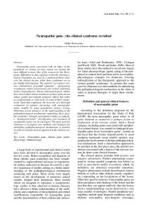
36 VOLTAGE GATED CALCIUM CHANNELS The plasma membranes of virtually all excitable cells, including neurons express voltage gated calcium channels (VGCC). VGCC’s are oligomeric protein complexes consisting of different central pore-forming α1 subunit isoforms (Table 6), surrounded by several auxiliary subunits (β, α2δ and γ) which serve to modulate the function of the α1 subunit (Castellano et al., 1993; Felix, 1999) (Figure 1B). The complex consists of four distinct domains (I-IV), each exhibiting six (S1-S6) helical membrane spanning regions. The S4 helices in each of the four domains are enriched with positively charged amino acid residues that allow the VGCC’s to detect changes in membrane potential. The selective hydrophilic pore is formed by the coalescence of the reentrant p-loops between S5 and S6 of each of the four domains. Plasma membrane depolarization triggers the opening of VGCC’s, resulting in the rapid influx of extracellular Ca2+ involved in various cellular processes including neuronal excitability and neurotransmitter release (Catterall, 2000; Cao, 2006) (Table 6). There are several distinct families of VGCC’s defined by various molecular, pharmacological, and genetic criteria (Tsien et al., 1988; Bean, 1989). In general, VGCC’s can be characterized by their activation range into two basic categories, highvoltage (HVA) and low-voltage (LVA) activated Ca2+ channels. The HVA Ca2+ channels open in response to large change in membrane potentials and include the CaV1(L) and CaV2 (P, Q, N, R) families of calcium channels. The LVA Ca2+ channels open in response to small changes in membrane potential and include the CaV3 (T) family of Ca2+ channels.
37 Ca2+ Channel
CaV1.1
α1 subunit
α1S
Gene
CACNA1S
Current
L
Blocker
Localization
dyhdropyridine
Skeletal muscle
CaV1.2
α1C
CACNA1C
L
dyhdropyridine
Cardiac muscle Neurons Endocrine cells
CaV1.3
α1D
CACNA1D
L
dyhdropyridine
Neurons, Endocrine cells
CaV1.4
α1F
CACNA1F
L
α1A
CACNA1A
P/Q
α1B
CACNA1B
N
CaV2.1 CaV2.2
CaV2.3
CaV3.1
α1E
α1G
CACNA1E
CACNA1G
R
T
CaV3.2
α1H
CACNA1H
T
CaV3.3
α1I
CACNA1I
T
Retina ω-agatoxin 1Va
Nerve terminals Dendrites
ω-conotoxinGVIA
SNX-482
Cell bodies, dendrites nerve terminals
nickel ethosuximide mibefradil
Cardiac muscle Skeletal muscle Neuron
mibefradil
Cardiac muscle Neuron Neuron
Table 6. Pharmacology and function of the α1 Ca2+ channel subunit.
Function Calcium homeostasis, gene expression and excitation – contraction coupling Excitationcontraction coupling, hormone secretion, gene regulation Hormone secretion and gene regulation Neurotransmitter release Neurotransmitter release, dendritic Ca2+ transients Neurotransmitter release, dendritic Ca2+ transients Ca2+ dependent AP’s neurotransmitter release, dendritic Ca2+ transients Action potential generation Rhythm pattern generation Action potential generation Rhythm pattern generation Rhythm pattern generation
38
A
B
Figure 1. Schematic of voltage-gated calcium channel. (A) Cartoon depicting the various subunits of the voltage-gated Ca2+ channel and (B) structural features of the central pore-forming α1 subunit. Figure reproduced from (Tedford and Zamponi, 2006).
39 High-voltage activated Ca2+ channel isoforms HVA Ca2+ channels activate in response to large changes in membrane potentials (Fox et al., 1987). Opening of some HVA Ca2+ channel types facilitates neurotransmitter release, excitation-contraction coupling, and secretion. The different HVA Ca2+ channels are described below. CaV 1 (L- type) family of Ca2+ channels L type currents are predominant in cardiac, smooth, skeletal muscles and brain. They are also present in endocrine cells, where they are known to play a role in hormone secretion (Milani et al., 1990). CaV 1 channels located on the cell body and dendrites of nerves regulate gene expression and integrate synaptic inputs (Bean, 1989). L type currents are distinguished by their high voltage dependence of activation, slower voltage dependent inactivation, relatively large single channel conductance, regulation by cAMP–dependent phosphorylation pathways, and sensitivity to dihydropyridines, phenylalkylamines and benzothiazepines (Reuter, 1983; Hadley and Lederer, 1991). CaV 2 (P/Q, N, R - type) family of Ca2+ channels CaV 2.1 (P/Q) Ca2+ channels are prominently expressed on the dendrites and pre-synaptic nerve terminal of neurons. Currents from the P – type Ca2+ channels were first recorded in the cerebellar Purkinje neuron (Llinas et al., 1989) and are characterized by high affinity binding of ω- agatoxin IVA (Mintz et al., 1992). In comparison the Q-type Ca2+ currents present in the cerebellar granule cells (Randall and Tsien, 1995) exhibit a lower affinity binding to ω- agatoxin IVA (Stea et al., 1994). P/Q-type channel currents are
40 primarily regulated by G protein coupled pathways (Hille, 1994) and are functionally involved in initiating synaptic transmission (Catterall, 2000). The CaV2.2 (N-type) Ca2+ channels are expressed exclusively in the nervous system, with primary afferent sensory neurons and the superficial layer of the dorsal horn showing high expression levels (Bourinet and Zamponi, 2005). N-type currents are distinguished by their intermediate voltage dependence and a rate of inactivation which is more negative and faster than L-type currents but more positive and slower than T-type currents. The cone-snail peptide, ω-conotoxin GVIA irreversibly inhibits CaV2.2 Ca2+ channels and has been used as a primary tool to distinguish N-type from other Ca2+currents. Modulation of CaV2.2 channel function is primarily through GPCR mediated pathways (Ikeda, 1996; Ikeda and Dunlap, 1999). Recently, CaV 2.2 channels have been implicated in pain signaling. In the superficial layer of the dorsal horn, CaV 2.2 Ca2+ channels regulate the release of the nociceptive neurotransmitters including glutamate and neuropeptides (Smith et al., 2002). Accordingly, CaV 2.2 channel knockout mice do not develop inflammatory or neuropathic pain associated mechanical hyperalgesia, suggesting a role for CaV2.2 channels in nociception (Kim et al., 2001; Saegusa et al., 2001). The CaV2.3 (R-type) Ca2+ channel currents are known to be resistant to most Ca2+ channel blockers. These channels have been thought to play a crucial role in regulating the release of neurotransmitters (Wu et al., 1998; Albillos et al., 2000). Also, they may
41 participate in Ca2+ influx in response to action potentials (Magee and Johnston, 1995; Sabatini and Svoboda, 2000). Low-voltage activated Ca2+ channels CaV3 family (T-type) Ca2+ channels The CaV3 family of calcium channels are activated by small depolarization of the cell membrane, hence commonly referred to as – low-voltage activated calcium channels. The CaV3 family comprises of three isoforms based on biophysical and pharmacological properties, namely CaV3.1 α1G, CaV3.2 α1H, and CaV3.3 α1I (Cribbs et al., 1998; PerezReyes et al., 1998).
The first recording of low-voltage activated calcium currents was
made in mouse neuroblastoma cell line (N1E-115) (Moolenaar and Spector, 1978). Ttype calcium channels are expressed in the nervous system (Carbone and Lux, 1984), the heart (Nilius et al., 1985), kidneys (Gordienko et al., 1994), endocrine organs, smooth muscles (Brueggemann et al., 2005) and sperm (Arnoult et al., 1996). They have been reported to be involved in the pathophysiology of cardiac arrhythmias (Satoh, 1995), sleep disorders (McCormick and Bal, 1997), epilepsy (Tsakiridou et al., 1995), nociception (Kim et al., 2003), and fertilization (Talavera and Nilius, 2006). Biologically, these channels modulate vital cellular functions including excitability (Chemin et al., 2002), secretion (Bhattacharjee et al., 1997), differentiation (Bijlenga et al., 2000), proliferation (Lory et al., 2006) and excitation-contraction coupling (Zhou and January, 1998).
42 T-type Ca2+ currents have several unique electrophysiological characteristics that separate them from the high-voltage activated calcium channels. T-type Ca2+ channels begin to activate after small depolarization of the cell membrane and exhibit rapidly inactivating transient currents in response to sustained depolarization. Upon repolarization, they close slowly resulting in the generation of tail currents with slow deactivation time constant. (Perez-Reyes, 2003). T-type Ca2+ channels open at small voltage ranges, but do not inactivate completely, resulting in window currents which may be essential for regulating intracellular Ca2+ concentration (Bijlenga et al., 2000). Role of voltage-gated Ca2+ channels in neuropathic pain Modulation and transmission of painful stimuli from the periphery to the CNS is mediated at the level of DRG by a variety of ion channels including the persistent Na+ channels, inward rectifying K+ channels and voltage gated Ca2+ channels (Woolf and Mannion, 1999; Julius and Basbaum, 2001). Recent studies have demonstrated that VGCC’s play an important role in pain signaling. VGCC’s modulate key aspects of pain transmission by regulating neuronal properties such as excitability, generation and propagation of action potential and release of nociceptive neurotransmitters in sensory neurons. Voltage activated
N-, T- and to some extent, P/Q-type Ca2+ channels are
established mediators of pain signaling (Zamponi et al., 2009), while the role of L- and R-type in nociception is uncertain (Altier and Zamponi, 2004; Cao, 2006; Gribkoff, 2006). The N-type Ca2+ channels control the release of glutamate and substance P at the
43 pre-synaptic nerve terminals in the dorsal horn of the spinal cord (Smith et al., 2002), thereby supporting pain transmission from the periphery to the CNS. Moreover, engineered mice lacking N-type channels demonstrate decreased behavioral response to neuropathic pain stimuli suggesting their role in nociception (Hatakeyama et al., 2001; Saegusa et al., 2001). Recent studies have demonstrated the existence of multiple isoforms of the N-type channel, derived from alternative splicing of N-type mRNA (Pan and Lipscombe, 2000; Lin et al., 2004). Among the multiple isoforms, the e37a isoform is markedly expressed in nociceptive DRG neurons and is specifically required for the onset and maintenance of thermal and tactile hyperalgesia (Altier et al., 2007). Neuromodulators/neurotransmitters
such
as
endogenous
opioids,
endo-
cannabinoids or GABA have been shown to regulate N-type calcium currents through G protein-coupled receptor signaling (Holz et al., 1986; Polo-Parada and Pilar, 1999). Ntype channel inhibition occurs, in some cases, through direct interaction of the Gβγ subunit with the intracellular loop between domains I and II (Figure 1) of the N-type α1 subunit (De Waard et al., 1997; Herlitze et al., 1997). Although the Gβγ subunits play a primary role in N-type Ca2+ channel regulation, there is evidence to suggest that other synapse associated proteins such as syntaxin1 may also regulate channel function (Stanley and Mirotznik, 1997). Unlike the HVA N-type channels, the LVA T-type Ca2+ channels regulate the excitability of the sensory neurons (Catterall, 2000; Perez-Reyes, 2003) and play a key role in the generation of acute peripheral nociceptive signals (Bourinet et al., 2005;
44 Nelson and Todorovic, 2006). T-type channel modulation of neuronal activity results in enhanced neurotransmission and amplification of sensory afferent signals, along with increased pain perception (Perez-Reyes, 2003; Todorovic and Jevtovic-Todorovic, 2007). There is increasing experimental evidence to support a role for T-type Ca2+ channels in the development of neuropathic pain associated with peripheral nerve injury (Dogrul et al., 2003) and, more recently, with painful diabetic neuropathy (Jagodic et al., 2007; Messinger et al., 2009b). T-type channels are typically resistant to modulation by conventional analgesic compounds. However, recent molecular, genetic and electrophysiological studies have described novel methods of T-type channel regulation that could have important implications in neuropathic pain management. Specifically, T-type channels are regulated by a number of cellular mechanisms including redox modulation. Reducing agents such as dithiothreitol and L-cysteine (L-cys) selectively enhance LVA calcium currents in small diameter DRG neurons. L-cys, DTT and other thiol containing L-cys analogs produce hyperalgesia when experimentally introduced into peripheral receptive fields. These agents are thought to sensitize C-type nociceptors that express T-type currents. Furthermore, agents that are capable of chelating zinc ions mimic and occlude the effects of L-cys and DTT whereas cysteine-modifying agents do not prevent the effects of reducing agents on T-currents in DRG neurons. These effects can be completely abolished by single-point mutations of histidine 191 to glutamine (H191Q) without affecting channel kinetics (Zamponi et al., 2009). The H191Q mutation disrupts high-
45 affinity zinc inhibition of CaV3.2 T-channel, suggesting that the effects of reducing agents may occur indirectly by dis-inhibition of the channels endogenous zinc ions. The CaV3.2 T-channel isoform that is predominately expressed in nociceptive neurons is inhibited by the activation of G protein-coupled receptor signaling involving selective interaction of Gβ2γ2 subunits to the channel α1 subunit (Wolfe et al., 2003; DePuy et al., 2006). The CaV3.2 T-channel isoform has also been reported to be modulated by various compounds such as nitrous oxide (Todorovic et al., 2001), phenytoin (Todorovic and
Lingle,
1998),
certain
neuroactive
steroids
(Pathirathna
et
al.,
2005),
endocannabinoids (Ross et al., 2009) and by the actin binding protein, Kelch-like 1 (Aromolaran et al., 2010). The exact molecular determinants of the T-type channel that is involved in this regulation is however, still unknown.
CHAPTER TWO FORCED-EXERCISE ALLEVIATES NEUROPATHIC PAIN IN EXPERIMENTAL DIABETES: EFFECTS ON VOLTAGE-GATED CALCIUM CHANNELS
ABSTRACT Exercise is now established as an integral adjunct to the management of diabetes. Diabetic polyneuropathy, a painful complication of diabetes, remains untreatable, emphasizing a critical need for improved therapeutic strategies. Recent evidence suggests that exercise may facilitate recovery of peripheral nerve function in the diabetic neuropathic patient. The mechanism by which exercise protects against peripheral nerve dysfunction in diabetes is unknown, but may involve correction of glucose-associated alterations of voltage-gated calcium currents (VGCC) in DRG neurons. Here, using a combination of behavioral and patch clamp methodology, we examined the functional consequences of exercise on VGCC in DRG neurons from streptozotocin (STZ)-induced diabetic rats. Compared to vehicle control, STZ-treated sedentary rats developed marked tactile hyperalgesia within two weeks of induction that was sustained for the 10-week duration of study. STZ-treated rats subjected to forced-exercise (treadmill, 5.4 km/week), by comparison, exhibited a significant delay in the onset of tactile hyperalgesia independent of changes in blood glucose control. Addition of the non-selective opiate receptor antagonist naloxone dose-dependently reversed the analgesic effect of forced46
47 exercise in STZ-treated rats. Compared to sedentary vehicle-treated rats, the doseresponse to naloxone in exercised animals was rightward shifted. Small diameter DRG neurons harvested from STZ-treated sedentary hyperalgesic rats exhibited a 2-fold increase in peak high-voltage activated (HVA) calcium current density with a 10 mV rightward shift; the low-voltage activated (LVA) calcium current component was similarly enhanced 2-fold. Forced-exercise attenuated the diabetes-associated increase in HVA peak current density. Moreover, the observed shift in HVA peak current density by diabetes was normalized with forced-exercise. Steady-state inactivation (SSI) properties of LVA calcium channels in DRG neurons from STZ-treated sedentary hyperalgesic rats demonstrated a significant rightward shift (+8 mV; V50 = -50.9 ± 0.6 mV) that was prevented by forced-exercise (V50 = -58.2 ± 1.4 mV; vehicle-treated control rats SSI V50 = -58.4 ± 0.9 mV). However, diabetes-associated increase in LVA current component was unaffected by forced-exercise. These findings demonstrate that acute hyperglycemichypoinsulinemic insult elicits marked tactile hyperalgesia in the STZ-diabetic sedentary rat by enhancing calcium influx in small diameter nociceptive DRG neurons. This may occur, in part, by a mechanism that increases LVA calcium channel availability and enhances both HVA and LVA Ca2+ channel currents. Forced-exercise was found to significantly delay the onset of tactile hyperalgesia largely by preventing diabetesassociated changes in HVA and LVA channel functions.
48 INTRODUCTION Poorly controlled diabetes often leads to impaired function of central, peripheral, and/or autonomic nerves (Emerick et al., 2005). As a result, nerve dysfunction associated with diabetes is best realized clinically as a heterogeneous disease that encompasses a wide range of neurologic abnormalities. Distal symmetrical polyneuropathy, the most common form of diabetic polyneuropathy, is particularly debilitating, affecting both large and small nerve fibers and altering a patient’s stability, sensorimotor function, gait, and quality of life (Menz et al., 2004). The development of neuropathic pain including hyperalgesia/allodynia, burning, tingling, and/or numbness sensations in a lengthdependent distribution, is prominent among diabetic patients and is strongly suggestive of small nerve fiber impairment (Tavee and Zhou, 2009). Approximately a third of patients with painful sensory neuropathy, and nearly half with otherwise idiopathic small fiber neuropathy, experience some form of impaired glucose tolerance or metabolic dysregulation (Singleton et al., 2001). Lifestyle intervention designed to improve blood glucose control through weight management with diet and exercise significantly reduced by 58% the incidence of type 2 diabetes in high risk patients (Knowler et al., 2002). In a small cohort of pre-diabetic patients, Smith et al. (Smith et al., 2006) observed that lifestyle intervention similarly improved impaired glucose tolerance with significant preservation of intra-epidermal nerve fiber density, increased foot sweat volume (autonomic tone), and decreased neuropathic pain. The cause of neuropathic pain associated with impaired glucose tolerance, or with frank diabetes, remains unclear. Early experimental studies suggest that
49 intermittent hyperglycemia damages sensory neurons and increases spontaneous C-fiber firing, resulting in neuropathic pain (Chen and Levine, 2001). The analgesic effect of exercise has been previously reported (Willow et al., 1980; Janal, 1996; O'Connor and Cook, 1999). Rats subjected to swimming exercise, demonstrate an attenuated response to inflammatory neuropathic pain (Kuphal et al., 2007). Mice subjected to treadmill running exhibited increased tail flick latencies to a heat stimulus (Blustein et al., 2006). Evidence supporting a protective effect of moderate aerobic exercise against the development of neuropathic complications in diabetic patients is mounting (Balducci et al., 2006), raising interest in aerobic exercise as a putative therapeutic intervention for the management of painful diabetic neuropathy. In a preliminary study, (Fisher et al., 2007) report that aerobic exercise statistically improves some measures of peripheral nerve function in diabetic patients with established lengthdependent neuropathy. The cellular mechanism(s) by which neuropathic pain develops in the diabetic patient is poorly understood, but may involve remodeling of voltage- or ligand-gated ion channels (Carbone and Lux, 1984; Nowycky et al., 1985; Cao, 2006) leading to abnormal enhancement of sensory neuron excitability (Ikeda and Dunlap, 2007). Voltage gated Nand T-type Ca2+ channels are established mediators of pain signaling and processing in afferent neurons (Zamponi et al., 2009). N-type Ca2+ channels are activated by large changes in membrane potential and regulate neurotransmitter release. By comparison, Ttype Ca2+ channels are activated by small changes in membrane potential, thereby
50 regulating the excitability of the sensory neuron (Catterall, 2000; Perez-Reyes, 2003) and playing a key role in the generation of acute peripheral nociceptive signals (Bourinet et al., 2005; Nelson and Todorovic, 2006). There is increasing experimental evidence to support a role for T-type Ca2+ channels in the development of neuropathic pain associated with peripheral nerve injury (Dogrul et al., 2003) and, more recently, with painful diabetic neuropathy (Jagodic et al., 2007; Messinger et al., 2009b). Given the potential application of moderate intensity exercise to be of therapeutic value for the management of painful nerve dysfunction in patients with DM, we investigated in this study the consequence of forced-exercise on voltage gated Ca2+ channel function in nociceptive (small-diameter) DRG neurons from streptozotocininduced hyperalgesic rats. A preliminary account of these findings have been previously reported (Shankarappa et al., 2007; Shankarappa et al., 2009). The present chapter addresses the proposed first and second specific aims of this dissertation.
51 MATERIALS AND METHODS
Animal care This study was conducted using protocols approved by the Edward Hines Jr. VA Hospital Institutional Animal Care and Use Committee in accordance with the principles of laboratory animal care (National Institutes of Health Publ. 86-23, 1985). All animals were housed in pairs, allowed standard rat diet and water ad libitum, and maintained on a 10h/14h light/dark cycle. Adult male Sprague-Dawley rats (initial body weight 200 g) were randomly divided into two treatment groups (vehicle-control or STZ-induced diabetes) and a subset of each group subjected to daily 1h-sessions of either exercise or sedentary inactivity for up to 10 weeks. Induction of experimental diabetes mellitus Streptozotocin (STZ) was used in this study to induce experimental DM, a well established animal model of painful diabetic neuropathy. Non-fasted ketamine (100 mg/kg)-xylazine (5 mg/kg) anesthetized rats received a single intraperitoneal injection of freshly prepared streptozotocin (STZ, 60 mg/kg body wt; Sigma-Aldrich, St. Louis, MO) dissolved in citrate buffer (100 mmol/l; pH 4.5). Non-diabetic control, gender and agematched rats received an equal volume of intraperitoneally injected, freshly prepared citrate buffer (100 mmol/l; pH 4.5). Non-fasting blood glucose levels were determined before vehicle or STZ was administered and at regular weekly intervals throughout the study. Blood samples were obtained by tail prick, and glucose content was quantified
52 using a calibrated commercial glucometer (MediSense Precision QID glucometer, Abbot Laboratories, Bedford, MA). STZ-treated rats exhibiting blood glucose levels