CME Neurology
How to spot mitochondrial disease in adults Michael J Keogh, ST3 at James Cook Hospital, Middlesbrough and academic clinical fellow, Northern Region; Patrick F Chinnery, professor of neurogenetics and director of the Institute of Genetic Medicine, Newcastle University; Wellcome Trust Senior Fellow in Clinical Science; honorary consultant neurologist, Newcastle upon Tyne Hospitals NHS Foundation Trust
Clinical Medicine 2013, Vol 13, No 1: 87–92
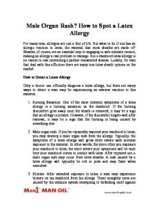
Clinical feature(s) suggestive of mitochondrial disease
Full multisystem enquiry and assessment
Is it a classical syndrome?
Yes
Numerous mitochondria are present in every nucleated cell in the body. They have a diverse role in cellular metabolism and are the principal source of adenosine triphosphate (ATP). ATP is produced through oxidative phosphorylation (OXPHOS) by the mitochondrial respiratory chain. Impairment of OXPHOS leads to cellular dysfunction and, eventually, cell death. As a result, mitochondrial disorders primarily affect tissues that have a high metabolic demand, such as the neural, muscular, cardiac, ocular and endocrine systems. Mitochondrial function is dependent on the interplay of mitochondrial DNA and the nuclear genome.2 Mitochondria contain their own DNA (mtDNA), which comprises 16,569 base pairs (bp) and encodes only 37 genes (compared with the 3.3 billion bp in, and thousands of genes encoded by, the nuclear genome). However, most of the approximately 1,500 mitochondrial proteins are coded by nuclear DNA and synthesised on cytoplasmic ribosomes.3 Therefore, abnormalities of mitochondrial proteins can result from defects in both mtDNA and nuclear DNA, and so cause maternally inherited, Mendelian (autosomal dominant, recessive or X-linked), or sporadic diseases (owing to de novo mutations, or recessive inheritance).
How to spot mitochondrial disease Although mitochondrial disease can present with a heterogeneous array of symptoms, there are certain key neurological clinical
© Royal College of Physicians, 2013. All rights reserved.
CMJ1301-CME_Chinnery.indd 87
Oligosymptomatic
Family history of neurological disease
Yes
Are there any features of a mitochondrial disease?
No
Mitochondrial disease less likely
Testing (see Fig 2)
Fig 1. Mechanisms of alerting the clinician to the presence of a mitochondrial disorder.
features that suggest the possibility of such disease (Fig 1; Table 1). Occasionally, these features form a ‘classic mitochondrial syndrome’ (Table 2). Although some patients fall neatly into a defined syndrome, many patients will be so-called ‘oligosymptomatic’, having one or two components of a syndrome (such as a young individual with an occipital stroke-like lesion, or mild nonfatigable ptosis without ophthalmoplegia), and are more problematic to reach a diagnosis for. These patients often also have several other features that, if present, further support evidence of a mitochondrial disease. A clinician should look and test for these additional feature if suspecting the diagnosis (Table 3). Non-neurological involvement in a patient with unexplained multisystem neurological disease is a ‘red flag’ for mitochondrial disease. Finally, and perhaps most challenging, are patients presenting with ‘non-specific presentations’ (such as epilepsy, diabetes or sensorineural deafness), who often have a multisystem disease that does not fit neatly into any particular category. In these circumstances, the diagnosis of a mitochondrial disorder is often only made in a tertiary referral centre with direct access to specialist laboratory investigations.
Specific clinical features in ‘suspected mitochondrial disease’ Key neurological features of mitochondrial disease are outlined in Table 1. Chronic progressive external ophthalmoplegia (CPEO) is the most common specific manifestation of mitochondrial disease, and is considered to be present in approximately 20% of patients presenting in adult life.4,5 It typically causes symmetrical limitation of all extraocular muscles and usually occurs in conjunction with ptosis.6 Other patients present with refractory seizure disorders, particularly associated with underlying stroke-like lesions (especially occipital)7 or fluctuating or progressive encephalopathy. Ataxia, peripheral neuropathy and proximal myopathy are common, and can lead to fatigue and myalgia in middle age as the presenting feature.8 Myoclonic jerks, optic atrophy and pigmentary retinopathy also suggest a mitochondrial aetiology. Neurological features, such as extrapyramidal symptoms (Parkinsonism and dystonia) and spasticity, are recognised but uncommon features of mitochondrial disease. Key extra-neurological features include sensorineural hearing loss developing in
87
1/16/13 11:03:34 AM
CME Neurology young adulthood, short stature, cardiomyopathy, diabetes and other endocrine disorders, often associated with gastrointestinal dysmotility (Table 3).
Classic ‘mitochondrial syndromes’ Several classic clinical syndromes of mitochondrial disease are detailed in Table 2, and have been reviewed elsewhere.9 Some mitochondrial syndromes, such as mitochondrial encephalomyopathy with lactic acidosis and stroke-like episodes (MELAS), are readily recognised when ‘full blown’, and suggest specific genetic causes; however, milder forms can be difficult to recognise, particularly in the early stages. Patients usually present after normal childhood development, before sensorineural deafness, diabetes and stroke-like episodes begin during their late teens.10 Genetic blood tests can be misleadingly negative (explained below). Leber’s hereditary optic neuropathy (LHON) has a highly stereotypic presentation, with progressive painless visual loss that affects men more than women. Once suspected clinically, LHON
Heteroplasmy refers to the presence of more than one type of mtDNA genome within a cell. Given that each cell has between 100 and 10,000 mitochondria, each with 2–10 copies of the mtDNA, a mixture of mutated and ‘wild-type’ DNA can exist within cells. Differences in the ratio of mutant and wild-type mtDNA (the ‘mutation load’) between individual family members, and between organs within the same individual, lead to a high degree of clinical heterogeneity. This also poses diagnostic challenges, because low levels of mutant mtDNA in sampled tissues (eg blood) can lead to false negative tests and must always be interpreted with caution. This is particularly the case for the m.3243A>G mutation (see above, in the context of MELAS).
is easy to diagnose with a simple blood test, because three mutations (m.3460G>A, m.11778G>A or m.14484T>C) are responsible for 90% of cases.11 Clear diagnostic criteria also exist for Kearns–Sayre syndrome, myoclonic epilepsy with ragged red fibres (MERRF) and mitochondrial neurogastrointestinal encephalopathy (MNGIE) (Table 2).
Family history and heteroplasmy Mitochondrial disorders can present with any form of inheritance pattern, and onethird of patients have a sporadic disorder.12 Therefore, the importance of taking a family history is to direct genetic tests and to determine potential disease in other family members. If a diagnosis of mitochondrial DNA disease is suspected (and especially if it might be a primary mtDNA disorder; ie there is no male–male transmission in the pedigree), then it is important to ask about subtle clinical features in additional family members.2 This is because different family members often have different phenotypes, owing in part to ‘mtDNA heteroplasmy’.
Defining the extent of the clinical phenotype Several investigations can help to build the ‘mitochondrial phenotype’. Mandatory investigations are an electrocardiogram (ECG), echocardiogram, fasting blood sugar and formal retinal assessment,
Table 1. Neurological features that should specifically prompt the clinician to consider a potential mitochondrial disorder. Neurological feature
Additional information
Syndrome
Ataxia
Highly variable severity and temporal relation with disease
KSS, NARP, POLG, MELAS, MERRF
CPEO
Can be isolated or on a spectrum with other symptoms
CPEO, CPEO+, KSS, MELAS, MNGIE, OPA1
Dementia
Variable age of presentation
KSS, Leigh, MELAS, MERRF
Encephalopathy
Can be highly variable and intermittent
KSS, MELAS, Leigh, MNGIE, MERRF
MRI features
Heterogeneous and include basal ganglia necrosis, strokelike lesions, calcification and leukodystrophy (unexplained)
KSS, Leigh, MELAS
Myoclonus
Subtle to frank myoclonus and generalised seizures
MERRF
Myopathy
Can be ocular, proximal, distal or generalised
CPEO+, KSS, MERRF, NARP, MNGIE
Optic atrophy
Can be clinically mild
LHON, OPA1, MERRF, HMSN VI
Peripheral neuropathy
Sensory, motor or both; usually axonal
MNGIE, NARP, MERRF, KSS, OPA1
Pigmentary retinopathy
From retinitis pigmentosa to ‘salt and pepper’ fundi
KSS, MELAS, NARP
Seizures
Often progressive, generalised and difficult to control
MERRF, Leigh, MELAS
SNHL
Often in younger patients
MELAS, MERRF, KSS
Stroke-like episodes
Predominantly occipital
MELAS
CPEO ⫽ chronic progressive external ophthalmoplegia; CPEO⫹ ⫽ chronic progressive external ophthalmoplegia plus; HMSN ⫽ hereditary motor and sensory neuropathy; KSS ⫽ Kearns–Sayre syndrome; LHON ⫽ Leber’s hereditary optic neuropathy; MERRF ⫽ mitochondrial encephalopathy with ragged red fibres; MELAS ⫽ mitochondrial encephalomyopathy with lactic acidosis and stroke-like episodes; MNGIE ⫽ mitochondrial neurogastrointestinal encephalopathy; NARP ⫽ neuropathy ataxia and retinitis pigmentosa; OPA ⫽ optic atrophy; POLG ⫽ polymerase gamma; SNHL ⫽ sensorineural hearing loss.
88
CMJ1301-CME_Chinnery.indd 88
© Royal College of Physicians, 2013. All rights reserved.
1/16/13 11:03:35 AM
CME Neurology because manifestations in these systems are often subclinical. Subsequent investigations might be necessary, depending on the phenotype and symptoms (Table 4). Blood lactate levels can be abnormal, especially during an acute metabolic crisis, although lactate levels are often normal in adults presenting with mitochondrial disease subacutely.
Specific mitochondrial investigations The diagnosis of mitochondrial disorders can range from the simple to awkwardly complex. In the first instance, the decision of whether to embark on this process depends on how strongly the phenotype is felt to represent a mitochondrial disorder.
Fig 2 provides an overview of the basic investigative approach. For a specific clinical syndrome, a noninvasive genetic test might be possible on blood, urine or buccal mucosa. However, if these tests return negative, or the clinical features in the first instance necessitate a muscle biopsy (eg CPEO), then this is most commonly undertaken. Other
Table 2. Classic mitochondrial phenotypes. Syndrome
Primary features
Secondary features
Inheritance
Common mutations
CPEO
Progressive external ophthalmoplegia and bilateral ptosis
Mild facial or proximal myopathy
S/M
95% sporadic; generally mtDNA deletions; more than 150 described
Kearns–Sayre
CPEO onset before the age of 20 with pigmentary retinopathy plus one of the following: CSF protein ⬎1 g/l, cerebellar ataxia and/or heart block
Dementia, diabetes mellitus, sensorineural deafness (50%) myopathy, hypoparathyroidism and dysphagia
S/M
Generally sporadic mtDNA deletions; over 100 described
MELAS
Stroke-like episodes (typically before the age of 40); encephalopathy with seizures and or dementia; mitochondrial myopathy on muscle biopsy
Recurrent headaches, recurrent vomiting, sensorineural deafness, short stature, diabetes mellitus, ataxia, pigmentary retinopathy and cardiomyopathy
M
3243A⬎G, 3271T⬎C, 3251A⬎G
MERRF
Myoclonus, seizures, encephalopathy and ataxia
Dementia, peripheral neuropathy, cervical lipomatosis, sensorineural deafness and optic atrophy
M
8344A⬎G, 8356T⬎C
NARP
Pigmentary retinopathy, ataxia and peripheral neuropathy
Dementia, SNHL, short stature, seizures, basal ganglia MRI changes and clinical overlap with Leigh’s syndrome
M
8993T⬎G/C
MNGIE
Gastrointestinal dysmotility, cachexia, peripheral neuropathy, ptosis and SNHL
GORD, vomiting, distal weakness and leukoencephalopathy on MRI
AR
Mutation in TYMP gene
OPA1
Painless visual loss or asymptomatic
SNHL, peripheral neuropathy and ataxia
AD
Over 200 mutations in OPA1 gene described
LHON
Subacute painless bilateral visual failure; more common in males (4:1)
Dystonia and cardiac arrhythmias
M
11778G⬎A, 14484T⬎C, 3460G⬎A
Leigh’s syndrome
Subacute relapsing encephalopathy with cerebellar and brainstem signs presenting during infancy
Overlap with NARP and basal ganglia changes on MRI
M
8993T⬎G/C
POLG
Sensory or cerebellar ataxia
Highly variable: ptosis, PEO, myopathy, myoclonus, epilepsy (continuum of conditions previously felt to be discrete diagnostic entities eg Alpers’ syndrome, MEMSA and arPEO or adPEO)
AR/AD
Numerous mutations in DNA Polymerase γ (POLG)
AD ⫽ autosomal dominant; AR = autosomal recessive; CPEO ⫽ chronic progressive eternal ophthalmoplegia; CSF ⫽ cerebrospinal fluid; GORD ⫽ gastrooesophageal reflux disease; LHON ⫽ Leber’s hereditary optic neuropathy; MERRF ⫽ maternal inheritance; MEERF ⫽ mitochondrial encephalopathy with ragged red fibres; MELAS ⫽ mitochondrial encephalomyopathy with lactic acidosis and stroke-like episodes; MEMSA ⫽ myoclonic epilepsy myopathy sensory ataxia; MNGIE ⫽ mitochondrial neurogastrointestinal encephalopathy; MRI ⫽ magnetic resonance imaging; NARP ⫽ neuropathy ataxia retinitis pigmentosa; OPA1 ⫽ optic atrophy type 1; PEO ⫽ progressive external ophthalmoplegia; POLG ⫽ polymerase gamma; S ⫽ sporadic; SNHL ⫽ sensorineural hearing loss; TYMP ⫽ thymidine phosphorylase.
© Royal College of Physicians, 2013. All rights reserved.
CMJ1301-CME_Chinnery.indd 89
89
1/16/13 11:03:35 AM
CME Neurology
Table 3. Non-neurological symptoms that clinicians should assess for and, if present, add to the suspicion of a mitochondrial disease. Category
Symptom
Cardiovascular
Hypertrophic cardiomyopathy with conduction deficit Cardiac conduction defect Cardiomyopathy with lactic acidosis Wolff–Parkinson–White syndrome
Endocrine
Diabetes Hypothyroidism Hypoparathyroidism Gonadal failure
Gastrointestinal
Severe dysmotility Pseudo-obstruction Unexplained cyclical vomiting Dysphagia Hepatic failure
General
Short stature Hypersensitivity to general anaesthesia Sudden death in family Recurrent miscarriages in patient or family Exercise intolerance
Ophthalmologic (in addition to Ptosis (nonfatigable) those already listed) Colour vision deficits Renal
Renal tubular disease (renal tubular acidosis and/or aminoaciduria) De Toni–Debré–Fanconi syndrome Nephrotic syndrome
organs are sometimes biopsied (eg liver or myocardium) if they dominate the clinical picture. Histochemical analysis might support a mitochondrial abnormality and guide subsequent biochemical and genetic testing. Thereafter, appropriate testing for mtDNA deletions, depletion, point mutations or nuclear gene screening will lead to a genetic diagnosis in most cases.13,14 However, in a substantial proportion of patients, a genetic diagnosis is not possible using National Health Service (NHS) services. Under these circumstances, it is helpful to engage an active research laboratory with an interest in the patient’s phenotype or biochemical abnormality. At present, three laboratories in the UK (in Newcastle, Oxford and London) have been awarded National Specialist Commissioning (NSC) funding to provide the Rare Mitochondrial Disorders Service for Adults and Children, which is a diagnostic and clinical service for patients with mitochondrial disease in England and Scotland.15 With the development of sequencing techniques, such as whole-exome sequencing, this process might change because investigation is likely to become much simpler, less invasive and, ultimately, available across the NHS.
Clinical management
Key points Mitochondrial DNA disorders are a common cause of inherited disease, affecting 1 in 5,000 of the UK population1 They should be considered in any complex multisystem disorder, especially those disorders in which neurological, ocular or endocrine features predominate Many patients will fit neatly into defined classic syndromes, but significant numbers are oligosymptomatic, or have an overlapping, poorly defined, phenotype If a mitochondrial disorder is suspected, it is vital to assess fully the clinical phenotype in the patient and their relatives with symptoms, paying particular attention to the non-neurological manifestations, because these can often be effectively managed Counselling depends on the underlying molecular basis of disease and might necessitate discussion with specialist centres There are currently no treatments known to modify the underlying disease process Investigation of mitochondrial disorders can be complex and might necessitate referral to one of the three commissioned groups forming the Rare Mitochondrial Disorders Service for Adults and Children (in Newcastle, Oxford and London) KEY WORDS: mitochondria, genetic, neurology, familial, metabolic, mitochondrial disease
90
CMJ1301-CME_Chinnery.indd 90
Genetic counselling for mitochondrial disorders is complex. For some mtDNA diseases, such as mtDNA deletions (which often occur as sporadic mutations in patients), the recurrence rate in offspring is low. Other mtDNA disorders are maternally inherited, such as LHON, and have empirical gender-specific recurrence risks that can be given to families. Heteroplasmic mutations are complex and the inheritance pattern can be difficult to predict. Nuclear DNA disorders causing mtDNA disorders are counselled according to their inheritance pattern (dominant, recessive or X-linked), and known clinical penetrance. Treatment options are also limited for patients, and management remains largely supportive.16 Prevention of secondary complications is best achieved by specialist teams with multidisciplinary resources.
© Royal College of Physicians, 2013. All rights reserved.
1/16/13 11:03:35 AM
CME Neurology
Table 4. Clinical investigations most commonly undertaken to investigate a potential mitochondrial disorder. Investigation
Potential abnormalities
CK
Raised in several mitochondrial myopathies, but often normal
Clinical examination
All systems with a particular focus on a neurological examination, including fundoscopy, cognitive assessment, features of ataxia, myopathy and neuropathy
CSF
Generally normal with exception of a potentially raised lactate; often used to exclude alternative diagnoses
Echocardiography
Assessment of ventricular wall thickness and function for a possible cardiomyopathy
Electrocardiography
Assessment of cardiac conduction defects and for features of left or right ventricular or hypertrophy
Electroencephalography
For ictal activity or features of an underlying encephalopathy
Electromyography and nerve conduction studies
For evidence of myopathy or peripheral neuropathy (sensory or motor)
Endocrine investigations
Fasting glucose, haemoglobin A1c, thyroid function tests and parathyroid hormone as an initial screen
Neuroimaging
MRI most useful to assess for cortical and subcortical abnormalities; CT occasionally more valuable to assess for basal ganglia calcification (present in approximately one in six patients)
Ophthalmological assessment
Optic atrophy, pigmentary retinopathy (retinitis pigmentosa, salt and pepper fundi), ptosis and extraocular movement disorder
Personal and family history
Full personal multisystem history with special focus on neurological, ocular, cardiac, endocrine and gastrointestinal symptoms; family history assessing for miscarriages, sudden death, death in infancy and neurological abnormalities
Plasma lactate
Generally raised but non specific and, if normal, does not exclude a mitochondrial disease
Urine dipstick and renal function blood test
Dipstick for blood and protein to suggest renal disease; blood tests for renal function
CK ⫽ creatine kinase; CSF ⫽ cerebrospinal fluid; CT ⫽ computerised tomography; MRI ⫽ magnetic resonance imaging.
High suspicion of mitochondrial disease
Defined and/or classic syndrome?
Yes
Positive: diagnosis
Non-invasive test available?
Yes
No
Negative
No
Common facets, such as epilepsy, heart failure and gastrointestinal dysmotility, are usually managed according to standard clinical practice, but these features can be challenging to manage in the context of multisystem complications, and long-term follow-up might be required, with ongoing surveillance.
References 1
Muscle biopsy for histochemistry +/- respiratory chain complex
Real-time PCR Long-range PCR (for mtDNA depletion) (mtDNA deletions)
PCR-RFLP (common mtDNA point mutations)
mtDNA sequencing (novel mutations)
Nuclear DNA sequencing (Sanger or nextgeneration) for known or novel mutations
Fig 2. A broad schematic of the investigatory process of suspected mitochondrial disease. mtDNA ⫽ mitochondrial DNA; PCR ⫽ polymerase chain reaction; RFLP = restriction fragment length polymorphism.
© Royal College of Physicians, 2013. All rights reserved.
CMJ1301-CME_Chinnery.indd 91
2
3
4
Schaefer AM, Taylor RW, Turnbull DM, Chinnery PF. The epidemiology of mitochondrial disorders: past, present and future. Biochim Biophys Acta 2004;1659:115–20. Cree LM, Samuels DC, Chinnery PF. The inheritance of pathogenic mitochondrial DNA mutations. Biochim Biophys Acta 2009;1792:1097–102. Smeitink J, van den Heuvel L, DiMauro S. The genetics and pathology of oxidative phosphorylation. Nat Rev Genet 2001;2:342–52. Petty RK, Harding AE, Morgan-Hughes JA. The clinical features of mitochondrial myopathy. Brain 1986;109:915–38.
91
1/16/13 11:03:35 AM
CME Neurology 5
6
7
8
9
Kirkman MA, Yu-Wai-Man P, Chinnery PF. The clinical spectrum of mitochondrial genetic disorders. Clin Med 2008;8:601–6. Richardson C, Smith T, Schaefer A, Turnbull D, Griffiths P. Ocular motility findings in chronic progressive external ophthalmoplegia. Eye (Lond) 2005;19:258–63. Goodfellow JA, Dani K, Stewart W et al. Mitochondrial myopathy, encephalopathy, lactic acidosis and stroke-like episodes: an important cause of stroke in young people. Postgrad Med J 2012;88:326–34. Pfeffer G, Chinnery PF. Diagnosis and treatment of mitochondrial myopathies. Ann Med 2011;early online:1–13. Taylor RW, Turnbull DM. Mitochondrial DNA mutations in human disease. Nat Rev Genet 2005;6:389–402.
10 Sproule DM, Kaufmann P. Mitochondrial encephalopathy, lactic acidosis, and strokelike episodes: basic concepts, clinical phenotype, and therapeutic management of MELAS syndrome. Ann N Y Acad Sci 2008;1142:133–58. 11 Yu-Wai-Man P, Griffiths PG, Chinnery PF. Mitochondrial optic neuropathies – disease mechanisms and therapeutic strategies. Prog Retin Eye Res 2011;30:81–114. 12 Chinnery PF. Inheritance of mitochondrial disorders. Mitochondrion 2002;2:149–55. 13 Chinnery PF. Could it be mitochondrial? When and how to investigate. Pract Neurol 2006;6:90–101. 14 Taylor RW, Schaefer AM, Barron MJet al. The diagnosis of mitochondrial muscle disease. Neuromuscular Disord 2004;14:237–45.
QUEEN SQUARE BALANCE COURSE ‘Dizziness: A Multidisciplinary Approach’ on 18-21 June 2013
www.uclh.nhs.uk/BalanceCourse Four-day course for health professionals working with dizzy patients. Attend one day or all four… see what interests you!
DAY 1 • Clinical history and bedside examination • Observe NHNN Consultants assess and discuss complex patients in Grand-Rounds fashion
DAY 2 • Clinical and diagnostic assessment • Practice bedside clinical tests, position manoeuvres and diagnostic assessments
DAY 3 • Multi-disciplinary management
15 Rare Mitochondrial Disease Service for Adults and Children, 2012. www.mitochondrialncg.nhs.uk [Accessed 3 January 2013]. 16 Pfeffer G, Majamaa K, Turnbull DM et al. Treatment for mitochondrial disorders. Cochrane Database Syst Rev. 2012;4:CD004426.
Address for correspondence: Prof PF Chinnery, Institute of Genetic Medicine, Newcastle University, Central Parkway, Newcastle-uponTyne, NE1 3BZ. Email:
[email protected]
RCP information
Change of address? If you receive Clinical Medicine, please inform us of any changes to your contact details. This helps us to maintain an accurate record of your account and avoids any problem with journal distribution. Fellows and members should contact the Membership Department: Email:
[email protected] Tel: +44 (0)20 3075 1362/1467 Institutional subscribers should contact Julie Dalton, Publications Department: Email:
[email protected] Tel: +44 (0)20 3075 1358 Fax: +44 (0)20 7486 5425
• Present case study to panel for feedback
DAY 4 • International vestibular clinical practice Visit website for more information or contact course organiser Kate Eykamp on Ph: 020 3448 3275, E:
[email protected]
92
CMJ1301-CME_Chinnery.indd 92
© Royal College of Physicians, 2013. All rights reserved.
1/16/13 11:03:35 AM