BioMol Concepts, Vol. 3 (2012), pp. 107–115 • Copyright © by Walter de Gruyter • Berlin • Boston. DOI 10.1515/bmc-2011-0058
Review
Mitochondrial DNA: a blind spot in neuroepigenetics Hari Manev*, Svetlana Dzitoyeva and Hu Chen The Psychiatric Institute, Department of Psychiatry, University of Illinois at Chicago, 1601 West Taylor Street, M/C912, Chicago, IL 60612, USA * Corresponding author e-mail:
[email protected]
Abstract Neuroepigenetics, which includes nuclear DNA modifications, such as 5-methylcytosine and 5-hydroxymethylcytosine and modifications of nuclear proteins, such as histones, is emerging as the leading field in molecular neuroscience. Historically, a functional role for epigenetic mechanisms, including in neuroepigenetics, has been sought in the area of the regulation of nuclear transcription. However, one important compartment of mammalian cell DNA, different from nuclear DNA but equally important for physiological and pathological processes (including in the brain), mitochondrial DNA has for the most part not had a systematic epigenetic characterization. The importance of mitochondria and mitochondrial DNA (particularly its mutations) in central nervous system physiology and pathology has long been recognized. Only recently have the mechanisms of mitochondrial DNA methylation and hydroxymethylation, including the discovery of mitochondrial DNA-methyltransferases and the presence and functionality of 5-methylcytosine and 5-hydroxymethylcytosine in mitochondrial DNA (e.g., in modifying the transcription of mitochondrial genome), been unequivocally recognized as a part of mammalian mitochondrial physiology. Here, we summarize for the first time evidence supporting the existence of these mechanisms and propose the term ‘mitochondrial epigenetics’ to be used when referring to them. Currently, neuroepigenetics does not include mitochondrial epigenetics – a gap that we expect to close in the near future. Keywords: amyotrophic lateral sclerosis (ALS); DNA methylation; DNMT1; DNMT3A; epigenetics; 5-hydroxymethylcytosine; mitochondria.
Introduction The earliest understanding of the functional role of epigenetic mechanisms relates to developmental genome regulation, e.g., silencing of gene expression involved in cell differentiation and in maintaining cell phenotypes during cell proliferation (1–6). Cellular chromatin, a structure composed of nuclear DNA (ncDNA) and nuclear proteins (including histones), has been the primary target of epigenetic research, which brought about major new concepts on the functionality of the genome
(7). These studies of chromatin remodeling comprise protein (e.g., histone) modifications and DNA modifications, such as the formation of 5-methylcytosine (5mC) and 5-hydroxymethylcytosine (5hmC). Whereas 5mC DNA modifications in the dinucleotide sequence CpG are generally considered transcriptional repressors [recent data suggest a more complex role for 5mC DNA, including stimulation of gene expression (8)], the functional role of 5hmC DNA modifications is currently unclear. Since the early days of developmental epigenetics, new ideas have emerged that imply epigenetic mechanisms are a bridge between the environment and lasting, sometimes heritable genome modifications. Hence, both the adaptive and harmful biological consequences of an individual’s bi-directional interaction with the environment are possibly best contained in that individual’s modified genome – the epigenome. Compared to a developmental role of epigenetics, considerations of epigenetic mechanisms as a modifiable functional system in postmitotic cells, such as neurons (i.e., neuroepigenetics) are more recent. In a series of articles published between 1974 and 1977, Boris Vanyushin and colleagues (9) provided the first direct evidence in support of the 1969 hypothesis (10) that DNA methylation of neuronal ncDNA functions as the epigenetic component of learning and memory. In these experiments, rats were trained (conditioned) to associate a light cue with food (a food reinforcement model). Brain samples (the cortex, hippocampus, and cerebellum) were collected from control and conditioned rats. For each brain region, neuronal nuclei were separated from the glial nuclei. ncDNA methylation was affected by the conditioning model in the cortex and the hippocampus but not in the cerebellum, and only in neuronal but not in glial ncDNA. In both affected brain regions, the learning model caused an increase of 5mC in neuronal ncDNA. This pioneering work received little attention and was soon forgotten. The same idea was resurrected 25 years later in a letter to the editor of the Journal of Theoretical Biology (11). Although no data were presented in support of the proposed hypothesis, which asked the question whether there was an epigenetic component in long-term memory, subsequent research has provided a clear positive response to this question (12). Moreover, recent advancements in the methodologies for the characterization of DNA methylation demonstrated the ease with which neuronal activity in the adult brain (e.g., in the hippocampus) is capable of modifying its DNA methylation landscape (13). Moreover, the same mechanisms appear to be involved not only in brain physiology but also in the pathobiological mechanism of neuropsychiatric disorders, e.g., schizophrenia (14–16). However, one important compartment of mammalian cell DNA, different from ncDNA but equally important for physiological and pathological processes (including in the brain), mitochondrial DNA (mtDNA) has for the most part escaped a systematic epigenetic characterization. The organization of
108 H. Manev et al.
mtDNA is different from the structural organization of ncDNA (17); most notably, mitochondria do not contain histones. Hence, epigenetic mechanisms, such as histone modifications, which are important for ncDNA, may not be applicable to mtDNA, stressing the importance of the mechanisms of 5mC and 5hmC formation for mtDNA.
Mitochondrial DNA mtDNA was discovered and first visualized through electron microscopy by Margit Nass-Edelson and Sylvan Nass in 1963 (18, 19). On the basis of the observed ultrastructural similarity of this intracellular organelle and the bacterial cells, the idea of endosymbiosis developed, which suggests that during evolution, bacterial cells became engulfed and modified to become eukaryotic organelles, such as mitochondria (20). Since the time of these original observations, enormous progress has been made in understanding the structure and function of mtDNA and about its role in human pathology, including neuropsychiatric disorders. Extensive reviews have summarized these achievements [e.g., (21–24)]. Briefly, mammalian mitochondria contain multiple copies of a maternally inherited genome, a 16,295–16,826 bp (25) circular double-stranded mtDNA that encodes 13 polypeptides, 2 ribosomal RNAs, and 22 transfer RNAs. The proteins encoded in mtDNA are all members of the oxidative phosphorylation (OXPHOS) complexes. Initially, it was believed that mtDNA is naked and thus vulnerable to damage. However, recent studies have established that mtDNA is protein coated and packaged into aggregates called nucleoids or mitochromosome. Mitochondrial transcription factor A (TFAM) is the most abundant component of the nucleoid and, similar to the action of histones on ncDNA, it plays a major role in the packaging and compaction of mtDNA (26–28). Current data indicate that, contrary to the previous belief, most nucleoids contain a single copy of mtDNA, supporting the proposal that nucleoids as a general rule do not exchange genomes with each other (26). Except for the 13 genes encoded by mtDNA, it appears that all other proteins (perhaps thousands) necessary for mitochondrial structure and function are encoded by ncDNA (29). As a consequence, regulation of transcription of mitochondrial genes is coordinated between these two genomes. A recent review has addressed the complex role of various mitochondrial proteins and the importance of mitochondrial nucleoid structure in the process of mtDNA transcription regulation (17). Briefly, the core machinery for physiological mitochondrial transcription regulation includes regulators of initiation, composed of three components, POLRMT (mitochondrial RNA polymerase), TFB2M (mitochondrial transcription factor B), and TFAM, plus several additional components that include the MTERF (mitochondrial terminator factor) family (23). Several nuclear transcription factors also are capable of binding mtDNA. It is believed that their role in mitochondria may be related to tissue-specific regulation of mtDNA expression and to apoptosis (programmed cell death) (30).
In postmitotic cells, such as neurons, mtDNA is prone to mutations and brain cells are known to have a higher degree of heteroplasmy (the presence of more than one type of mtDNA within a cell) than cells in rapidly dividing tissues. Generally, mtDNA mutations have been proposed as the source of regional ancient variants that evolutionarily permitted humans to adapt to differences in their energetic environments and as a cause of disease. The latter includes deleterious germline line mutations causing mitochondrial diseases, and somatic mutations that accumulate with age and may cause aging-associated disorders (31). Both mtDNA mutations and deregulation of mtDNA gene expression have been recognized as the basis for a number of human disorders (24).
mtDNA cytosine methylation Although as long ago as the late 1940s (32, 33), numerous studies had confirmed the presence of a substantial amount of 5mC in vertebrate ncDNA [including in the brain (34)], no significant functional role was assigned to this DNA modification for quite some time. A uniquely powerful argument for dismissing the important functionality of vertebrate DNA methylation, which is particularly abundant during development, was raised in 1985 (6). It was pointed out that Drosophila (fruit fly) had no proven DNA methylation, but nevertheless, like vertebrates, is capable of accomplishing sophisticated developmental pathways. The argument was then made that if Drosophila could accomplish its complicated differentiation without DNA methylation, how could vertebrate development use DNA methylation as an important gene regulator? In spite of this conceptual obstacle, the current understanding of the functional role of DNA methylation in the regulation of gene expression has evolved dramatically. In vertebrate ncDNA, a methyl group is added to the 5 position of the base cytosine to generate 5mC by the action of three DNA-methyltransferases (DNMTs) – DNMT1 (the maintenance enzyme) and DNMT3A and DNMT3B (which have the capacity to methylate DNA de novo). Ultimately, it was confirmed that similar to vertebrates, Drosophila utilizes DNA methylation. It turned out that various species, including insects, accomplish DNA methylation by expressing and utilizing different types of DNMTs; for example, Drosophila expresses only an insect isoform of DNMT2 (35). Compared with that of ncDNA, the history of mtDNA methylation research is marked by even more controversy. Ten years after she discovered mtDNA, Nass published an extensive report on the first characterization of the mammalian mtDNA methylation (36). She found that compared with ncDNA, mtDNA was generally undermethylated and that the only methylated base in mtDNA was 5mC. A couple of months later, a brief methodological report (37) challenged these findings, suggesting that the observed 5mC might have been an artifact. For years, the methodological issues and the ‘Drosophila argument’ had slowed mtDNA epigenetic research. Nevertheless, a group of researchers persisted in investigating animal mtDNA methylation. In the early 1970s, they
Mitochondrial epigenetics
found evidence for DNMT activity in a mitochondrial fraction from loach embryos and hypothesized that animal mtDNA may be methylated (38). Hence, they investigated ncDNA and mtDNA extracted from beef heart (39) and found 5mC in both. In a subsequent study (40), these scientists looked for evidence for the presence of epigenetic machinery (i.e., DNMTs) in mammalian mitochondria. In addition to characterizing 5mC in mtDNA of various species (including fish, birds, and mammals), they described significant differences in the DNMT activity of enzymes isolated from mitochondria and nuclei of a rat liver. The two enzymes differed in specificity in methylating the same DNA substrate, suggesting that mitochondria contain a particular DNMT isoform. No further progress was made in this field until 2011 when an isoform of mammalian DNMT1 was discovered that contains a mitochondrial targeting sequence, i.e., mtDNMT1 (41), and evidence was found that under certain conditions DNMT3A becomes associated with mitochondria (42). The early studies of mtDNA methylation did not take into the account the distribution of 5mC in mtDNA sequences, particularly in CpG dinucleotides. The first study that addressed this issue (43) found that in mouse mtDNA, 5mC occurred exclusively at the CpG dinucleotide sequence. Furthermore, an assay based on the use of methylation-sensitive endonucleases revealed that different sites of mtDNA are methylated to different extents, suggesting that mtDNA methylation is a non-random event possibly involved in the regulation of mtDNA expression. In addition, this study concluded that the CpG dinucleotide sequence was under-represented in mouse mtDNA. Subsequent work (25) has confirmed that the CpG dinucleotide is pervasively under-represented in all animal mitochondria, while it is relatively abundant in fungal and plant mtDNA. It is possible that this CpG deficiency in mammalian mtDNA could explain why the methylation of mtDNA had typically been dismissed or had not been captured by techniques designed to measure the DNA methylation status of ncDNA, including in the brain (13). In cell cultures, the methylation status of mtDNA extracted from human fibroblasts decreased with culture age, but only in fibroblasts obtained from young donors and not in fibroblasts of old donors (44). Like nearly 1200 mitochondrial proteins that are encoded by ncDNA (29), both mtDNMT1 and DNMT3A are also nuclear-encoded genes. The mitochondrial targeting sequence for DNMT1 has been identified for several mammalian species, including rat, mouse, and human (41). In the National Center for Biotechnology Information database, rat DNMT1 has only one entry, NM_053354. On the other hand, mouse DNMT1 has four variants: NM_001199431 variant 1, NM_010066 variant 2, NM_001199432 variant 3, and NM_001199433 variant 4. A DNA sequence upstream from the reported ATG translation initiation codon of rat DNMT1 and mouse DNMT1 variants 1 and 2 are identical, except for a few nucleotide mismatches. The mouse sequence has two additional in-frame ATG codons, 135 and 159 nt upstream from the reported ATG translation initiation codon. The rat sequence has one in-frame ATG codon, 159 nt upstream from the reported ATG translation initiation codon, but the triplet at position 135 has a T nucleotide substitute at the third position AT(T); it is unclear whether this
109
is a sequencing error. For human DNMT1, two variants are reported: NM_001130823 variant 1 and NM_001379 variant 2. Both have three additional in-frame ATG codons, 186, 303, and 459 nt upstream from the reported ATG translation initiation codon. Thus far, mtDNMT1 has been studied only in mouse and human cells in vitro (41). Hence, it was demonstrated that in a human cell line, HCT116, the expression of mtDNMT1 is regulated by factors that respond to oxidative stress, i.e., peroxisome proliferator-activated receptor-γ coactivator α (PGC1α) and nuclear respiratory factor 1 (NRF1). An interaction of PGC1α with the transcription factor NRF1 typically up-regulates the expression of a number of ncDNA-encoded mitochondrial genes and plays a role in the regulation of mitochondrial biogenesis (45). Under the basal conditions, the mtDNMT1 transcript makes up about 2% of the total DNMT1. The transfection of HCT116 cells with PGC1α and NRF1 significantly increases mtDNMT1 expression. In addition, a preferential up-regulation of mtDNMT1 mRNA relative to the total mRNA was observed in cells deficient for the transcription factor p53 (41). Hence, the first glimpse is emerging of the existence of a signaling pathway involved in the epigenetic regulation of mtDNA. Upon mtDNMT1 mRNA expression, the translated mtDNMT1 protein, which contains both the DNMT1 catalytic domain and the mitochondrial targeting sequence, is imported into the mitochondria where it is present in the mitochondrial matrix, bound to mtDNA. The interactions of mtDNMT1 with mtDNA appear to be CpG dependent and particularly evident in the D-loop control region. Furthermore, this mtDNA-bound mtDNMT1 is active in modifying the transcription of the mitochondrial genome. A recent antibody-based study (42) has demonstrated the presence of a DNMT3A immunoreactive protein in mitochondria of human and mouse brain and spinal cord, particularly in the motor neuron mitochondria. Furthermore, DNMT3A immunoreactivity was found in mitochondria of the NSC34 mouse cell line. Using a procedure for overexpressing the labeled mutant form of DNMT3A, these authors confirmed that this protein localizes not only to the nucleus and the cytosol but also to mitochondria. An interesting observation from these studies is that an overexpression of DNMT3A activity increases DNA methylation and leads to cell death (apoptosis) that can be prevented by DNMT inhibitors. Furthermore, both DNMT1 and DNMT3A were increased in the mitochondria of neurons of patients with amyotrophic lateral sclerosis (ALS). ALS, also known as Lou Gehrig’s disease, is a debilitating disorder characterized by a progressive degeneration of motor neurons. These authors (42) proposed that DNMT3A (and possibly also DNMT1) are up-regulated in ALS motor neurons, and that the increased DNMT activity and elevated DNA content of 5mC, possibly also in mtDNA, may be an important component of ALS pathobiology and a putative target for drug development.
mtDNA cytosine hydroxymethylation For a long time, the presence of 5hmC in mammalian ncDNA had either been disputed or was considered a product of
110
H. Manev et al.
non-physiological DNA oxidation. The concept of oxidative damage to nuclear and mtDNA has been particularly attractive as the basis for pathobiological mechanisms involved in aging-associated neurodegeneration and functional impairment. A typical biomarker of oxidative DNA damage is the presence of the oxidized base 8-hydroxy-2′-deoxyguanosine (8-OHdG) in DNA samples. A detailed analysis of ncDNA and mtDNA extracted from postmortem brain regions of control subjects and subjects with Alzheimer ’s disease (46) and subjects with cognitive impairment (47) showed elevated levels of not only 8-OHdG but also other bases, including 5-hydroxycytosine in the DNA (particularly mtDNA) of Alzheimer ’s and cognitively impaired subjects. Data about 5hmC were not reported in these studies. Only as recently as 2009 was the presence of 5hmC in the ncDNA of a normal mammalian cell (i.e., in the absence of 8-OHdG) firmly established (48). Furthermore, it was reported that 5hmC is particularly abundant in neuronal nuclei, e.g., ncDNA from cerebellar Purkinje neurons. Consequently, 5hmC has emerged as a significant topic of interest in neuroepigenetics. Several studies using different assays have confirmed and characterized the widespread presence of 5hmC in ncDNA from various brain regions (49–53). It was noted that the abundance of 5hmC in ncDNA is brain region specific (49, 52) and that it is affected by brain development (49, 50) and brain aging (53). Significant progress regarding the identification of the pathways involved in the synthesis of 5hmC and its putative role in the regulation of specific promoters and enhancers has been made in studies of embryonic stem cells (54–56). A strong impetus to explore this field was provided by the discovery that ten-eleven translocation (TET) proteins (TET1, TET2, TET3) are dioxygenases that catalyze the hydroxylation of 5mC to 5hmC (54, 55). As of yet, only one report has described the presence of 5hmC in mammalian mtDNA (41). However, as no data are available on the presence of TET proteins in mitochondria, it cannot be ascertained whether mitochondrial 5hmC is a product of the same enzymatic reaction that takes place in the nucleus.
Functional consequences of epigenetic mtDNA modifications Studies in epigenetics, including neuroepigenetics (57), have revealed bidirectional physicochemical and functional interactions between the mechanisms for chromatin remodeling (typically executed by histone modifications) and ncDNA modifications (exemplified by actions of DNMTs and DNA demethylases on CpG cytosine molecules). Although mitochondria are devoid of histones (the key proteins of the chromatin), various other mitochondrial proteins are likely taking the role of nuclear histones in forming the mitochondrial correlate of chromatin, the mitochromosome (nucleoid). Hence, TFAM is a main constituent of the mitochromosome, besides mtDNA, and plays a major role in the packaging and compaction of mtDNA (26). Experiments in human cell lines have demonstrated that alterations in mitochondrial
TFAM protein content significantly change the structure of the mitochromosome and determine the exposure of mtDNA to the action of enzymes, such as mitochondrial DNMTs. Hence, protein-protein and protein-mtDNA interactions appear to play a role in mtDNA methylation. In these experiments, exogenous bacterial DNMTs had been artificially expressed in human cells (the presence of endogenous mtDNMT1 was not considered in these studies) (58). It was observed that DNMTs had different accessibility to different sites on the mtDNA, on the basis of the levels of protein occupancy. Interestingly, oxidative stress, which has been suggested to stimulate the expression of endogenous mtDNMT1 in human cells (41), decreased the ability of exogenously added bacterial DNMTs to methylate mtDNA in these cells (58). Possibly, the oxidative stress-induced up-regulation of the endogenous mtDNMT1 may have had methylated mtDNA and thus prevented the exogenous bacterial mtDNMTs from causing additional mtDNA methylation. Currently, data are emerging that directly demonstrate a modulatory role for mtDNMT1 and epigenetic modifications in the regulation of mitochondrial transcription. Thus, in mammalian cells in vitro, an increase in mtDNMT1 suppressed the expression of NADH dehydrogenase subunit 6 (ND6), the only protein-coding gene on the light strand of mtDNA. At the same time, this increase in mtDNMT1 stimulated the expression of ND1, a protein-coding gene on the heavy strand of mtDNA (41). The exact nature of the mechanism by which 5mC in mtDNA leads to a differential modification of mitochondrial transcription requires further elucidation. For example, the suppression of ND6 expression by increased mtDNA methylation may reflect a similar mechanism that leads to 5mC-mediated transcription suppression in the ncDNA (41). Furthermore, these authors proposed that the opposite effect on ND1 expression could involve an interaction of MTERF1 with 5mC in CpG dinucleotides and/or its interaction with the mtDNA-bound mtDNMT1 protein molecules. The emerging evidence for mitochondrial localization of DNMT3A suggests that both DNMT3A and DNMT1 may be involved in neuronal cell death (particularly in ALS) (42). Further research is needed to elucidate whether and how mtDNA methylation participates in neurodegeneration and neuroprotection. Although 5hmC DNA modifications have now been confirmed both in mammalian ncDNA and mtDNA, the functional consequences of this epigenetic mark is still under investigation. One possibility is that 5hmC may serve as an intermediate structure for the removal of 5mC in CpG dinucleotides of a gene-regulatory DNA region and thereby an indirect regulator of gene expression. This 5mC removal could occur by passive dilution through the presence of 5hmC, which impairs remethylation in dividing cells (hence, possibly in dividing mitochondria), or by active demethylation by enzymes that include the TET family and the activation-induced deaminase/ apolipoprotein B mRNA-editing enzyme complex (59, 60). Alternatively, like 5mC, 5hmC could modify gene expression by directly interacting with regulatory proteins. For example, 5mC-mediated transcriptional repression requires the binding of 5mC-binding proteins, particularly the methyl-CpG
Mitochondrial epigenetics
binding domain (MBD) family and the Uhrf family. While hydroxylation of 5mC interferes with DNA binding by MBD proteins (and consequently might prevent the MBD-mediated chromatin remodeling), 5hmC is recognized by Uhrf equally well as 5mC (61). Whether the above 5hmC-related mechanisms are operative in mitochondria, and if so, whether their functional consequences are the same in ncDNA and mtDNA, remains to be elucidated.
Mitochondrial epigenetics Above, we have summarized current evidence for the existence of epigenetic mechanisms capable of modifying the mitochondrial genome, and we suggest that this evidence justifies the use of the term ‘mitochondrial epigenetics’ to delineate epigenetic events in the mitochondria. In the past, the term mitochondrial epigenetics has been used solely in reference to the observed ability of mitochondria to participate in the modulation of epigenetic mechanisms in the nucleus (62). Hence, it is believed that mitochondria are capable of modifying chromatin remodeling and the methylation of ncDNA, e.g., by modulating the generation of S-adenosylmethionine (SAM), the methyl donor in DNA and histone methylation through the mitochondrial folate metabolism. Furthermore, it was shown that changes in the mtDNA copy number in a cell directly change the methylation pattern of a number of nuclear genes (63). Thus, various methods for depleting mtDNA from cultured cell lines resulted in the aberrant CpG island methylation (both hypoand hypermethylation) of a number of genes. These aberrations were partially restored by repletion of mtDNA. The exact mechanism of mtDNA-mediated epigenetic regulation of ncDNA is currently unknown. Whereas a role for mitochondria and mitochondrially modulated SAM levels has been explored with respect to ncDNA methylation, no data are available on the role of mitochondria and endogenous SAM modifications in the regulation of mtDNA methylation, e.g., as a modifier of mitochondrial epigenetic mechanisms and mitochondrial functioning. Instead, epigenetic contributions to mitochondrial functioning have been restricted to epigenetic regulation of ncDNA-encoded regulatory and maintenance mitochondrial genes. A disruption of these mechanisms has been proposed as a pathobiological basis for epigenetic diseases (62). On the other hand, the differential ncDNA methylation status of nuclear-encoded mitochondrial genes has been identified as a physiological mechanism, i.e., a factor that determines tissue-dependent mitochondrial functions (64). These authors analyzed the DNA methylation status of 899 ncDNA-encoded mitochondrial genes in brain, liver, and heart tissues and found that 636 of these genes carry clear tissue-dependent and differentially methylated regions (T-DMRs). In the brain, genes with hypomethylated T-DMRs were characterized by the enrichment of the target genes of specific transcription factors, such as CCAAT/enhancer-binding protein α (CEBPA) and signal transducers and activators of transcription factor 1 (STAT1) (64).
111
The coordinated expression of nuclear and mitochondrial genes appears to be particularly important during cell division and mitochondrial biogenesis. Considering that the biogenesis of mitochondria can be independent of the cell cycle, as exemplified by mitochondria in postmitotic cells, such as neurons (which typically do not proliferate), it is important to stress that even in these cells, the transcription of the mitochondrial genome is coordinated with the transcription of ncDNA-encoded mitochondrial genes (and possibly also non-mitochondrial genes). There are several layers of interactions that could account for this coordination. To this end, epigenetic mechanisms have been considered a way for mitochondria to influence ncDNA methylation and nuclear transcription or as a feedback loop whereby altered ncDNA methylation of nuclear-encoded genes (e.g., transcription factors) may influence mitochondrial transcription. As yet, no consideration has been given to the possibility that a common epigenetic mechanism, say DNA methylation, can simultaneously affect both ncDNA and mtDNA and thus coordinate the transcription from these two substantially different cellular (e.g., neuronal) genomes. Focusing on mitochondrial epigenetics as a novel component of neuroepigenetics may pave the way for a better understanding of this and numerous other mitochondrial mechanisms important for brain physiology and pathology.
Expert opinion Recent methodological advances in characterizing epigenetic modifications, such as DNA methylation and DNA hydroxymethylation have advanced multiple fields of biological sciences, including neuroscience. Neuroepigenetics is emerging as the leading field in molecular neuroscience. Historically, a functional role for epigenetic mechanisms, including in neuroepigenetics, has been sought in the area of the regulation of nuclear transcription. The importance of mitochondria and mtDNA (particularly the mutation of mtDNA) in central nervous system (CNS) physiology and pathology has long been recognized. However, only recently have mechanisms of mtDNA methylation and hydroxymethylation, including the discovery of mtDNMT1 and the presence and the functionality of 5mC and 5hmC in mtDNA (e.g., in modifying mtDNA transcription), been unequivocally recognized as a part of mammalian mitochondrial physiology. Here, we summarize for the first time evidence supporting the existence of these mechanisms and we propose that the term ‘mitochondrial epigenetics’ can be used when referring to them. In spite of substantial research efforts aimed at clarifying the nuclear neuroepigenetic mechanisms in CNS development and aging, and the nuclear neuroepigenetic basis of neuropsychiatric disorders and drug effects in the CNS (e.g., the long-term effects of drugs of abuse), no research has been currently reported addressing the possibility of an epigenetic regulation of mtDNA in the CNS. We expect that in the near future, this gap will be closed and that we will witness the emergence of mitochondrial epigenetics as an important component of neuroepigenetics.
112 H. Manev et al.
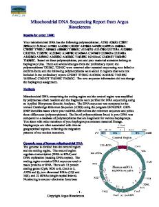
Outlook Recent convincing evidence regarding the presence and functionality of 5mC and 5hmC modifications of mammalian mtDNA is bound to stimulate interest in mitochondrial epigenetics. Currently, available data show that these modifications may be driven by the expression of a particular type of mammalian mtDNMT1 and possibly by DNMT3A. It is expected that in the future, characterization of mtDNMT1 and DNMT3A, e.g., the pattern of their cell-type-specific and tissue-specific expression (including in the CNS) and the understanding of the pathways involved in the regulation of mtDNMT1 expression vs. the total DNMT1 expression, will bring about significant progress and open new directions of epigenetic research. Regulation of 5hmC is particularly important for neuroepigenetics (the first conclusive evidence for a significant 5hmC presence in mammalian ncDNA was found in neurons). Hence, it is expected that research directed toward understanding the action of TET enzymes and other factors involved in 5hmC formation and possibly in DNA demethylation (65) will shift in focus from ncDNA-only to both ncDNA and mtDNA (Figure 1). In addition, evidence is emerging that another mechanism of posttranslational gene regulation, the pathway of RNA interference mediated by microRNAs, which interacts tightly with epigenetic mechanisms, may be operative in mitochondria as well (66). A therapeutic (i.e., pharmacological) targeting of epigenetic mechanisms is likely to become important in mitochondrial
Nc mtDNMT1 DNMT3A
DNMTs
ncDNA
TETs
5mC 5hmC
?
and
Mt
Histones 5mC 5hmC
mtDNA and
TFAM
? Chromatin remodeling
Nucleoid remodeling
Figure 1 A speculative model of mitochondrial epigenetic mechanisms. ncDNA encodes for proteins (e.g., DNMTs, TETs, mtDNMT1) involved in epigenetic DNA modifications occurring both in the nucleus (Nc) and in the mitochondrion (Mt). Both the maintenance and the de novo DNMTs lead to formation of 5mC in ncDNA, whereas mtDNMT1 and possibly DNMT3A synthesizes 5mC in mtDNA. TET enzymes are involved in 5hmC formation (and possibly DNA demethylation) in the nucleus. The exact mechanism of 5hmC formation in mtDNA is currently unclear. Additional epigenetic mechanisms in the nucleus involve chromatin remodeling (mostly through histone modifications). It is not known whether similar mechanisms (e.g., targeted at the TFAM protein) are operative in the mitochondrion, i.e., whether epigenetic mechanisms could lead to nucleoid remodeling.
epigenetics. No such research has been reported yet, although it is known that drugs used to target nuclear epigenetic mechanisms, such as the histone deacetylase inhibitor valproic acid, are capable of triggering significant mitochondrial side effects (67). The concept of epigenetherapy is rapidly evolving (68), and it is likely that in the near future it would include mitochondria. To this end, mitochondria may offer certain advantages for achieving therapeutic target specificity. For example, these organelles possess complex protein import machinery (69). This machinery and other properties of mitochondria, such as the large membrane potential across the inner membrane, could be targeted to selectively direct bioactive molecules (70), e.g., modifiers of mtDNMT1 and DNMT3A activity, to mitochondria. Finally, novel and better tools for mapping the mtDNA pattern of 5mC and 5hmC distribution could be developed. Combined with translational clinical studies, these tools may lead to the discovery of certain types of epigenetically modified mtDNA as biomarkers (e.g., in samples obtained from blood cells) for neuropsychiatric disorders. Ultimately, we expect that within a few years, mitochondrial epigenetics will cease to be a blind spot in epigenetic research, including in neuroepigenetics.
Highlights • Mammalian mitochondria contain multiple copies of a maternally inherited genome, a circular double-stranded mtDNA that encodes 13 proteins, 2 ribosomal RNAs, and 22 transfer RNAs. • Mitochondria do not contain histones; however, mtDNA is protein coated (TFAM is the most abundant) and packaged into aggregates called nucleoids or mitochromosome. • Except for the 13 proteins encoded by mtDNA, other proteins (perhaps thousands) necessary for mitochondrial structure and function are encoded by ncDNA. • Mammalian ncDNA expresses an isoform of DNMT1 (enzyme involved in DNA methylation and formation of 5mC) that contains a mitochondrial targeting sequence, i.e., mtDNMT1. • mtDNMT1 protein (and possibly DNMT3A) is imported into the mitochondria where its interactions with mtDNA appear to be CpG dependent and particularly evident in the D-loop control region. • The CpG dinucleotide is pervasively under-represented in mammalian mtDNA, which could explain why the methylation of mtDNA has typically been dismissed or has not been captured by techniques designed to measure the 5mC status of ncDNA. • mtDNMT1 is active in modifying the transcription of the mitochondrial genome, but the full functional implications of mtDNA 5mC are yet to be elucidated. • DNMT1 and DNMT3A appear to be associated with mitochondria in the motor neurons of patients with amyotrophic lateral sclerosis where they may contribute to DNA methylation-mediated cell death. • mtDNA contains 5hmC, but the mechanism of its formation in mitochondria is not yet understood.
Mitochondrial epigenetics
• Currently, neuroepigenetics does not include mitochondrial epigenetics – a gap that we expect to close in the near future.
Acknowledgments This research was supported in part by the National Institutes of Health (NIH) grant R01AG015347 from the National Institute on Aging (NIA). NIH and NIA had no role in the preparation, review, or approval of the manuscript. The content is solely the responsibility of the authors and does not necessary represent the official views of the NIA. The authors declare no conflict of financial and other interest.
References 1. Riggs AD. X inactivation, differentiation, and DNA methylation. Cytogenet Cell Genet 1975; 14: 9–25. 2. Holliday R, Pugh JE. DNA modification mechanisms and gene activity during development. Science 1975; 187: 226–32. 3. Manes C, Menzel P. Demethylation of CpG sites in DNA of early rabbit trophoblast. Nature 1981; 293: 589–90. 4. Sanford JP, Chapman VM, Rossant J. DNA methylation in extraembryonic lineages of mammals. Trends Genet 1985; 1: 89–93. 5. Li E, Beard C, Jaenisch R. Role for DNA methylation in genomic imprinting. Nature 1993; 366: 362–5. 6. Ehrlich M. The controversial denouement of vertebrate DNA methylation research. Biochemistry (Mosc) 2005; 70: 568–75. 7. Hake SB, Allis CD. Histone H3 variants and their potential role in indexing mammalian genomes: the “H3 barcode hypothesis”. Proc Natl Acad Sci USA 2006; 103: 6428–35. 8. Wu H, Coskun V, Tao J, Xie W, Ge W, Yoshikawa K, Li E, Zhang Y, Sun YE. Dnmt3a-dependent non-promoter DNA methylation facilitates transcription of neurogenic genes. Science 2010; 329: 444–8. 9. Guskova LV, Burtseva NN, Tushmalova NA, Vanyushin BF. Level of methylation of nuclear DNA of neurons and glia in the cerebral cortex of rats and its changes during conditioning. Dokl Biol Sci 1977; 233: 153–6. 10. Griffith JS, Mahler HR. DNA ticketing theory of memory. Nature 1969; 223: 580–2. 11. Holliday R. Is there an epigenetic component in long-term memory? J Theor Biol 1999; 200: 339–41. 12. Day JJ, Sweatt JD. Epigenetic mechanisms in cognition. Neuron 2011; 70: 813–29. 13. Guo JU, Ma DK, Mo H, Ball MP, Jang MH, Bonaguidi MA, Balazer JA, Eaves HL, Xie B, Ford E, Zhang K, Ming GL, Gao Y, Song H. Neuronal activity modifies the DNA methylation landscape in the adult brain. Nat Neurosci 2011; 14: 1345–51. 14. Singer-Sam J. An epigenetic role in schizophrenia. Schizophr Bull 1991; 17: 365. 15. Tremolizzo L, Carboni G, Ruzicka WB, Mitchell CP, Sugaya I, Tueting P, Sharma R, Grayson DR, Costa E, Guidotti A. An epigenetic mouse model for molecular and behavioral neuropathologies related to schizophrenia vulnerability. Proc Natl Acad Sci USA 2002; 99: 17095–100. 16. Akbarian S, Ruehl MG, Bliven E, Luiz LA, Peranelli AC, Baker SP, Roberts RC, Bunney WE Jr, Conley RC, Jones EG, Tamminga CA, Guo Y. Chromatin alterations associated with down-regulated metabolic gene expression in the prefrontal cor-
113
tex of subjects with schizophrenia. Arch Gen Psychiatry 2005; 62: 829–40. 17. Rebelo AP, Dillon LM, Moraes CT. Mitochondrial DNA transcription regulation and nucleoid organization. J Inherit Metab Dis 2011; 34: 941–51. 18. Nass MM, Nass S. Intramitochondrial fibers with DNA characteristics. I. Fixation and electron staining reactions. J Cell Biol 1963; 19: 593–611. 19. Nass S, Nass MM. Intramitochondrial fibers with DNA characteristics. II. Enzymatic and other hydrolytic treatments. J Cell Biol 1963; 19: 613–29. 20. Sagan L. On the origin of mitosing cells. J Theor Biol 1967; 14: 255–74. 21. Wallace DC. Why do we still have a maternally inherited mitochondrial DNA? Insights from evolutionary medicine. Annu Rev Biochem 2007; 76: 781–821. 22. Falkenberg M, Larsson NG, Gustafsson CM. DNA replication and transcription in mammalian mitochondria. Annu Rev Biochem 2007; 76: 679–99. 23. Shutt TE, Shadel GS. A compendium of human mitochondrial gene expression machinery with links to disease. Environ Mol Mutagen 2010; 51: 360–79. 24. Greaves LC, Reeve AK, Taylor RW, Turnbull DM. Mitochondrial DNA and disease. J Pathol 2012; 226: 274–86. 25. Cardon LR, Burge C, Clayton DA, Karlin S. Pervasive CpG suppression in animal mitochondrial genomes. Proc Natl Acad Sci USA 1994; 91: 3799–803. 26. Kukat C, Wurm CA, Spåhr H, Falkenberg M, Larsson NG, Jakobs S. Super-resolution microscopy reveals that mammalian mitochondrial nucleoids have a uniform size and frequently contain a single copy of mtDNA. Proc Natl Acad Sci USA 2011; 108: 13534–9. 27. Kanki T, Nakayama H, Sasaki N, Takio K, Alam TI, Hamasaki N, Kang D. Mitochondrial nucleoid and transcription factor A. Ann N Y Acad Sci 2004; 1011: 61–8. 28. Uchiumi T, Kang D. The role of TFAM-associated proteins in mitochondrial RNA metabolism. Biochim Biophys Acta 2011; [Epub ahead of print]. 29. Mootha VK, Bunkenborg J, Olsen JV, Hjerrild M, Wisniewski JR, Stahl E, Bolouri MS, Ray HN, Sihag S, Kamal M, Patterson N, Lander ES, Mann M. Integrated analysis of protein composition, tissue diversity, and gene regulation in mouse mitochondria. Cell 2003; 115: 629–40. 30. Psarra AM, Sekeris CE. Nuclear receptors and other nuclear transcription factors in mitochondria: regulatory molecules in a new environment. Biochim Biophys Acta 2008; 1783: 1–11. 31. Wallace DC. Mitochondrial DNA mutations in disease and aging. Environ Mol Mutagen 2010; 51: 440–50. 32. Hotchkiss RD. The quantitative separation of purines, pyrimidines, and nucleosides by paper chromatography. J Biol Chem 1948; 175: 315–32. 33. Wyatt GR. Occurrence of 5-methylcytosine in nucleic acids. Nature 1950; 166: 237–8. 34. Vanyushin BF, Nemirovsky LE, Klimenko VV, Vasiliev VK, Belozersky AN. The 5-methylcytosine in DNA of rats. Tissue and age specificity and the changes induced by hydrocortisone and other agents. Gerontologia 1973; 19: 138–52. 35. Krauss V, Reuter G. DNA methylation in Drosophila – a critical evaluation. Prog Mol Biol Transl Sci 2011; 101: 177–91. 36. Nass MM. Differential methylation of mitochondrial and nuclear DNA in cultured mouse, hamster and virus-transformed hamster cells. In vivo and in vitro methylation. J Mol Biol 1973; 80: 155–75.
114
H. Manev et al.
37. Dawid IB. 5-methylcytidylic acid: absence from mitochondrial DNA of frogs and HeLa cells. Science 1974; 184: 80–1. 38. Vanyushin BF, Kiryanov GI, Kudryashova IB, Belozersky AN. DNA-methylase in loach embryos (Misgurnus fossilis). FEBS Lett 1971; 15: 313–6. 39. Vanyushin BF, Kirnos MD. The nucleotide composition and pyrimidine clusters in DNA from beef heart mitochondria. FEBS Lett 1974; 39: 195–9. 40. Vanyushin BF, Kirnos MD. Structure of animal mitochondrial DNA (base composition, pyrimidine clusters, character of methylation). Biochim Biophys Acta 1977; 475: 323–36. 41. Shock LS, Thakkar PV, Peterson EJ, Moran RG, Taylor SM. DNA methyltransferase 1, cytosine methylation, and cytosine hydroxymethylation in mammalian mitochondria. Proc Natl Acad Sci USA 2011; 108: 3630–5. 42. Chestnut BA, Chang Q, Price A, Lesuisse C, Wong M, Martin LJ. Epigenetic regulation of motor neuron cell death through DNA methylation. J Neurosci 2011; 31: 16619–36. 43. Pollack Y, Kasir J, Shemer R, Metzger S, Szyf M. Methylation pattern of mouse mitochondrial DNA. Nucleic Acids Res 1984; 12: 4811–24. 44. Shmookler Reis RJ, Goldstein S. Mitochondrial DNA in mortal and immortal human cells. Genome number, integrity, and methylation. J Biol Chem 1983; 258: 9078–85. 45. Scarpulla RC. Metabolic control of mitochondrial biogenesis through the PGC-1 family regulatory network. Biochim Biophys Acta 2011; 1813: 1269–78. 46. Wang J, Xiong S, Xie C, Markesbery WR, Lovell MA. Increased oxidative damage in nuclear and mitochondrial DNA in Alzheimer ’s disease. J Neurochem 2005; 93: 953–62. 47. Wang J, Markesbery WR, Lovell MA. Increased oxidative damage in nuclear and mitochondrial DNA in mild cognitive impairment. J Neurochem 2006; 96: 825–32. 48. Kriaucionis S, Heintz N. The nuclear DNA base 5-hydroxymethylcytosine is present in Purkinje neurons and the brain. Science 2009; 324: 929–30. 49. Münzel M, Globisch D, Brückl T, Wagner M, Welzmiller V, Michalakis S, Müller M, Biel M, Carell T. Quantification of the sixth DNA base hydroxymethylcytosine in the brain. Angew Chem Int Ed Engl 2010; 49: 5375–7. 50. Song CX, Szulwach KE, Fu Y, Dai Q, Yi C, Li X, Li Y, Chen CH, Zhang W, Jian X, Wang J, Zhang L, Looney TJ, Zhang B, Godley LA, Hicks LM, Lahn BT, Jin P, He C. Selective chemical labeling reveals the genome-wide distribution of 5-hydroxymethylcytosine. Nat Biotechnol 2011; 29: 68–72. 51. Davis T, Vaisvila R. High sensitivity 5-hydroxymethylcytosine detection in Balb/C brain tissue. J Vis Exp 2011; pii: 2661. 52. Jin SG, Wu X, Li AX, Pfeifer GP. Genomic mapping of 5hydroxymethylcytosine in the human brain. Nucleic Acids Res 2011; 39: 5015–24. 53. Chen H, Dzitoyeva S, Manev H. Aging-related alterations of the DNA base 5-hydroxymethylcytosine are brain-region- and celltype-specific. Program No.773.16. 2011 Neuroscience Meeting Planner. Washington, DC: Society for Neuroscience, 2011. Online.
54. Tahiliani M, Koh KP, Shen Y, Pastor WA, Bandukwala H, Brudno Y, Agarwal S, Iyer LM, Liu DR, Aravind L, Rao A. Conversion of 5-methylcytosine to 5-hydroxymethylcytosine in mammalian DNA by MLL partner TET1. Science 2009; 324: 930–5. 55. Iyer LM, Tahiliani M, Rao A, Aravind L. Prediction of novel families of enzymes involved in oxidative and other complex modifications of bases in nucleic acids. Cell Cycle 2009; 8: 1698–710. 56. Szulwach KE, Li X, Li Y, Song CX, Han JW, Kim S, Namburi S, Hermetz K, Kim JJ, Rudd MK, Yoon YS, Ren B, He C, Jin P. Integrating 5-hydroxymethylcytosine into the epigenomic landscape of human embryonic stem cells. PLoS Genet 2011; 7: e1002154. 57. Riccio A. Dynamic epigenetic regulation in neurons: enzymes, stimuli and signaling pathways. Nat Neurosci 2010; 13: 1330–7. 58. Rebelo AP, Williams SL, Moraes CT. In vivo methylation of mtDNA reveals the dynamics of protein-mtDNA interactions. Nucleic Acids Res 2009; 37: 6701–15. 59. Guo JU, Su Y, Zhong C, Ming GL, Song H. Hydroxylation of 5-methylcytosine by TET1 promotes active DNA demethylation in the adult brain. Cell 2011; 145: 423–34. 60. Bhutani N, Burns DM, Blau HM. DNA demethylation dynamics. Cell 2011; 146: 866–72. 61. Frauer C, Hoffmann T, Bultmann S, Casa V, Cardoso MC, Antes I, Leonhardt H. Recognition of 5-hydroxymethylcytosine by the Uhrf1 SRA domain. PLoS One 2011; 6: e21306. 62. Wallace DC, Fan W. Energetics, epigenetics, mitochondrial genetics. Mitochondrion 2010; 10: 12–31. 63. Smiraglia DJ, Kulawiec M, Bistulfi GL, Gupta SG, Singh KK. A novel role for mitochondria in regulating epigenetic modification in the nucleus. Cancer Biol Ther 2008; 7: 1182–90. 64. Takasugi M, Yagi S, Hirabayashi K, Shiota K. DNA methylation status of nuclear-encoded mitochondrial genes underlies the tissue-dependent mitochondrial functions. BMC Genomics 2010; 11: 481. 65. Nabel CS, Kohli RM. Molecular biology. Demystifying DNA demethylation. Science 2011; 333: 1229–30. 66. Bandiera S, Rüberg S, Girard M, Cagnard N, Hanein S, Chrétien D, Munnich A, Lyonnet S, Henrion-Caude A. Nuclear outsourcing of RNA interference components to human mitochondria. PLoS One 2011; 6: e20746. 67. Finsterer J, Segall L. Drugs interfering with mitochondrial disorders. Drug Chem Toxicol 2010; 33: 138–51. 68. Husso T, Turunen MP, Parker N, Yla-Herttuala S. Epigenetherapy, a new concept. BioMol Concepts 2011; 2: 127–34. 69. Mokranjac D, Neupert W. Thirty years of protein translocation into mitochondria: unexpectedly complex and still puzzling. Biochim Biophys Acta 2009; 1793: 33–41. 70. Smith RA, Hartley RC, Murphy MP. Mitochondria-targeted small molecule therapeutics and probes. Antioxid Redox Signal 2011; 15: 3021–38.
Received October 24, 2011; accepted December 1, 2011
Mitochondrial epigenetics
Hari Manev graduated from the Zagreb Medical School in Croatia (MD and PhD), and completed his postdoctoral studies in neuroscience and neuropharmacology at Georgetown University in Washington, DC. He is currently a tenured Professor of Pharmacology at the University of Illinois at Chicago. His previous appointments include research institutes (Institute Ruder Boškovi c´, Zagreb, Croatia; FIDIA-Georgetown Institute for the Neurosciences, Washington, DC; and the Allegheny-Singer Research Institute, Pittsburgh, PA), he has worked in the pharmaceutical industry (FIDIA SpA, Abano Terme, Italy), and in academia (Georgetown University, Washington, DC, and the Medical College of Pennsylvania and Hahnemann University – later named Allegheny University of the Health Sciences, Pittsburgh, PA). His research interests include brain aging, neurotoxicity and neuroprotection, circadian mechanisms, mechanisms of action of psychiatric treatments and addiction, and epigenetic mechanisms in the central nervous system. Svetlana Dzitoyeva (Dzitoeva) graduated from North Ossetian State University (MS) and the Koltzov Institute for Developmental Biology, Russian Academy of Sciences, Moscow, Russia (PhD). She completed postdoctoral studies in molecular biology at the University of Illinois at Chicago (UIC), Chicago, IL. Currently, she is a Senior Research Specialist in Health Sciences in The Psychiatric Institute (UIC). In recent years, she has developed Drosophila models and techniques for neuropharmacological studies. Her current research interest includes epigenetic mechanisms in mammalian brain and mechanisms of action of neuropharmacological treatments.
115
Hu Chen graduated in China from Shandong Traditional Chinese Medicine University (BS), Zhejiang Traditional Chinese Medicine University (MS), and the Medical College, Fudan University (PhD). He completed postdoctoral studies in neuroscience at the University of Illinois at Chicago (UIC) and studies in pharmacology at the University of New Mexico, Albuquerque, NM. Currently, he is a Research Specialist in Health Sciences in The Psychiatric Institute (UIC). His research interests include mechanisms of and role for the central nervous system action of the inflammatory enzyme 5-lipoxygenase and epigenetic regulation of genes involved in addiction and brain aging.
Copyright of Biomolecular Concepts is the property of De Gruyter and its content may not be copied or emailed to multiple sites or posted to a listserv without the copyright holder's express written permission. However, users may print, download, or email articles for individual use.