HEART VALVE DISEASE You’ve recently been feeling a bit dizzy and have been suffering from occasional chest pains. You’ve found yourself slightly short of breath and have definitely been more tired than usual. You’re worried that you’re retaining fluid. You’ve sensibly decided to seek reassurance from your doctor, who’s presently listening to your heart with a stethoscope Valves are small but critically important components of your heart. They help to make sure that blood keeps flowing in the right direction. Normally, the valves open and shut rapidly and completely, providing the lub-dub, lub-dub sound heard through the stethoscope. When your heart valves stop working properly, blood flow through the heart is disrupted leading to a swish-swish, swish-swish sound. Your doctor has been trained to make the distinction between both of these sounds. Some people are born with heart valve disease brought about by congenital defects. Valves may be the wrong size, or have flaps (also known as leaflets) that are badly formed or that aren’t attached properly to the ring of tissue supporting the valve (known as the annulus). Often, these defects can be corrected at birth or later in life. Other people are born with healthy valves but later develop a type of heart valve disease known as acquired valve disease. There are many possible causes of acquired valve disease, including infection (e.g. endocarditis) or systemic diseases (e.g. rheumatoid arthritis). The result can be a tight, rigid valve that limits forward blood flow (stenotic valve), or a leaky valve that does not close tightly, permitting back flow (regurgitant valve). Occasionally people have both sorts of valve disease (mixed valve disease). Valve disease can also be caused by heart attacks, coronary artery disease, heart muscle disease, an aortic aneurysm (a severe widening of the aorta), hypertension, connective tissue diseases and, less often, tumours, certain drugs or radiation therapy. In India, rheumatic heart disease is the major cause of valvular heart disease in children and young adults. Rheumatic heart disease is caused by rheumatic fever which is caused by streptococcal throat infection in children. Chronic rheumatic heart disease remains the leading cause of stenotic or regurgitant valve and valve replacement in children and young adults. After a patient with valve disease has undergone physical examination and further tests, doctors may recommend valve replacement. Worldwide, many heart valve operations are performed each year, most commonly on the mitral and aortic valves. Valve replacement surgery is performed on a daily basis. Figure 1. A picture of a heart that has been sliced horizontally to show the valves
ANATOMY AND FUNCTION OF THE HEART Your heart is an amazing organ. It is located under the rib cage between the lungs. The heart is shaped like an upside down pear and is about the same size as a clenched fist. This muscular organ continuously pumps five to six litres of blood per minute through blood vessels to all parts of the body. The heart has four chambers and is divided into a left and a right side by a muscular wall (septum). Each side of the heart is further divided into a top chamber (atrium), which receives blood from the veins, and a larger bottom chamber (ventricle), which pumps blood into the arteries. The atria and ventricles work together, contracting and relaxing to pump blood out of the heart. This contracting and relaxing causes blood to move from the atria to the ventricles, resulting in the opening and closing of the valves. This produces the distinctive sound of the heartbeat that your doctor has been trained to listen for. This cycle of contraction and relaxation happens some 70+ times per minute, or about 104,000 times a day. Over the course of an average lifetime, the heart can beat up to 2.5 billion times. Figure 2. Picture showing the different parts of the heart. The left and right sides of the heart are named according to their arrangement in the body, whilst drawings of the heart usually show the organ from the front, which is why this diagram shows the left side of the heart on the right-hand side and vice versa
BLOOD FLOW THROUGH THE HEART There are four valves within the heart. These valves open to allow blood to flow forward into the next chamber or vessel, and close to prevent blood from flowing back. Blood flow begins in the right side of the heart. The veins carry blood that is low in oxygen to the right atrium. From the right atrium, blood passes into the right ventricle via the tricuspid valve. Blood is then pumped from the right ventricle into the pulmonary artery via the pulmonary valve, and enters the lungs where it receives oxygen. From the lungs, the now oxygen-rich blood reenters the heart on the left side through the pulmonary veins, flowing into the left atrium, and passes into the left ventricle through the mitral valve. From here blood is pumped into the aorta via the aortic valve and circulated around the body. Figure 3. Diagrams of the heart showing the direction of blood flow through the valves into the heart chambers
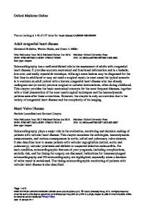
VALVE REPLACEMENT AND REPAIR OPTIONS Depending on exactly what’s wrong, diseased heart valves may be repaired or replaced. Different techniques can be used to repair a valve depending upon the problem you have. Based on the type of repair that is needed, surgeons will consider valvuloplasty or annuloplasty. These types of procedures are mainly used at the base of the mitral valve. Annuloplasty is a common procedure that involves inserting a ‘repair-ring’. This is applicable to the patients with valvular regurgitation when the structure of the valve is not severely diseased. The advantages of valvuloplasty and valve repair over the valve replacement are avoidance of anticoagulation and mandatory frequent INR (International Normalized Ratio) testing thereafter. Further, it preserves the function of the heart. However due to the continued rheumatic activity, most of these patients will require valve replacement later at some stage of their life. Your surgeon will decide whether receiving a repair-ring or an artificial (prosthetic) valve is the best option. Even if you receive a repair-ring, the guidelines presented in this booklet can still apply to you. If a heart valve cannot be repaired then the surgeon will have to remove the diseased valve and replace it with a prosthetic valve. There are two types of prosthetic valve used for replacement: artificial (mechanical) valves and tissue (bioprosthetic) valves. Figure 4. Annuloplasty ring (A), mechanical valve (B) and stented bioprosthetic valve (C). Mechanical valve A mechanical valve is carefully designed to perform the function of your own heart valve and is generally made of a material called pyrolytic carbon. Like the annulus of your own heart, the mechanical valve has a ring to support the valve leaflets. The mechanical valve opens and closes with each heartbeat, permitting proper blood flow through the heart. Figure 5. Mechanical replacement valves can be inserted at the mitral (A) or aortic (B) valve position Bioprosthetic valve A bioprosthetic or tissue valve is a valve of either porcine (pig) or bovine (cow) tissue origin that has been carefully prepared for use in the human heart. Tissue valves can be either stented or stentless. A stent is a frame that supports the new valve to maintain its shape. Stented valves can be used to replace all valves. Stentless valves are most commonly used for replacement of the aortic valve. Figure 6A. Stented bioprosthetic replacement valves are usually inserted in the mitral or aortic valve positions (both are shown here).
WHAT KIND OF VALVE IS SUITABLE FOR YOU Issues with mechanical valves The patients with a mechanical valve have to take lifelong anticoagulation medicines (warfarin or acitrom) and undergo repeated INR (International Normalised Ratio) testing to keep the INR in the desired range specific for the mitral or aortic position. The anticoagulant ‘thins’ the blood to prevent formation of dangerous blood clots on or around your new heart valve. The INR value indicates how long it takes the blood to clot. There are inherently possible complications of mechanical valves and the anticoagulation management. Some examples of mechanical valve complications include valvular dysfunction, thrombosis and embolism (blood clot) and disc dislodgement. Fortunately, the rate of these incidences happening is low. Abnormally high INR may result in bleeding in various body parts including brain. This will require stopping the anticoagulant medicine, hospitalization and the patient may need transfusions of plasma and blood products. Abnormally low INR could result in the formation of blood clots which may obstruct the movement of your mechanical valve and this could lead to a fatal outcome. Taking oral anticoagulants during pregnancy can adversely affect the fetus. Anticoagulation is also preferably avoided in patients with hypertension, diabetes mellitus, osteoporosis, and senility. Issues with bioprosthetic valves Are bioprosthetic valves free of problems? Far from it. Degeneration and calcification of bioprosthetic valves over a period of time make reoperation necessary and this has its own morbidity and mortality. The current generation bioprosthetic valves last for approximately 15 years or more. Also, early postoperative incidence of thromboembolism (formation of blood clot in vessels that break loose and plug other vessels) is the same as in mechanical valves, though later it decreases favorably. The cost of reoperation in the future would be the same as a patient with a mechanical valve who would spend on anticoagulation and INR tests during that period. Are there then any advantages of bioprosthetic valves? The blood flow across the bioprosthetic valve mimics that of the natural valve more than the flow across the mechanical valve. Also most patients will not need to take lifelong oral anticoagulation. Lifestyle How important is lifestyle? Anticoagulation is ill-advised in people involved in contact and/or highly competitive sports, nomadic living or in professions where risk to others’ lives is enormous in events of sudden anticoagulant related event e.g. train drivers, pilots. Conversely if such people are required to take anticoagulation, lifestyle or career modification is advised.
The pregnant females are required to stop oral anticoagulation and switch to parenteral anticoagulation in the first and third trimester. Anticoagulant taken in the first few weeks of pregnancy could adversely affect the fetus. Age Bioprosthetic valves are known to degenerate rapidly in children while their anticoagulation management in this group of patient is indeed difficult. However, there are certain positions whereby bioprosthetic valves are preferred substitutes, namely in the tricuspid and pulmonary valves, where clots can form easily if a mechanical valve is used. Bioprosthetic valves are also used in children undergoing mitral or aortic valve replacement when their follow up is difficult or adequate anticoagulation cannot be guaranteed due to lack of education, poverty, and inaccessibility of appropriate medical care. Though bioprosthetic valves are known to degenerate faster in the young, there are certain advantages that must be weighed carefully against the need for anticoagulation with mechanical valve. Bioprosthetic valves are preferred particularly in young females desirous of child bearing as these patients do not need to take anticoagulants. Should the bioprosthetic valves be preferred in middle-aged and elderly? There are several advantages for this choice. Avoiding anticoagulation in these patients would mean lesser anticoagulation related accidents. Procedures such as dental, orthopedic, urological or gynecological procedures can be safely performed with appropriate medical management. Also, no major dietary or lifestyle modification is necessary in these elderly patients. There are also significant advantages of using bioprosthetic valves in patients from rural and remote areas, patients who are poor, ill educated with severely limited or virtually inaccessible medical care. Multivalvular disease Multivalvular disease presents special situations. When two or more valves are affected and it becomes necessary to replace only the one predominantly diseased valve, bioprosthetic valves may be preferable as it would avoid anticoagulation related problems. If both or all of the affected valves need to be replaced, the presence of compromised left ventricular function limiting the long term survival of the patient should tilt the balance in favor of bioprosthetic valves. Choice Ultimately, the choice of the valve prosthesis must take into consideration every individual’s age, gender and lifestyle, the presence of any concomitant illness, the pathology of the native valvular disease and accessibility to medical care. Both types of valve have advantages, and your physician will select the valve that best suits your needs and anatomy.
YOUR LIFE AFTER HEART VALVE SURGERY Many people who receive a new heart valve are able to lead a more active and fulfilling life than before surgery. As a recipient of a new heart valve you will need to be actively involved in caring for your heart. By understanding how to maintain your health and fitness, your wellbeing and quality of life can be improved. Medication After a mechanical valve replacement (and sometimes after a tissue value replacement), your doctor will prescribe a medication called an anticoagulant to be taken daily for life. Patients with bioprosthetic valves must take anticoagulants for initial three months after surgery. The right level of anticoagulant for you will be calculated by your doctor and closely checked by blood tests. Patients receiving anticoagulation should check their INR initially every week for first month after starting the anticoagulant and consult the treating physician or surgeon. Your doctor will suitably advise you to adjust the dose of anticoagulant to achieve the expected INR value. Generally, the INR should be maintained at 2 to 2.5 for the aortic valve replacement patients and 3 to 3.5 for mitral or double (mitral and aortic) valve replacement patients. Expected INR values are achieved by gradually adjusting the dose of anticoagulation up or downwards so that the patient neither clots easily nor bleeds excessively. High INR values resulting in excessive overt or covert bleeding may compel the doctor to hospitalize the patient, administer medicines to counter the effects of anticoagulant and stop or reduce the dose of the anticoagulant until such time that INR returns to the desirable range. If pregnancy is suspected or confirmed, it is very important that you inform your doctor as oral anticoagulant must be avoided in the first 6 to 8 weeks of pregnancy to avoid harm to the fetus. Your doctor will administer alternative methods to anticoagulation to you and advice you on the course of action for the rest of your pregnancy. Though INR is fairly reliable and comparable, it still has its limitations. The presence of atrial fibrillation, large left atrium, left ventricular dysfunction will call for more anticoagulation while hepatic and renal disorders, congestive cardiac failure will make it necessary to decrease the anticoagulant dosage. Excessive green leafy vegetable diet, fatty foods and chronic alcohol consumption will reduce the anticoagulant activity. Concurrent medications e g painkillers could prolong INR and induce bleeding. Please report to the doctor immediately in case you notice bleeding from gums after brushing, blood in sputum, pink or red urine, black stools, unexplained swelling, excessive bruising, coffee ground vomiting, headache, abdominal or joint pain, breathlessness, visual disturbances, paralysis, convulsions, coldness of a limb, fever and excessive menstrual bleeding or a missed period in females. Make sure that you always follow your prescription and the recommendations provided by the doctor who is treating you.
Dental and other procedures If you are undergoing any dental or surgical procedure, bacteria can enter your bloodstream. This can result in an infection, such as endocarditis, occurring in the tissue surrounding your new valve. After you have received a mechanical heart valve replacement or a tissue heart valve replacement and are receiving anticoagulant medication, you need to notify your doctor or dentist that you have had heart valve surgery before going for any surgical procedure or intervention (e.g. cataract surgery). Your doctor will help you make adjustments in anticoagulation and infective endocarditis prophylaxis as best defence against infection. Your doctor may also advise you on diet, as food and alcohol can affect how well the medications work. Exercise and diet to nurture your heart By progressively increasing your physical activity through exercise, your strength and stamina will improve. This will help you to train your heart muscle to pump more efficiently. Aerobic exercise, such as walking, swimming, cycling or performing regular work-outs at the gym can help improve your cardiovascular fitness. Follow your doctor’s advice regarding exercise after heart valve surgery. A heart-healthy diet is always recommended, including for people who have had valve surgery. You should limit your intake of saturated fats and oils and consume a low-fat diet. Ask your doctor to help you develop a heart-healthy diet. If you experience fluid retention after surgery, this can overload your heart and make it work less efficiently. To prevent fluid retention, your doctor may advise you to follow a number of different strategies including consuming a low sodium diet. It is important that you follow the recommendations of the doctor who is treating you. Keep an eye out for change If you notice any sudden weight gain, or swelling of your ankles, legs, hands or stomach, or unusual shortness of breath, you should report this to your doctor. You should also inform your doctor if any unusual bleeding events occur. Long-term health of your heart Long-term management of your health requires your full participation. Your doctor will work with you to check the function of your new heart valve. The doctor will carry out tests when required and discuss and answer any questions you have about your health. Remember to follow any guidelines you are given for diet, exercise and medications, and to keep scheduled appointments.
SOME FREQUENTLY ASKED QUESTION How long does a prosthesis last? Studies have shown that a mechanical heart valve, which is made of pyrolytic carbon, will not wear out in your lifetime. However, there are clinical situations such as blood clots or infections that may require your valve to be replaced. On the other hand, a bioprosthetic valve, which is made of biological tissue, may have a shorter life than a mechanical valve. The symptoms of a failing valve may be the same as those you had before surgery: shortness of breath, dizziness, chest pain, fatigue and fluid retention. If any of these symptoms occur you must notify your doctor. Can an artificial heart valve be repaired? In general, an artificial heart valve is not repaired but replaced. Whether a replacement valve can be repaired by surgery depends on the reason for the repair. If a bioprosthetic valve has become hardened because of a build-up of calcium deposits, or if a mechanical valve is affected by the formation of clots or excessive tissue, it would probably be better to replace it with a new valve. If you have any concerns about your heart valve, your surgeon would be the best person to ask. What happens if I am exposed to a metal detector, magnetic resonance imaging (MRI) or electronic equipment? The amount of metal used in mechanical and bioprosthetic heart valves is very small. Normally, if you pass through a metal detector it shouldn’t set off the alarm. Metal detectors will not harm your heart valve. Mechanical heart valves are made of materials that will not be affected by computed tomography (CT) scans, X-rays or magnetic resonance imaging equipment. Additional information regarding the safety of MRI use is available from the device manufacturer for the personnel performing your test. However, if you have had a valve replacement, it is important that you let your doctor know before any diagnostic testing, even if the test is not heart related. Other electronic devices and equipment, including mobile phones and microwave ovens, will not affect your new heart valve. Will the mechanical valve make a noise? Some patients have said that they hear a clicking sound during quiet or restful times. If you hear this sound there is no need to be worried. The clicking is actually the sound of the mechanical valve closing. Many people do not even hear the clicking as the sound is affected by individual anatomy and physiology. If you have a new valve that produces a different sound, you will quickly become accustomed to this new sound. After a while, it will eventually grow unnoticeable. And, as studies have shown, this sound should not affect your quality of life. For those patients who can hear the clicking sound, they should familiarize themselves with the sound of the metallic prosthesis. Loss or muffling of this clicking sound may suggest obstruction to the valve. In such cases, please consult your doctor immediately and they will examine you and perhaps will investigate with fluoroscopy or echocardiography to confirm or rule out any obstruction to the valve and then advise you appropriately.
Other things to note High fever in patients with prosthetic valves may be due to infective endocarditis and requires prompt investigation. Inform your doctor and if Endocarditis is the cause of the symptoms, appropriate antibiotic treatment would be advised by the treating doctor. Bruises, cuts or any injuries should also be promptly attended to prevent any infection.
Remember that you are an important member of your healthcare team Heart-health management is a team effort and you are the key player in the team. Your doctor will prescribe your medication and manage other medical problems. Other team members, including nurses, dietitians, pharmacists and exercise specialists will help you to successfully achieve a healthy heart. But it is up to YOU to take your medications make dietary changes, live a healthy lifestyle, keep your future appointments and be an active member of the team. The following list of points will help you maintain a healthy heart: • Take all your medication as prescribed • See your doctor regularly to check that the anticoagulant in your body is at the correct level • Follow a heart-healthy diet • Follow an approved exercise program • Try to control the stress in your life • Report any signs of fluid retention or infection to your doctor • Inform your dentist and doctors that you have an artificial heart valve – they may prescribe additional medication before a dental or medical procedure • Enjoy your rejuvenated heart!
Patient’s Record for INR Management. Date
INR
Daily Dosage(mg)
Patient’s Record for INR Management.
Comments
Signature
Date
INR
Daily Dosage(mg)
Comments
Signature
Comments
Signature
Patient’s Record for INR Management.
Date
INR
Daily
Dosage(mg)