Original Article
Evidence of Woven Bone Formation in Heart Valve Disease Masoud Mirzaie, MD,1 Michael Schultz, MD, PhD,2 Peter Schwartz, PhD,2 Marlon Coulibaly, MD,1 and Friedrich Schöndube, MD1
The pathogenesis of acquired cardiac valve disease still remains a matter of controversy. In this work, scanning electron and polarised light microscopic investigations in addition to energy dispersive X-ray microanalyses (EDAX) were carried out on explanted human aortic and mitral valves to determine the morphology and element composition of calcified areas in valvular lesions. Biopsies were taken from aortic valves removed from 28 male patients (average age, 75±1 years) and 46 females (68±3 years) and from mitral valves obtained from 18 male patients (72±3 years) and 8 females (71±6 years). By means of scanning electron microscopy, multiple foci of calcified areas were identified. Endothelial cells in these areas appeared swollen and displayed reduced cell-cell contacts. The calcium deposits were separated from the adjacent tissue by layers of collagen fibers. Often a layer of woven bone tissue separated intravalvular inclusions from hyperplastic collagen fibers. Using EDAX analysis, calcium and phosphorus were detected in these valvular lesions. The major finding of our study is the presence of woven bone tissue in explanted cardiac valves, which may result from pathological strains or mechanical overloading of the collagen fibers. (Ann Thorac Cardiovasc Surg 2003; 9: 163–9) Key words: woven bone, human heart valves
Introduction Dystrophic calcification is the most common pathological finding in surgically explanted heart valves. Recently, several reports described bone formation in surgically excised heart valves and suggested an unexpected process of tissue repair.1-3) The exact pathomechanisms of acquired cardiac valve disease are still unknown, although the calcification process of biological valves has been well investigated. Mechanical stress-induced factors are regarded to induce the calcification of biological valves.4-6) In this report, we describe the morphology of degenerative changes in explanted aortic and mitral valves by usFrom 1Department of Thoracic, Heart and Vascular Surgery, The University Clinics Göttingen, and 2Institute for Anatomy, University of Göttingen, Germany Received September 13, 2002; accepted for publication November 9, 2002. Address reprint requests to Masoud Mirzaie, MD: Department of Thoracic, Heart and Vascular Surgery, The University Clinics Göttingen, Robert-Koch-Str. 40, D-37075 Göttingen, Germany.
Ann Thorac Cardiovasc Surg Vol. 9, No. 3 (2003)
ing polarised light microscopy in addition to conventional electron microscopical techniques. We were especially interested in identifying bony structures mimicking valve calcification.
Materials and Methods Tissue biopsies Tissue samples from human aortic and mitral valves were taken from patients undergoing routine valve replacement operations at the Department of Thoracic, Heart and Vascular Surgery. From each valve one leaflet was removed for further study. Biopsies were taken from 74 aortic and 26 mitral valves. The aortic valves were removed from 28 male patients (average age, 75±1 years) and 46 females (68±3 years) and the mitral valves were taken from 18 males (72±3 years) and 8 females (71±6 years). The study population included patients suffering from aortic stenosis (n=38, 51.4%) and aortic insufficiency (n=36, 48.6%). There were 17 cases of mitral insufficiency (65.4%) and 9 cases (34.6%) of mitral stenosis. Table 1
163
Mirzaie et al.
Table 1. Baseline characteristics of the study population
Valve pathology Stenosis exclusively Insufficiency/mixed
Aortic valve disease (n=74)
Mitral valve disease (n=26)
38 (51.3%) 36 (48.6%)
9 (34.6%) 17 (65.4%)
Age (mean±SD) Gender Male Female NYHA I/II III/IV Concomitant diseases CHD Syncope Embolism Heart failure Hypertonia Endocarditis Rheumatic fever Concomitant CABG Isolated AVR AVR+CABG AVR+MVR
n=18 n=8
n=28 n=46
Male Female
72±9 71±8
75±11 68±8 13 (17.5%) 61 (82.4%)
5 (19.2%) 21 (80.7%)
42 (56.7%) 13 (17.5%) 2 (2.7%) 19 (25.6%) 53 (71.6%) 4 (5.4%) 2 (2.7%)
14 (53.8%) 0 1 (3.8%) 9 (34.6%) 12 (46.1%) 0 0
32 42 9
Isolated MVR 12 MVR+CABG 14
Continuous variables are presented as mean ± standard deviation. Categorical variables are presented as an absolute percentage. NYHA, New York Heart Association; CHD, coronary heart disease; CABG, coronary artery bypass grafting; AVR, aortic valve replacement; MVR, mitral valve replacement
summarises the basic data of the study patients. In comparison, postmortal tissue samples from 3 male patients (average age, 78.5 years) and 3 female patients (average age, 73.3 years) who died primarily from noncardiac causes, were explanted and investigated. Scanning electron microscopy In order to reveal the surface morphology of the explanted valves, scanning electron microscopy (SEM) was performed. Specimens from valve leaflets were fixed for 6 h in a solution containing 2.5% glutaraldehyde and 0.2 M cacodylate. Samples were then dehydrated in an ascending series of alcohol. After critical point drying, all samples were sputtered with gold-palladium. Samples were visualised using the digital scanning microscope DSM 960 (Carl Zeiss, Oberkochen, Germany). Polarised light microscopy To evaluate the presence of woven bone tissue in calcified areas of pathologically altered aortic and mitral
164
valves, polarised light microscopy was performed. To prepare thin ground sections from undecalcified materials for polarised light microscopy, a special technique was established based upon the method of plastination developed by Hagens et al.7) and modified for histological purposes by Schultz and Drommer.8) Samples were dehydrated using ascending concentration steps of alcohol, put into methyl chloride as an intermediate solution for the exchange of substances and embedded in epoxy resin Biodur®. Unstained thin ground sections (30, 50 and 70 애m) were prepared and viewed in transmitted plane and in polarised light using a quartz plate red first order, equipped with photo documentation.9,10) Energy dispersive X-ray microanalysis (EDAX) In order to determine the element content in explanted human aortic and mitral valves, the specimens were examined by means of EDAX. The samples were fixed for 6 h in a solution containing 2.5% glutaraldehyde and 200 mM cacodylate, dehydrated in an ascending series of al-
Ann Thorac Cardiovasc Surg Vol. 9, No. 3 (2003)
Bony Structures in Heart Valve Disease
a
b
c
Fig. 1. Scanning electron micrographs of explanted aortic and mitral valves. Multiple tears in the leaflet tissue parallel to the edge of the leaflet (a, aortic valve, male patient, 74 years old). The tears showed an endothelial cover (b, aortic valve from the same patient as in Fig. 1a). Calcified material predominantly accumulated in the center of the leaflet and along its attachments to the aortic and mitral rings (c, mitral valve, female patient, 68 years old).
cohol and dried using a critical point drier. The specimens were then sputtered with carbon and irradiated with electrons in a digital scanning microscope (DSM 960, Carl Zeiss). The X-rays emitted from the samples were measured by energy dispersion using an EDS system (Chromeritz, Leipzig, Germany) coupled to the microscope.
Results Scanning electron microscopical findings The tissue biopsies from the human aortic and mitral valves examined showed various types of endothelial lesions which have already been described in detail.11) In all of the explanted valves, we observed multiple tears in the leaflet tissue oriented parallel to the edge of the leaflet (Fig. 1a). At higher magnification, these areas usually exhibited an endothelial cover (Fig. 1b). Calcified material was predominantly accumulated in the center of the leaflet and along its attachments to the aortic and mitral rings (Fig. 1c). The endothelial cells in these regions were ultrastructurally altered and, furthermore, loosely bound to each other (Fig. 2a). The mineralised
Ann Thorac Cardiovasc Surg Vol. 9, No. 3 (2003)
material was frequently surrounded by a layer of collagen fibres. When the calcium deposits were removed, it was seen that the collagen fibres were arranged in several layers (Fig. 2b, c). Polarised light microscopical findings Recent techniques employing polarised light microscopy are potentially useful to evaluate the presence of woven bone tissue. Tissue samples from normal aortic valves revealed no pathological alterations. Even though bundles of collagen fibres were present, no morphological changes were detected (Fig. 3). When unstained thin ground sections of pathologically altered samples were viewed in transmitted plane light, the intravalvular localised inhomogeneous inclusions represented as a secondary substance of yellowish color (Fig. 4a). In regular light, the character of this substance could be described as fibrous resembling natural woven bone. By high magnification (×620), bundles of collagen fibres were detected (Fig. 4b). Using the quartz plate red first order as a compensator, the bundle of collagen fibres were clearly identified by their blue colors (Fig. 4c).
165
Mirzaie et al.
a
b
c
Fig. 2. Ultrastructurally altered endothelial cells loosely bound to each other (a, mitral valve, female patient, 69 years old). The calcium deposits after removal of the upper tissue layer (b. aortic valve, female patient, 71 years old). The mineralised material was clearly surrounded by several layers of collagen fibres (c, aortic valve from the same patient in Fig. 2b).
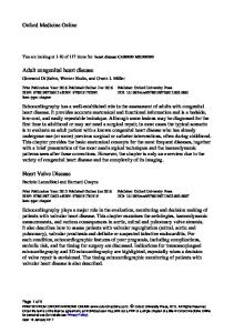
Results of the energy dispersive X-ray microanalysis Energy dispersive X-ray microanalysis (EDAX) was carried out to determine the element content in various endothelial lesions of the explanted aortic and mitral valves. With the exception of degenerative lesions, no calcium signals were detected in the extracellular matrix of valvular alterations. However, mineralised deposits were frequently observed in areas resembling bone-like tissue features. These areas contained calcium and phosphorus as demonstrated by characteristic EDAX patterns (Fig. 5).
Discussion In this paper we report on our systematic SEM and polarised light microscopical investigations as well as on the EDAX data on explanted human aortic and mitral valves. In all the explanted valves, we detected uniform changes in the endothelium and the basement membrane. The endothelial cells often showed hyperplasia with loose binding to each other. Rarely, an endothelial layer was completed. The loss of endothelial cells may expose the extracellular matrix, which obviously sets various patho-
166
logical processes in motion.12-15) The increased activation of matrix metalloproteinases in pathologically altered human cardiac valves emphasises the crucial role of the extracellular matrix in the development of this disease.15) The longitudinal tearing which we frequently found parallel to the edge of the leaflet suggests that mechanical factors regulate the integrity of the endothelial coverage in human aortic and mitral valves. Similar tearing was already discovered by Ishihara et al. in explanted biological valves prostheses and in native valves in the pulse duplicator and appeared as a result of inadequate strain with damaged collagen fibres.16) In explanted human aortic and mitral valves, we identified predominantly longitudinal tearing, which was regarded to result from inadequate strain on the leaflet. There was no significant correlation with regard to duration of the disease, age or sex in the study population. In a consecutive study on pathological altered aortic and mitral valves we intend to evaluate if there is a correlation between the aforementioned factors and collagen malformation. The causes of acquired cardiac valve disease are still largely unknown. In addition to hemodynamic parameters,
Ann Thorac Cardiovasc Surg Vol. 9, No. 3 (2003)
Bony Structures in Heart Valve Disease
a
b
c Fig. 3. Polarised light microscopical findings of explanted aortic and mitral valves showing woven bone formation (aortic valve, female patient, 72 years old). In normal light view, bundles of collagen fibres could be detected as a yellow layer (a, arrow). In polarised light, the white layer consisted of collagen fibres (b, arrow). Using quartz plate red first order, the collagen fibres were seen as light blue lines, no morphological changes were detected (c, arrow).
a
b
c Fig. 4. Aortic valve, male patient, 74 years old. Inhomogeneous inclusions located in the interior of the valve were covered by a yellow layer as determined in normal light view (a). In polarised light, amorphous masses (arrow) surround the yellow layer, which turned out to resemble characteristic features of woven bone tissue. The white layer adjoining bony structures consisted of collagen fibres (b). Using quartz plate red first order, the bundle bone tissue appeared as a blue-green layer and the collagen fibres were seen as light blue lines (c).
Ann Thorac Cardiovasc Surg Vol. 9, No. 3 (2003)
167
Mirzaie et al.
Fig. 5. Results of the energy dispersive X-ray microanalysis. Mineralised deposits containing calcium and phosphorus as demonstrated by characteristic EDAX patterns were frequently observed in areas resembling the bone-like tissue lesions.
altered expression of matrix proteins, increased calcium metabolism in dialysis patients, inherited malformation of the aortic valve (e.g. the bicuspid aortic valve) and unphysiological mechanical strains have all been addressed as pathophysiologically relevant factors.17-20) The major finding of our study is the presence of woven bone tissue in explanted cardiac valves. Dytrophic calcification first described by Mönckeberg is the most common pathological finding in surgically explanted valves.21) Virchow recognised already that the mineralisation of the walls of arteries in atherosclerosis is a process of ossification and not only a process of calcification.22) Several case reports and clinical studies have identified bone proteins in ossified areas. In particular, the extracellular bone morphogenetic proteins BMP2 and BMP4 were detected in diseased heart valves.1-3,21-24) Mohler et al. demonstrated in a study on 324 consecutive explanted aortic and mitral valves the presence of dystrophic calcification.25) Dystrophic calcification is a passive process in degenerating connective tissue, whereas heterotopic ossification is an active process of abnormal tissue repair. The exact pathophysiologic mechanisms of heterotopic ossification and the origin of bone cells in ossified valves are unknown. The studies on the pathophysiology of heterotopic enchondral ossification in atherosclerotic plaque of arterial walls showed that osteoprogenitor cells resemble microvascular pericytes.26,27) Myofibroblast-like cells, situated throughout the fibrosal layer of cardiac valves and cultured in vitro, are capable of phenotypic
168
differentiation into osteoblast-like cells.25,28) Many authors therefore suggest the existence of a population of ossifying cells in both aorta and cardiac valves.25,26,28-30) Taking into consideration the fact that in recent studies enchondral ossification was detected in its early stages, we identified the presence of woven bone tissue in the explanted cardiac valves. During the process of desmal ossification, collagen is produced which can easily be diagnosed by light microscopic examination using polarised light. The newly formed primitive woven bone separates intravalvular inclusions from surrounding collagen fibres.10) Our study also demonstrated the utility of EDAX analysis to identify crystals of calcium and phosphorus. The detection of woven bone tissue suggests that inadequate strain favours the mineralisation of valve tissue. The altered mechanical environment and the associated abnormal hemodynamic flow conditions may induce proliferative stimuli for the endothelium resulting in a combination of degenerative and hyperplastic responses. Pathological strains on the leaflet finally result in the development of primary bundle bone-like tissue with mineralised deposits containing calcium and phosphorus. Thus, the use of polarised light microscopy in combination with EDAX analysis permits a reliable and sensitive technique to identify areas of desmal ossification within highly affected valves. The findings of present and prior studies document the complex nature regarding the ossification of acquired cardiac valve disease. Further studies employing biochemical and biophysical techniques may reveal the pathological basis of the underlying bony development and will help to understand the contribution of calcification in the context of acquired cardiac valve disease.
Conclusions In degenerative valve disease, calcified areas showed characteristic morphological features of woven bone formation. Pathologically altered heart valves appear to exhibit distinct stages of desmal osteogenesis.
References 1. Feldman T, Glagov S, Carroll JD. Restenosis following successful balloon valvuloplasty: bone formation in aortic valve leaflets. Cathet Cardiovasc Diagn 1993; 29: 1–7. 2. Fernandez Gonzalez AL, Montero JA, Martinez Monzonis A, Gil O, Alemany P. Osseous metaplasia and hematopoetic bone marrow in a calcified aortic
Ann Thorac Cardiovasc Surg Vol. 9, No. 3 (2003)
Bony Structures in Heart Valve Disease
valve. Tex Heart Inst J 1997; 24: 232. 3. Arumugam SB, Sankar NM, Cherian KM. Osseous metaplasia with functioning marrow in a calcified aortic valve. J Cardio Surg 1995; 10: 610–1. 4. Miller DC, Stinson EB, Oyer PE, et al. The durability of porcine xenograft valves and conduits in children. Circulation 1982; 66: 1172–85. 5. Rocchini AP, Weesner KM, Heidelberger K, Keren D, Behrendt D, Rosenthal A. Porcine xenograft valve failure in children: an immunologic response. Circulation 1981; 64 (Suppl II): 162–71. 6. Round JM. Plasma calcium, magnesium, phosphorus, and alkaline phosphatase levels in normal British schoolchildren. Br Med J 1973; 3: 137–40. 7. von Hagens G. Impregnation of soft biological specimens with thermostetting resins and elastomers. Anat Rec 1979; 194: 247–55. 8. Schultz M, Drommer R. Möglichkeiten der Präparatherstellung aus dem Gesichtsschädelbereich für die makroskopische und mikroskopische Untersuchung unter Verwendung neuer Kunststofftechniken. Exp Mund-Kiefer-Gesichts-Chir 1983; 28: 95–7. 9. Schultz M. Methoden der Licht- und Elektronenmikroskopie. In: Knusmann R ed.; Anthropologie, Handbuch der vergleichenden Biologie des Menschen 1,1. Stuttgart/New York: G. Fischer, 1988; pp 698–730. 10. Harasaki H, Hanano H, Tanaka J, Tokunaga K, Torius M. Surface structure of the human cardiac valve. A comparative study of normal and diseased valves. J Cardiovasc Surg (Torino) 1978; 19: 281–90. 11. Mirzaie M, Schultz M, Schwartz P, Schöndube F. Structural alterations in acquired aortic and mitral valves diseases as revealed by scanning and transmission electron microscopical investigations. Ann Thorac Cardiovasc Surg 2002; 8: 24–30. 12. Chambers BJ, Klein NW, Conrad SH, et al. Reproduction and sera embryotoxicity immunization of monkeys with the laminin peptides YIGSR, RGD, and IKVAV. Proc Natl Acad Sci U S A 1995; 92: 6818–22. 13. Corcoran ML, Kibbey MC, Kleinman HK, Wahl LM. Laminin SIKVAV peptide induction monocyte/macrophage prostaglandin E2 matrix metalloproteinase. J Biol Chem 1995; 270: 10365–8. 14. Grant DS, Kinsella JL, Fridman R, et al. Interaction of endothelial cell with a laminin A chain peptide (SIKVAV) in vitro and induction of angiogenic behavior in vivo. J Cell Physiol 1992; 153: 614–25. 15. Iwamoto Y, Nomizu M, Yamada Y, Ito Y, Tanaka K, Sugioka Y. Inhibition of angiogenesis, tumor growth and experimental metastasis of human fibrosarcoma cells HAT 1080 by a multimeric form of the laminin sequence Tyr-Ile-Gly-Ser-Arg (YIGSR). Br J Cancer 1996; 73: 589–95. 16. Ishihara T, Ferrans VJ, Jones M, Cabin HS, Roberts
Ann Thorac Cardiovasc Surg Vol. 9, No. 3 (2003)
WC. Calcific deposits developing in a bovine pericardial bioprosthetic valve 3 days after implantation. Circulation 1981; 63: 718–23. 17. Chanda J, Kuribayashi R, Abe T. Pathogenesis of calcification of native and bioprosthetic valves is different. Circulation 1997; 96: 3790–2. 18. Deiwick M, Glasmacher B, Baba HA, et al. In vitro testing of bioprostheses: influence of mechanical stresses and lipids on calcification. Ann Thorac Surg 1998; 66: S206–11. 19. Kuboki K. Clinicopathologic study of congenital bicuspid aortic valve in the aged. J Cardiol 2000; 35: 287–96. (in Japanese) 20. London GM, Pannier B, Marchais SJ, Guerin AP. Calcification of the aortic valve in the dialysed patient. J Am Soc Nephrol 2000; 11: 778–83. 21. Mönckeberg JG. Der normale histologische Bau und die Sklerose der Aortenklappen. Virchows Archiv Pathol Anat Physiol 1904; 176: 472–514. 22. Virchow R. Cellular pathology: as based upon physiological and pathological histology. (translated by Frank Chance, 1971. an unabridged and unaltered republication of original English translation) New York: Doves, 1863; pp 404–8. 23. Mohler ER 3rd, Adam LP, McClelland P, Graham L, Hathaway DR. Detection of osteopontin in calcified human aortic valves. Arterioscler Thromb Vasc Biol 1997; 17: 547–52. 24. O’Brien KD, Kuusisto J, Reichenbach DD, et al. Osteopontin is expressed in human aortic valvular lesions. Circulation 1995; 92: 2163–8. 25. Mohler ER, Gannon F, Reynolds C, Zimmerman R, Keane MG, Kaplan FS. Bone formation and inflammation in cardiac valves. Circulation 2001; 103: 1522– 8. 26. Boström K, Watson KE, Horn S, Wortham C, Herman IM, Demer LL. Bone morphogenetic protein expression in human atherosclerotic lesions. J Clin Invest 1993; 91: 1800–9. 27. Willette RN, Gu JL, Lysko PG, Anderson KM, Minehart H, Yue T. BMP-2 gene expression and effects on human vascular smooth muscle cells. J Vasc Res 1999; 36: 120–5. 28. Bobik A, Agrotis A, Kanellakis P, et al. Distinct patterns of transforming growth factor-웁 isoform and receptor expression in human atherosclerotic lesions: colocalization implicates TGF-웁 in fibrofatty lesion development. Circulation 1999; 99: 2883–91. 29. Proudfoot D, Skepper JN, Shanahan CM, Weissberg PL. Calcification of human vascular cells in vitro is correlated with high levels of matrix Gla protein and low levels of osteopontin expression. Arterioscler Thromb Vasc Biol 1998; 18: 379–88. 30. Watson KE, Demer LL. The atherosclerosis-calcification link? Curr Opin Lipidol 1996; 7: 101–4.
169