Pathophysiology 12 (2005) 233–242
Evolutionary aspects and muscular properties of the trunk—Implications for human low back pain Nadja Schilling a,∗ , Dirk Arnold b , Heiko Wagner c , Martin S. Fischer a b
a Institute of Systematic Zoology und Evolutionary Biology, Friedrich-Schiller-University, Erbertstr. 1, 07743 Jena, Germany Institute of Pathophysiology and Pathobiochemistry, Motor Research Group, Friedrich-Schiller-University, Erfurter Str. 35, 07740 Jena, Germany c Institute of Sport Science, Department Motion Science, Friedrich-Schiller-University, Seidelstr. 20, 07749 Jena, Germany
Received 25 March 2005; received in revised form 12 May 2005; accepted 5 September 2005
Abstract Considerations about back pain, its aetiology, pathogenesis and therapy often argue that low back pain is the “price” that humanity has to pay for the upright body posture and the bipedal mode of locomotion. In fact, there are only few species that have evolved an obligate bipedal locomotion (e.g. kangaroos). Surprisingly, there are only minor morphological adaptations in humans clearly connected to the upright body posture (e.g. the habitual lumbar lordosis). The overall organization of the body axis has been evolved in quadrupedal animals and was more or less unchanged suitable for the human bipedal mode of locomotion. Up to now, the assumed uniqueness of human’s trunk does not explain the frequency of back problems. Because of minor macroscopic differences between humans and other mammals, and even primates, we started to take a closer look at the paravertebral musculature. A three-dimensional investigation of muscle’s fibre type distribution was undertaken on laboratory rats. Serial sections from the caudal thoracic and lumbar regions of the back were analysed, and the fibre type distribution pattern in all paravertebral muscles was described. Comparisons to other species imply more general characters of the fibre type distribution in mammals. Established concepts of human back muscle function were exposed to be valid for quadruped mammals as well. Muscle properties predicted by a biomechanical model based on human’s anatomy (Wagner et al., this issue) were confirmed by results of the current study on a small mammal. Therefore, we propose only minor differences from the observed pattern in human back muscles. © 2005 Elsevier Ireland Ltd. All rights reserved. Keywords: Bipedality; Back muscles; Fibre type distribution; Enzyme histochemistry; Rat
1. Introduction Humans differ from other mammalian and even primate species due to their upright body posture. Surprisingly, there are only few adaptations of the postcranial skeleton to bipedality, e.g. orientation of the femoral head and lumbar lordosis. The overall organization of the trunk and its musculature evolved millions of years ago in quadrupedal species and apparently this body axis was suited also for a bipedal mode of locomotion through relatively minimal changes. Especially, the topography of back muscles in humans is nearly identical to that found in other mammals. Despite ∗
Corresponding author. Tel.: +49 3641 949157; fax: +49 3641 949142. E-mail address:
[email protected] (N. Schilling).
0928-4680/$ – see front matter © 2005 Elsevier Ireland Ltd. All rights reserved. doi:10.1016/j.pathophys.2005.09.005
the long tradition of comparative anatomy, the intramuscular parameters have been largely ignored. For instance, the fibre type distribution pattern and possible changes due to different demands in upright body posture have not been looked at in detail. The bipedal mode of locomotion is often considered as one of the reasons for back problems like low back pain. Many concepts have been developed in the past in order to understand the function and dysfunction of the human back in more detail [1–3]. All those concepts suffer from analysing the human back under the assumption of the uniqueness and singularity of the human postcranial anatomy. But the human trunk is not an ahistorical structure. Humans like every other organism bear traces of 2 billion years of evolutionary development. The human back has
N. Schilling et al. / Pathophysiology 12 (2005) 233–242
234
had to fulfil the functional demands, but it is also highly constrained by the physical properties of its components, e.g. muscle, bone, and connective tissue, and its evolutionary history. Such interdependences between the evolutionary background of a given structure, its function and the physical properties of its components have been described as a socalled ‘adaptive triangle’ [4,5]. That is, analyses of the human back and its problems should keep all three aspects in mind. To light up a bit the evolutionary heritage of human’s back, we will first review some of the key events in the evolution of the body axis from basic vertebrates to humans.1 Secondly, we will present the three-dimensional fibre type distribution pattern in the paravertebral muscles of the laboratory rat in order to contribute to the discussion of the physical properties of back muscles in mammals and their relevance in human back pain. 1.1. Evolution of the trunk in vertebrates In all primarily aquatic vertebrates (e.g. fishes), lateral undulation of the body axis is the dominant mode of locomotion (Fig. 1a). The spine consists of more or less similar, arch shaped skeletal elements (Arcualia) and is regionalized only in a trunk and a tail region. The axial musculature is ordered by somites in serial, i.e. segmental units, e.g. myomeres. These myomeres are separated by the Septum horizontale into epaxial and hypaxial parts innervated by the dorsal and ventral rami of the spinal nerves, respectively. With the transition to land, the gravitational forces now acted in a right angle to the direction of locomotion. In tetrapods, e.g. salamanders or lizards, lateral undulation persists, but axial propulsion is combined through legged locomotion. The limbs act as anchors to support undulatory movements of the body. The axial skeleton in tetrapods consists of a slightly mobile cervical region, a trunk region possessing ribs, a sacral region formed originally by one vertebra, and a tail region. The general differentiation of axial musculature in epaxial and hypaxial parts was maintained in basal tetrapods, e.g. salamanders. Whereas the segmental organization persisted in the epaxial portion, hypaxial muscles fused to form polysegmental, multiinnervated muscle tracts spanning over more than one segment—the rectus, the obliquus, and the transversus system (Fig. 1b). In amniotes, e.g. reptiles, birds, mammals, the epaxial musculature was also reorganized and fused to long, polysegmental muscle tracts. Three major epaxial muscles tracts have evolved into the transversospinalis, the longissimus, and the iliocostalis system (Fig. 1c). But the original segmental organization of epaxial and hypaxial muscles is comprehensible in the innervation pattern of back muscles (Fig. 2). In therian mammals (marsupials, placentals), body propulsion during locomotion is still gained by axial and leg movements. Axial motions occur in all three rotary degrees of 1
Parts are modified from [6–8].
freedom depending upon the gait (Fig. 1d). Lateral undulations combined with rotations around the body axis (‘tilting’) are observed in symmetrical gaits as either walk or trot. During asymmetrical gaits such as gallop or halfbound, extensive spine movements in the sagittal plane contribute a decisive amount to body propulsion [9–12]. In general, spinal movements are the effect of small intervertebral movements that add up to an observable body motion. Only in mammals, do rib free regions such as the lumbar region occur. The axial skeleton is regionalized into a highly mobile cervical region, a thoracic region bearing ribs, a lumbar region, a sacral region (formed by three vertebrae), and a tail. The major organization made up of three epaxial muscle tracts persisted in mammals and enabled the high mobility of the vertebral column especially within in horizontal and sagittal planes. Especially, the longissimus and the iliocostalis tracts were strengthened and gained importance in axial motion. Profound modifications took place in the subvertebral musculature, former limb muscles were shifted onto the trunk and became antagonists of the epaxial musculature (m. quadratus lumborum, m. psoas major and minor) together with other hypaxial muscles. Even the bipedal gait in humans is characterized by a consistent use of axial and leg movements in body propulsion. A dominant component in axial movement is the rotary motion around the cranio-caudal body axis (‘tilting’ in lower tetrapods, torsion). Lateral and sagittal bending movements still exist in humans (i.e. lateral flexion, ante- and retroflexion, respectively), but they are not longer as relevant for body propulsion as in other tetrapod animals. Torsions are driven by the muscle-tendon complexes of the body wall which are orientated diagonal crosses and result in a hyperboloid body form with a waist. Amplitudes of torsional movements depend on walking speed and this implies the use of resonance mechanisms [13,14]. Structural differences of the axial skeleton between humans and other primates deal only with the cross-sectional shape of the thorax and lumbar region. The thorax in humans is flattened in the dorso-ventral direction. In the lumbar region of humans, the number of vertebrae is reduced in comparison to non-hominoid primates (baboons: 9), but is greater than the number found in other hominids (gorilla, chimpanzee: 3–4) [15]. But the shortening of the lumbar region is not solely due to the reduction of the number of vertebrae but it is also due to a reduction in the height of the vertebrae themselves [16,17]. This broadening of the lumbar vertebrae is also seen in other hominoid primates as for instance in gibbons and seems to be connected to the bridging behaviour during brachiating [18] or to the higher vertical loads acting during climbing [19]. Whereas the mammalian sacrum is originally composed of three sacral vertebrae, it consists of about five or six in hominid primates. Adaptations, such as the lumbar lordosis or the promontorium, were often correlated to the bipedal, upright locomotion of humans, but they are habitual phenomenons and develop simply during postnatal development with the beginning of
N. Schilling et al. / Pathophysiology 12 (2005) 233–242
235
Fig. 1. Summary of major steps of the evolutionary development of the body axis in vertebrates with respect to the main mode of locomotion, preferred body axis of axial motion, regions of the axial skeleton, and the overall organization of axial musculature. Epaxial and hypaxial musculature are indicated by light and dark greys, respectively.
Fig. 2. Caudal thoracic and lumbar region of the spinal cord in the laboratory rat. Innervation pattern of epaxial (light grey), hypaxial muscles (dark grey), and the skin (white) by spinal nerves.
236
N. Schilling et al. / Pathophysiology 12 (2005) 233–242
walking. Similar adaptations were obtained after a training period for bipedal walking in macaques [20,21]. To summarize, unique, anatomical adaptations clearly connected to the bipedal upright mode of locomotion in humans are missing (with the exception of the habitual lordosis). Most of the characters under discussion were already present in hominid primates and are only more accentuated in humans. Therefore, the macroscopic topography of human’s back does not serve as an explanation for low back pain. The causes may be due, on the other hand, to behavioural aspects, i.e. habits in daily life or may be found in the intramuscular parameters as for instance the appropriateness of metabolic profile of a given muscle in comparison to its function or use. 1.2. Muscle fibre type distribution pattern Whereas a large number of studies have been undertaken on fibre type distribution in mammalian limb muscles [22–32], only a restricted amount of information is available for paravertebral muscles. Usually, only certain regions of the back were studied especially when using biopsies [33–40]. The first more detailed investigation of the three-dimensional muscle fibre type distribution pattern was undertaken recently on the pika, a small lagomorph [41]. Since metabolic profiles are related to muscle function, fibre type distribution combined with muscle’s topography can serve as an indicator of function [42,43]. Limb muscles involved mainly in fast, powerful or propulsory movements were shown to differ in their fibre type distribution pattern from muscles sustaining the body posture by slow but continuous activity. In general, muscle fibres of different types can be spread over the whole muscle belly forming a socalled ‘salt and pepper pattern’ or they can be accumulated in distinct regions (‘regionalised’). Such regionalisations were described in the leg muscles that are responsible for maintaining the limb posture continuous against gravitation (‘antigravity muscles’) [24,26,28,29,32,42–46]. In those muscles, oxidative fibres were set together around large intramuscular tendons or close to the bones and these regions were shown to be active independently from the rest of the muscle belly [43,47,46]. In axial musculature, regionalisations of tonic muscles fibres close to myosepta were described in a basal vertebrate, the Atlantic hagfish [48]. The first more detailed study done on a small mammal, including a statistical analysis of fibre sizes and its relation to their distribution pattern, pointed to a regionalisation of oxidative fibres within the erector spinae muscle complex near the vertebral column, as well [41]. The aim of this study was to describe the muscle fibre type distribution pattern in the paravertebral muscles of the laboratory rat. As the last 7 ± 1 presacral vertebrae (independent from their affiliation to thoracic or lumbar region) were involved in the bending movement in small mammals during locomotion, the caudal thoracic and lumbar part were investigated [49]. Characteristics of the fibre type distribution pattern will be discussed in its relevance to human back pain.
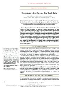
2. Material and methods Female adult laboratory rats (Rattus norvegicus, HanWistar; 229 g and 286 g) were used for this study. The animals were obtained from our own university breeding colony. All procedures were in accordance with the guidelines of animal health care. Animals were sacrificed by an overdose of chloroform. Skin, fat, and fore limbs were removed immediately by severing all muscles connecting to the trunk as well as the clavicle. The hind limbs were exarticulated from the hip joint and pelvic musculature was removed. The sternum was removed by severing all ribs in their middle without injuring axial muscles. After the dissection, the backs were cut into two parts between the 10th and the 11th thoracic vertebrae. Only the caudal parts, reaching from the last two thoracic vertebrae over all six lumbar vertebrae to the iliosacral joint were investigated (levels of intervertebral joint T11/12 to L6/S1). The complete samples including the vertebral column in order to preserve the topographical relationships between the muscles and their intramuscular architecture were quick-frozen in liquid nitrogen cooled isopentan. Serial sections were made using a kryostat microtome (SLEE MTE, D-knife, 12 m) and processed for oxidative capacity and muscle fibre type characterisation using the alkaline combination reaction based on Ziegan’s protocol [50, for details see 32]. Type-I muscle fibres (slow twitch, oxidative) were stained blue, type-IIa (fast twitch, oxidativeglycolytic) dark brown, and type-IIb (fast twitch, glycolytic) light brown. Serial sections were analysed using a Zeiss® microscope (Stemi SV 11). Drawings were made at the level of the intervertebral joints and at the centre of each vertebra (Fig. 3). The fibre type distribution pattern of m. multifidus, m. longissimus lumborum medialis and lateralis, m. quadratus lumborum medialis and lateralis, and m. psoas minor as well as mm. semispinales, interspinales, intermammillares, intertransversarii and rotatores were investigated at the selected cranio-caudal levels and illustrated schematically by different colours. The mm. intermammillares, intertransversarii and rotatores were dealt with as one unit, because they could not be clearly distinguished from one another at each serial section. Towards the caudal direction, additional muscles were included as far as possible (m. psoas major, m. sacrococcygeus dorsalis lateralis and ventralis lateralis and m. gluteus medius, profundus and superficialis). Furthermore, macroscopic dissections were made in order to comprehend the topography of the paravertebral muscles [51,52]. The nomenclature of the muscles follows the textbook of the laboratory rat [51].
3. Results The individuals under study were very similar in their overall distribution pattern of the different fibre types. Therefore, only one serial section was selected for the drawings in
N. Schilling et al. / Pathophysiology 12 (2005) 233–242
237
Fig. 3. Fibre type distribution pattern in the paravertebral musculature of the laboratory rat at different cranio-caudal levels in the thoracic (T), lumbar (L), and sacral (S) region.
Fig. 3. As the distribution pattern is dominated by the proportion of oxidative and glycolytic fibres whereas the percentage of oxidative-glycolytic fibres depended on that of the other types [41], the following description was focussed on the distribution of oxidative and glycolytic fibres only. Nearly all muscles showed a heterogeneous distribution of fibre types both in cranio-caudal direction and within muscle’s cross-section (Fig. 3). Only m. psoas minor presented a homogenous fibre type distribution at nearly all levels and over its cross-section. When looking at the overall pattern, the cross-sectional area of regionalisations with higher percentages of oxidative fibres seems to be caudal reduced. But, the percentage of oxidative fibres within a given region increased in the caudal direction. The highest percentage of oxidative fibres was observed in deep and short monosegmental muscles close to the vertebral column (mm. intertransversarii, intermammillares, rotatores and interspinales) (Fig. 3). Mm. interspinales were almost
completely composed of oxidative fibres especially in the caudal direction. Due to their complex spatial topographical arrangement, intermammillares, intertransversarii and rotatores muscles could not always be recognized clearly as individual muscles. But, derived from their anatomical relationship the higher proportion of oxidative fibres close and dorsally placed to the hypapophyses most likely belonged to mm. intermammillares. Mm. rotatores covered mm. intermammillares dorsally and contained also a high percentage of oxidative fibres but in some levels less than intermammillares muscles (level L5–L6 in Fig. 3). More ventrally situated regions, lateral from the vertebrae most likely represented the mm. intertransversarii. These regions contained less oxidative fibres than those located more dorsally but still a higher percentage than other paravertebral muscles. The m. multifidus was located between the hypapophyses and spinous processus. A higher proportion of the fibres were oxidative close to the vertebral column, whereas
238
N. Schilling et al. / Pathophysiology 12 (2005) 233–242
predominantly glycolytic fibres were observed near the superficial parts (e.g. level L6 in Fig. 3). In most superficial parts of m. multifidus, oxidative fibres were even absent. The highest percentage of glycolytic fibres was observed in the m. psoas major. The psoas major muscle was completely free of oxidative fibres to its origin and their proportion increased slightly towards the caudal region their proportion in the central region of the muscle belly (level L6–L6/L6–S1 in Fig. 3). The m. psoas minor was also predominantly composed of glycolytic fibres, but oxidative fibres were regularly scattered over the muscle’s cross-section (‘salt and pepper pattern’). A slight increase in the proportion of oxidative fibres was observed in the caudal direction, especially in central parts of the muscle. The m. longissimus lumborum medialis and lateralis also contained a high proportion of glycolytic fibres, but a few oxidative fibres were spread over the muscle belly. Whereas pars medialis and lateralis were clearly distinguishable in superficial regions due to the large intramuscular tendon, they became indistinguishable in deeper parts. In the thoracic and cranial lumbar regions, more oxidative fibres were found in the dorsal parts of the muscle especially in pars medialis. The percentage of oxidative fibres dropped under 10% in caudal direction (from L2/3 in Fig. 3). More ventral parts were almost free of oxidative fibres. Between the third and the sixth lumbar vertebrae, a noticeable oxidative region was found in the deep and central region of m. longissimus lumborum. The percentage of oxidative fibres increased up to level L5/6 and decreased more caudally. Towards the iliosacral joint, the continuation of the m. longissimus lumborum medialis was formed by the m. sacrococcygeus dorsalis lateralis. At this level, the muscle consisted predominantly of glycolytic muscle fibres. The mm. semispinales consisted predominantly of glycolytic muscles fibres. But in comparison to the longissimus lumborum muscles, the mm. semispinales contained more oxidative fibres that spread over the cross-sectional area. In the thoracic and cranial lumbar regions, a higher percentage of oxidative fibres was found. Within the lumbar region, the muscle group became thicker and the percentage of oxidative fibres decreased. Only a few of them were scattered over the muscles. A distinct regionalisation of oxidative fibres was found in the m. quadratus lumborum lateralis. The oxidative fibres were arranged in the central parts of the muscle belly and near the intramuscular tendons. This regionalisation was most extensive in cranial i.e. thoracic parts of the serial section (over 60% oxidative fibres). In the caudal direction, the percentage of the oxidative fibres and the extension of the oxidative region was decreased. The m. quadratus lumborum medialis contained only few oxidative fibres, which were distributed homogeneous over the cross-section. Towards the hip, in addition to the m. gluteus medius, profundus and superficialis and the m. sacrococcygeus ventralis lateralis emerged. Although no prediction can be made of the three-dimensional fibre type distribution pattern within the
whole muscle belly, a conspicuous regionalisation of oxidative fibres deep and close to the ilium was observed in the m. gluteus medius and profundus (level L6 and L6/S1 in Fig. 3). Especially the gluteus profundus muscles contained very high percentages of oxidative fibres. The m. gluteus superficialis and the m. sacrococcygeus ventralis lateralis consisted predominantly of glycolytic fibres with only few oxidative fibres spread over the muscle cross-section.
4. Discussion 4.1. Three-dimensional distribution pattern of fibre types In general, the muscles under study presented a highly three-dimensional distribution pattern of fibre types both in cranio-caudal and in transversal direction, i.e. all muscles were highly diverse in their spatial distribution of fibre types. The same heterogeneous distribution pattern was previously described for paravertebral muscles in the pika [41] and the rat [53], but also for limb muscles of diverse mammals [23,32,46,54–56] as well as for m. rectus abdominis in rats [57]. This poses the question of the representativeness of the biopsy results. If the fibre type distribution is so highly diverse from cranial to caudal, proximal to distal and even from deep to superficial directions, how can a single, local sample be representative for a whole muscle belly. Therefore, conclusions based on biopsies should be interpreted with great care as these samples may not reflect the conditions of the entire muscle belly. 4.2. Concepts of back muscle function The current study resulted in clear differences of fibre type distribution pattern between diverse paravertebral muscles. These differences are suggested to be connected to the diverse functions of the muscles. Different concepts on back muscles function were suggested in the past especially for humans. According to a certain anatomical position, superficial fibre direction and activity, various muscles were suggested to stabilise or mobilise the vertebral column. Stabiliser muscles were described as being monosegmental, deep, working eccentrically, able to control the range of motion [58] and maintain segmental stability [59]. They were also named as local muscles [1] and later divided into local and global stabilisers [2]. In humans, m. multifidus and mm. rotatores and interspinales were suggested to fulfil the named functions [2,59]. Described properties imply a large portion of oxidative, non-fatiguing, slow fibres in local stabilisers. Mobility muscles were presented as being bi- or polysegmental, superficial, working concentrically and produce power for large ranges of motion [58]. These muscles correspond to the global muscles [1] or global mobiliser [2]. The m. iliocostalis was thought to be best suited to act as a global mobiliser. These muscles should then be composed predominantly of glycolytic, fast fibres.
N. Schilling et al. / Pathophysiology 12 (2005) 233–242
4.3. Fibre type distribution and back muscle function Remarkable high percentages of fatigue resistant, oxidative fibres were found in the deep and especially monosegmental muscles of the intertransversal, the spinal and the transversospinal group in the rat. Therefore, but also inferred from their anatomical position the mm. interspinales, intertransversarii and rotatores are best suited to maintain segmental stability and resist continuous small intervertebral movements. Based on enzyme-histochemical investigations, a postural function was also assumed for the mm. intertransversarii in the rabbit [60]. Comparable high proportions of oxidative fibres were also described also for the mm. intermammillares and mammillocaccessorii in the Japanese macaque [38] and the mm. multifidi, interspinales and intertransversarii in the cat [34] and the pika [41]. Low percentages of oxidative fibres were found in the m. psoas minor and the m. longissimus lumborum of the rat. A high percentage of glycolytic fibres in the m. sacrospinalis (m. longissimus lumborum and iliocostalis) has been reported in the rat [61], the cat [34], the rabbit [60], the pika [41], the sheep [62], the dog [26], the Japanese macaque [38], the Rhesus macaque [63] and humans [37,64]. In the rat, a segregation of oxidative fibres was described from the second to the sixth lumbar vertebrae in superficial parts and in the deep medial part at the level of the fifth lumbar vertebra in the lateral longissimus muscle [61]. The dorsal, superficial accumulation of oxidative fibres, described in the current study, was less distinctive but obvious to see around the thoraco-lumbar transition. However, the deep and striking regionalisation of oxidative fibres described by Schwartz-Giblin et al. was confirmed by the results of the current study. In the pika, the same distribution pattern with more oxidative fibres in dorsal and superficial parts up to the second lumbar vertebra, but also a clear regionalisation in the medial and deep part between the second and the third lumbar vertebrae was found in the sacrospinalis muscle (note, the pika has only four lumbar vertebrae) [41]. In the cat, as well, a higher percentage of type I fibres was described for the central region of the longissimus muscles near the intermuscular septum [34]. The same cranio-caudal distribution pattern as described above was found in primates, but not found in the other species such as mice, cats, or dogs and surprisingly also not in rats [35]. Despite this, early results from studies on mice refute this observation (Hesse, personal communication). Generally, the fibre composition of the m. longissimus lumborum and the m. sacrospinalis, respectively make them suitable to generate large forces and speed for wide ranges of movement. Kojima also investigated the epaxial muscles in the rat, but only at the height of the forth lumbar vertebra [53]. He reported higher percentages of oxidative fibres especially in the dorsal parts of the lateral longissimus lumborum muscle in comparison to more ventral regions [53]. The medial longissimus lumborum muscle contained about
239
10% of oxidative fibres in all muscle regions investigated [53]. But inferred from dissections in the current study, the pars medialis of the longissimus lumborum muscle in Kojima’s study corresponds to the mm. semispinales and the m. longissimus lumborum lateralis represents the medial as well as the lateral part of the longissimus lumborum. In regard to the misleading nomination, the described fibre composition was confirmed by the results of the current study. The m. quadratus lumborum lateralis and the m. multifidus were conspicuous in the current study because of their accumulation of oxidative fibres close to intramuscular tendons and the vertebral column, respectively. In the pika, the same regionalisation within the quadratus lumborum muscle was observed [41]. Due to its function as extensor of the pleural cavity and stabiliser of the posterior ribs during inspiration [65], the deep oxidative region was suggested to be activated separately from the rest of the muscle belly to fulfil this continuous function during respiration [41]. This may also be true in the rat. In the current study, a clear decrease in the proportion of oxidative fibres was observed from deep to superficial parts in the m. multifidus. The superficial regions contained predominantly glycolytic muscle fibres. The same was reported earlier for the transversospinal group at the height of the forth lumbar vertebra [53]. In the rabbit, the m. multifidus was described as ‘white muscle’ [60]. For humans, no differences in the fibre type composition were found between the multifidus, the longissimus or the iliocostalis muscles [37,64]. The observed metabolic profile in the rat points to a superficial sample collecting in the rabbit and the human. In the pika, the same increase of oxidative fibres towards deeper regions was found [41]. But the pika has a thick m. spinalis which extends to the iliosacral joint and superimposes the multifidi muscles. A deep oxidative regionalisation within the spinalis muscles was also found in the Japanese macaque [38]. Interestingly, a comparable fibre type distribution pattern was found in analogous topographical position within the cross-section of the back and thus independent from the anatomical classification as spinalis or multifidus muscles. Therefore, the fibre type distribution pattern is suggested to be influenced more by the topography i.e. the functional demands in a given position than by its anatomical classification. However, the m. quadratus lumborum and the m. multifidus, but also the mm. semispinales were composed of both, oxidative and glycolytic fibre types and therefore well suited to act as global stabiliser and control the range of movement. The m. psoas major, the m. gluteus superficialis, medius and profundus and the mm. sacrococcygeus dorsalis lateralis and ventralis lateralis emerged towards the hip region. Close to its origin, the psoas major muscle was complete free of oxidative fibres and only in the more caudal parts some more oxidative fibres were found in the central region of the muscle. Together with the psoas minor muscles, which also contained
240
N. Schilling et al. / Pathophysiology 12 (2005) 233–242
a very high percentage of glycolytic fibres, the psoas muscles are well suited by their metabolic profile to produce the high forces for large ranges of motion [1,2]. A comparable fibre type composition with high proportions of glycolytic fibres was described earlier for the psoas major muscle in the pika [41], the tree-shrew and the lesser bushbaby [66]. The gluteus superficialis and the sacrococcygeus muscles were composed mainly of glycolytic fibres in the rat, but the gluteus medius and the gluteus profundus muscles attracted attention due to their conspicuous oxidative regionalisation dorso-lateral to the ilium. High percentages of oxidative fibres in deep regions of the gluteus medius and especially in the gluteus profundus muscles were also described in the sheep [67] and in the pig [68]. 4.4. Fibre type composition and self-stability Fibre type composition i.e. ratio of fast to slow twitch fibres influences the properties and the force production characteristics of a given muscle [69]. The shape of the force–velocity relation depends on the ratio of slow to fast twitch fibres, but also on physiological cross-sectional area and length of the muscle belly. The force–velocity relation is highly important for the mechanical stability of the musculoskeletal systems [70]. The concept of mechanical stability, without the need of sensory feedback, has been termed selfstability [70,71]. In addition to the force–velocity relation, there are many other properties of muscles supporting selfstability, e.g. force-length relation, oblique muscle orientation. In order to analyse the self-stabilising behaviour of antagonistic trunk muscles, a biomechanical model for the human spine was worked out (Wagner et al., in this issue). The model consisted of a pair of antagonistic Hill-type muscles, their geometric arrangement with respect to the spine and the instantaneous centre of rotation. Using sensitivity analyses, the influence of different muscles properties on stability was investigated i.e. fibre type composition and their geometry. Simulations showed that the stability of the spine model against lateral perturbations depended on the percentage of fast twitch fibres, on the geometrical arrangement of muscles but also on the position of the centre of rotation. The higher the proportion of fast twitch fibres in a muscle the more stabile was the system. Muscles containing a high number of fast twitch fibres were able to stabilise the system by a more parallel organization. The predicted muscle properties were confirmed by the results of the current study. From this, we assume that m. psoas major and minor and m. longissimus lumborum are best suited to fulfil a self-stabilising function by their high percentage of glycolytic, fast twitch fibres and their geometrical arrangement. The mm. rotatores were shown to be stabilisers of the system by their more oblique orientation and in the current study by their high percentage of oxidative, slow twitch fibres. Those muscles were able to determine the instantaneous centre of rotation of the spine.
5. Conclusions The most remarkable feature of humans is the upright body posture with a bipedal mode of locomotion. This bipedal mode of locomotion evolved more than 6 million years ago in our hominid ancestors [72,73]. The approximated body size of that fossil hominid was much smaller than that of recent human beings (less than 1.40 m). Considerations about the evolution of bipedality and its consequences in human pathologies should keep in mind, especially that the upright body posture evolved in individuals of only about one-third the body weight of recent representatives. The overall organization of the human trunk is very old and neither fossils nor comparison to recent close relatives led to unique characters adapted to bipedality (with the exception of the habitual lordosis). Most characteristics of the human trunk were only emphasized during the human evolution, i.e. the tetrapod mammalian trunk construction was obviously suitable to be used also for the bipedal mode of locomotion without much change. As the macroscopic topography of the human back does not differ greatly from that of other mammals as often assumed, its ‘uniqueness’ does not serve as an explanation for low back pain. Explanations may be found rather in our all daily life’s behaviour, intermuscular coordination, or intramuscular parameters. However, it should always be taken into account, that the human back is not an ahistorical structure, and it represents as well as any other structure a compromise between its functional demands, its evolutionary heritage and the physical properties of its components. Constraints of physical properties of the material (e.g. muscles, connective tissue, and bone), its organization (former segmental) and its ontogenetic development (interdependences, timing and order of processes) have strong influences on a structure. The classification of back muscles [2], thought as typical and specific for humans was confirmed by the metabolic profiles investigated on quadruped mammals. The concept of local and global stabiliser, as well as global mobiliser was shown to be transferable to small mammals. We therefore propose it as a general concept of mammalian back muscle function. To summarize, the mm. interspinales, intermammillares, intertransversarii and rotatores appear to be best suited to act a local stabiliser. The m. multifidus, mm. semispinales, and the m. quadratus lumborum are suggested as global stabiliser because of their metabolic profile composed of oxidative and glycolytic fibres. The m. longissimus lumborum and the m. psoas major and minor are capable of producing forces for large motions and suggested to act as global mobiliser. The described fibre type distribution pattern was surprisingly similar between the species studied up to now and is therefore suggested to be a more general characteristic for mammals and possible also for humans. We hypothesize that only minor differences exist in the fibre type distribution pattern between humans and other mammalian species due to the alteration
N. Schilling et al. / Pathophysiology 12 (2005) 233–242
of the preferred body axis use. Surprisingly, the hypothesised functions of the diverse mammalian back muscles due to their metabolic profile were confirmed by simulations with a biomechanical model adapted from human’s anatomy. The predicted muscle properties were verified by the current study. Consequently, animal models are suitable for specific questions in human pathology and particularly with regard to back dysfunctions.
Acknowledgements We thank I. Weiß, S. Moritz and L. Al Kuwaiti for their technical assistance. E. Watts thoroughly revised the manuscript. The study was supported by the Centre for Interdisciplinary Prevention of Diseases related to Professional Activities funded by the Friedrich-Schiller-University Jena and the Berufsgenossenschaft Nahrungsmittel und Gastst¨atten Erfurt (Germany).
[14]
[15]
[16] [17] [18]
[19]
[20]
References [1] A. Bergmark, Stability of the lumbar spine, Acta Orthopaedica Scand. 230 (1989) 1–54. [2] S.G.T. Gibbons, M.J. Comerford, Strength versus stability. Part 1. Concept and terms, Orthopaedic Division Rev. (2001) 21–27. [3] M.M. Panjabi, The stabilizing system of the spine. Part II. Neutral zone and instability hypothesis, J. Spinal Disorders 5 (1992) 390–397. [4] A. Seilacher, Arbeitskonzept zur Konstruktionsmorphologie, Lethaia 3 (1970) 393–396. [5] S.J. Gould, Structure of Evolutionary Theory, Belknap Press, Cambridge/London, 2002, pp. 1–1433. [6] M.S. Fischer, Bewegungsappart: Postcraniales Skelett und Muskulatur, in: W. Westheide, R. Rieger (Eds.), Spezielle Zoologie, Teil 2: Wirbel-oder Sch¨adeltiere, Spektrum Verlag, Heidelberg, 2004, pp. 44–53. [7] M.S. Fischer, N. Schilling, F¨ur ein evolutionsbiologisches Verst¨andnis des R¨uckenschmerzes, in: R. Grieshaber, W. Schneider, H.-Ch. Scholle (Eds.), Kongressband 10. Erfurter Tage “Pr¨avention von arbeitsbedingten Gesundheitsgefahren und Erkrankungen”, Monade Verlag, Leipzig, 2004, pp. 69–84. [8] M.S. Fischer, H.F. Witte, Evolution of vertebrate locomotory systems, in: F. Pfeiffer, T. Zielinska (Eds.), Walking: Biological and Technological Aspects. CISM Courses and Lecture Notes, Springer Verlag, Wien, 2004, pp. 51–79 (course no. 272). [9] F.A.J. Jenkins, S.M. Camazine, Hip structure and locomotion in ambulatory and cursorial carnivores, J. Zool. (London) 181 (1977) 351–370. [10] M.S. Fischer, R. Lehmann, Application of cineradiography for metric and kinematic study of in-phase gaits during locomotion of pika (Ochotona rufescens, Mammalia: Lagomorpha), Zoology 101 (1998) 148–173. [11] N. Schilling, M.S. Fischer, Kinematic analysis of treadmill locomotion of tree shrews, Tupaia glis (Scandentia: Tupaiidae), Int. J. Mamm. Biol. 64 (1999) 129–153. [12] M.S. Fischer, N. Schilling, M. Schmidt, D. Haarhaus, H.F. Witte, Basic limb kinematics of small therian mammals, J. Exp. Biol. 205 (2002) 1315–1338. [13] H. Witte, M.S. Fischer, M. Schmidt, S. Gruber, O. Ludwig, R. Hackert, N. Schilling, D. Voges, H. Hoffmann, H. Preuschoft, Human
[21]
[22]
[23]
[24]
[25]
[26]
[27] [28]
[29]
[30]
[31]
[32]
241
bipedality: mechanical preconditions and morphological adaptations, Cour. Forschungsinstitut Senckenberg 243 (2003) 25–33. H. Witte, N. Schilling, H. Hoffmann, R. Hackert, D. Voges, K.E. Lilje, M. Schmidt, M.S. Fischer, Der Rumpf wird vom Menschen und von anderen S¨augetieren systematisch f¨ur die Fortbewegung genutzt, in: R. Grieshaber, W. Schneider, H.-Ch. Scholle (Eds.), Kongressband 8. Erfurter Tage Pr¨avention von arbeitsbedingten Gesundheitsgefahren und Erkrankungen, Monade Verlag, Leipzig, 2002, pp. 291–304. A.H. Schultz, Vertebral column and thorax, in: H. Hofer, A.H. Schultz, D. Starck (Eds.), Primatologia IV/5, vol. 4, Karger, Basel, 1961, pp. 1–66. A.H. Schultz, The skeleton of the chimpanzee, in: G.H. Bourne (Ed.), The Chimpanzee, vol. 1, Karger, Basel, 1969, pp. 50–103. L. Shapiro, Functional morphology of indrid lumbar vertebrae, Am. J. Phys. Anthrop. 98 (1995) 323–342. M. Cartmill, K. Milton, The lorisiform wrist and the evolution of ‘brachiating’ adaptations in the Hominoidea, Am. J. Phys. Anthrop. 47 (1977) 249–272. W.L. Jungers, Scaling of the hominoid locomotor skeleton with special references to lesser apes, in: H. Preuschoft, D. Chivers, W. Bockelman, N. Creel (Eds.), The Lesser Apes, Edinburgh University Press, Edinburgh, 1984, pp. 146–169. S. Hayama, M. Nakatsukasa, Y. Kunimatsu, Monkey performance: the development of bipedalism in trained Japanese monkeys, Acta Anatomica Nippon 76 (1992) 169–185. H. Preuschoft, S. Hayama, M.M. G¨unther, Curvature of the lumbar spine as a consequence of mechanical necessities in Japanese macaques trained for bipedalism, Folia Primatol. 50 (1988) 42– 58. J.M. Stein, H.A. Padykula, Histochemical classification of individual skeletal muscle fibers of the rat, Am. J. Anat. 110 (1962) 103– 123. M.A. Ariano, R.B. Armstrong, V.R. Edgerton, Hindlimb muscle fiber populations of five mammals, J. Histochem. Cytochem. 21 (1973) 51–55. T.C. Collatos, V.R. Edgerton, J.L. Smith, B.R. Botterman, Contractile properties and fiber type compositions of flexors and extensors of elbow joint in cat: implications for motor control, J. Neurophysiol. 40 (1977) 1292–1300. D.W. Sickles, C.A. Pinkstaff, Comparative histochemical study of prosimian primate hindlimb muscles I. Muscle fiber types, Am. J. Anat. 160 (1981) 175–194. R.B. Armstrong, C.W. Saubert, H.J. Seeherman, C.R. Taylor, Distribution of fiber types in locomotory muscles of dogs, Am. J. Anat. 163 (1982) 87–98. R.B. Armstrong, R.O. Phelbs, Muscle fiber type composition of the rat hindlimb, Am. J. Anat. 171 (1984) 259–272. J.E. Brasseur, R.L. Curtis, J.W. Mellender, A.A. Rimm, J.L. Melvin, A.R. Sulaiman, Systematic distribution of muscle fiber types in the medial gastrocnemius of the laboratory mouse: a morphometric analysis, Anat. Rec. 218 (1987) 396–401. K.G. Braund, K.A. Amling, J.R. Mehta, J.E. Steiss, C. Scholz, Histochemical and morphometric study of fiber types in ten skeletal muscles of healthy young adult cats, Am. J. Vet. Res. 56 (1995) 349–357. A. Suzuki, Differences in distribution of myofiber types between the supraspinatus and infraspinatus muscles of sheep, Anat. Rec. 242 (1995) 483–490. I. Fuentes, A.R. Cobos, L.A.G. Segade, Muscle fibre types and their distribution in the biceps and triceps of the rat and rabbit, J. Anat. 192 (1998) 203–210. F. von Mering, M.S. Fischer, Fibre type regionalization of forelimb muscles in two mammalian species, Galea musteloides (Rodentia, Caviidae) and Tupaia belangeri (Scandentia, Tupaiidae), with comments on postnatal myogenesis, Zoomorphology 119 (1999) 117–126.
242
N. Schilling et al. / Pathophysiology 12 (2005) 233–242
[33] M.W. Fidler, R. Jowett, J.D.G. Troup, Myosin ATPase activity in multifidus muscle from cases of lumbar spinal derangement, J. Bone Joint Surg. 57 (1975) 220–227. [34] H. Carlson, Histochemical fiber composition of lumbar back muscles in the cat, Acta Physiol. Scand. 103 (1978) 198–209. [35] I. Yokoyama, Analyses of the fibre composition of the lumbar back muscles in mammals, Nippon Seikeigeka Gakkai Zasshi 56 (1982) 579–594. [36] D.M. Ford, K.M. Bagnall, K.D. McFadden, D.C. Reid, A comparison of muscle fiber characteristics at different levels of the vertebral column in the rhesus monkey, Acta Anat. 126 (1986) 163–166. [37] A. Thorstensson, H. Carlson, Fibre types in human lumbar back muscles, Acta Physiol. Scand. 131 (1987) 195–202. [38] R. Kojima, M. Okada, Distribution of muscle fibre types in thoracic and lumbar epaxial muscles of Japanese macaques (Macaca fuscata), Folia Primatol. 66 (1996) 38–43. [39] A.F. Mannion, G.A. Dumas, J.M. Stevenson, R.G. Cooper, The influence of muscle fiber size and type distribution on electromyographic measures of back muscle fatigability, Spine 23 (1998) 576–584. [40] K.S. Gellman, J.E.A. Bertram, J.W. Hermanson, Morphology, histochemistry, and function of expaxial cervical musculature in the horse (Equus caballus), J. Morphol. 251 (2002) 182–194. [41] N. Schilling, Characteristics of paravertebral muscles—fibre type distribution pattern in Ochotona rufescens (Mammalia: Lagomorpha), J. Zool. Syst. Evol. Res. 43 (2005) 38–48. [42] R.E. Burke, Motor units: anatomy, physiology, and functional organization, in: J.M. Brookhart, V.B. Mountcastle (Section Eds.), Handbook of Physiology Section I—The Nervous System II. Motor Control, Part I, vol. 10, American Physiological Society, Bethesda, 1981, pp. 345–422. [43] H.C. Scholle, N.P. Schumann, F.H.W. Biedermann, D.F. Stegeman, R. Graßme, K. Roeleveld, N. Schilling, M.S. Fischer, Spatiotemporal surface EMG characteristics from rat triceps brachii muscle during tradmill locomotion indicate selective recruitment of functionally distinct muscle regions, Exp. Brain Res. 138 (2001) 26–36. [44] R.B. Armstrong, Properties and distribution of the fiber types in the locomotory muscles of mammals, in: K. Schmidt-Nielsen, L. Bolis, C.R. Taylor (Eds.), Comparative Physiology of Primitive Mammals, Cambridge University Press, Cambridge, 1980, pp. 243–254. [45] J. Lexell, K. Henriksson-Larson, M. Sj¨ostr¨om, Distribution of different fiber types in human skeletal muscles, Acta Physiol. Scand. 117 (1983) 115–122. [46] M.S. Fischer, Kinematics, EMG, and inverse dynamics of the therian forelimb—a synthetic approach, Zool. Anz. 238 (1999) 41–54. [47] C.M. Chanaud, C.A. Pratt, G.E. Loeb, Functionally complex muscles of the cat hindlimb V. The roles of histochemical fiber-type regionalization and mechanical heterogenity in differential muscle activation, Exp. Brain Res. 85 (1991) 300–313. [48] P.R. Flood, S.J. Mathisen, A third type of muscle fibre in the parietal muscle of the atlantic hagfish Myxine glutinosa, Z. Zellforsch. 58 (1962) 638–640. [49] N. Schilling, R. Hackert, M.S. Fischer, Vertebral column movements of small mammals during locomotion, Zoology 102 (1999) 44. [50] J. Ziegan, Kombination enzymhistochemischer Methoden zur Faserdifferenzierung und Beurteilung der Skeletmuskulatur, Acta Histochem. 65 (1979) 34–40. [51] R. Hebel, M.W. Stromberg, The Anatomy of the Laboratory Rat, The Williams and Wilkins Company, Baltimore, 1976, pp. 1–174. [52] E.E. Brink, D.W. Pfaff, Vertebral muscles of the back and tail of the albino rat, Brain Behav. Evol. 17 (1980) 1–47. [53] R. Kojima, Distribution of muscle fiber types in the rat lumbar epaxial muscles, Bull. Saitama Med. Sch. Jun. Coll. 9 (1998) 7–16.
[54] J. Polgar, M.A. Johnson, D. Weightman, D. Appleton, Data on fibre size in 36 human muscles. An autopsy study, J. Neurol. Sci. 19 (1973) 307–318. [55] S. Hansen, J.H. Cutts, W.J. Krause, J.H. Cutts, Distribution of fiber types in thirty-seven muscles of Didelphis virginiana, Anat. Anz. 164 (1987) 153–158. [56] J.S. McIntosh, M. Rinquist, E.M. Schmidt, Fiber type composition of monkeys forearm muscle, Anat. Rec. 211 (1985) 403–409. [57] T. Hijikata, H. Wakisaka, T. Yohro, Architectural design, fiber-type composition, and innervation of the rat rectus abdominis muscle, Anat. Rec. 234 (1992) 500–512. [58] B. Goff, The application of recent advances in neurophysiology to Miss R Rood’s concept of neuromuscular facilitation, Physiotherapy 58 (1972) 409–415. [59] M.M. Panjabi, K. Abumi, J. Duranceau, T. Oxland, Spinal stability and intersegmental muscle forces: a biomechanical model, Spine 14 (1989) 194–199. [60] K.D. McFadden, K.M. Bagnall, M. Mahon, D.M. Ford, Histochemical fiber composition of lumbar back muscles in the rabbit, Acta Anat. 120 (1984) 146–150. [61] S. Schwartz-Giblin, L. Rosello, D.W. Pfaff, A histochemical study of lateral longissimus muscle in rat, Exp. Neurol. 79 (1983) 497– 518. [62] B. Peinado, R. Latorre, J.M. Vaquez-Auton, A. Poto, G. Ramirez, O. Lopez-Albors, F. Moreno, F. Gil, Histochemical skeletal muscle fibre types in the sheep, Anat. Histol. Embryol. 33 (2004) 236–243. [63] K.M. Bagnall, D.M. Dord, K.D. McFadden, B.J. Greenhill, V.J. Raso, A comparison of vertebral muscle fiber characteristics between human and monkey tissue, Acta Anat. 117 (1983) 51–57. [64] J. Rantanen, A. Rissanen, H. Kalimo, Lumbar muscle fiber size and type distribution in normal subjects, Eur. Spine J. 3 (1994) 331–335. [65] W. Boyd, H. Blincoe, J.C. Hayner, Sequence of action of the diaphragm and quadratus lumborum during breathing, Anat. Rec. 151 (1965) 579–582. [66] D.W. Sickles, C.A. Pinkstaff, Comparative histochemical study of prosimian primate hindlimb muscles II. Populations of fiber types, Am. J. Anat. 160 (1981) 187–194. [67] A. Suzuki, H. Tamate, Distribution of myofiber types in the hip and the thigh musculature of sheep, Anat. Rec. 221 (1988) 494–502. [68] A. Suzuki, K. Watanabe, R. Konno, S. Ohwada, Distribution of myofiber types in the hip and thigh musculature of pigs, Anim. Sci. J. 70 (1999) 519–525. [69] W. Herzog, Muscle, in: B.M. Nigg, W. Herzog (Eds.), Biomechanics of the musculo-skeletal system, John Wiley & Sons, Chichester, 1994, pp. 154–190. [70] H. Wagner, R. Blickhan, Stabilizing function of skeletal muscles: an analytical investigation, J. Theor. Biol. 199 (1999) 163–179. [71] R. Blickhan, H. Wagner, A. Seyfarth, Brain or muscles? in: S.G. Pandalai (Ed.), Recent Research Developments in Biomechanics, vol. 1, Trivandrum, India, 2003, pp. 215–245. [72] B. Senut, M. Pickford, D. Gommery, P. Mein, K. Cheboi, Y. Choppens, First hominid from the Miocene (Lukeino Formation, Kenya), CR Acad. Sci. Paris Sci. Terre Planetes 332 (2001) 137–144. [73] M. Brunet, F. Guy, D. Pilbeam, H.T. Mackaye, A. Likius, D. Ahounta, A. Beauvilain, C. Blondel, H. Bocherens, J.-R. Boisserie, L. de Bonis, Y. Coppens, J. Dejax, C. Denys, P. Duringer, V. Eisenmann, G. Fanone, P. Fronty, D. Geraads, T. Lehmann, F. Lihoreau, A. Louchart, A. Mahamat, G. Merceron, G. Mouchelin, O. Otero, P.P. Campomanes, M. Ponce de Leon, J.-C. Rage, M. Sapanet, M. Schuster, J. Sudre, P. Tassy, X. Valentin, P. Vignaud, L. Viriot, A. Zazzo, C. Zollikofer, A new hominid from the Upper Miocene of Chad, Central Africa, Nature 418 (2002) 145–151.