Differential Diagnosis of Chronic Low Back Pain James J. Lehman, DC, MBA, FACO Associate Professor of Clinical Sciences University of Bridgeport College of Chiropractic Director Community Health Care Education
Learning Objective
• Identify injured and painful tissues through careful assessment and intelligent use of neuromusculoskeletal testing and document the findings of chronic low back pain.
Learning Objective • Implement the scientific method and integrate the use of an evaluation protocol practiced by evidence-based and patient-centered chiropractic physicians in order to perform a differential diagnosis.
Opening Statement …
• Diagnosis is the key to successful treatment!
Musculoskeletal Disabilities • The leading causes of disability in people in their working years are musculoskeletal conditions.
•
Russell IJ. A new journal. J Musculoskeletal Pain 1(1):1-7. 1993.
Lower Back Pain • How do you differentiate the types of tissues that may be involved with a chief concern of low back pain?
Active Learning Task • Form groups of 3-5 learners • Select a spokesperson • Organize a clinical thought process that would enable you to determine the pain generators with a chronic low back pain patient • Describe your physical examination process for a patient without organic disease but with a neuromusculoskeletal condition.
Focused History of Low Back Pain • • • • • • • • •
Location Mechanism of injury New condition Onset Provocative and palliative Quality of pain Referred or radiating pain Severity Timing and treatment
Focused Neuromusculoskeletal Examination • • • • •
Observation/Inspection Palpation Range of motion Special tests/orthopedic tests Neurological examination – 3 part peripheral nervous system exam – CNS examination – Cranial nerve examination – Mental status
Definition of an Orthopedic Test • Most often a provocative maneuver that reproduces the patient’s chief concern pain with stretching, contracting, and/or compressing in order to identify the involved tissues.
Low Back Pain Case • Patient strained lower back unloading a truck, which required lifting heavy boxes, twisting and placing boxes on flats three years ago. • Lower back pain persists on a daily basis and increases with bending and twisting. • Rest reduces the constant dull ache and/or intermittent sharp, stabbing pains located over the areas of the right lumbar spine, SIJ, buttocks and anterior thigh. • Medications and hot showers reduce the pain.
Low Back Pain Differential Diagnosis • Please list 5 differential diagnoses • List the physical examination procedures that you would use to rule-in and rule-out your differential diagnoses.
Differential Diagnosis • Chronic pain syndromepost-traumatic • Myofascial pain syndrome • Lumbar facet syndrome • Degenerative disc disease • Degenerative joint disease
Spinal Muscle Strain
• • • •
Cramps Knots Spasms Dull ache
Myofascial Trigger Point Characteristics
Myofascial Trigger Point Palpation • Localized pain with palpation • Active trigger point may produce referred pain
Myofascial Pain Syndrome Referred Pain • Paresthesias • Crawling sensation (formication) • Dull or deep ache • Myotogenous • Myotomal
Joint Pain Zygapophyseal or Facet Joint • Sharp pain on motion • Constant dull or deep ache • Source of chronic low back pain •
Manchikanti L, et al. Prevalence of facet joint pain in chronic spinal pain of cervical, thoracic, and lumbar regions. BMC Musculoskelet Disord. 2004; 5: 15.
Dorsal Ramus of Spinal Nerve
• Primary division of a posterior ramus of a spinal nerve has three branches
Sclerotogenous Referred Pain • Resembles radiating pain but it is a referred deep, dull ache from bone, ligaments and joints •
Ivanusic JJ. The evidence for the spinal segmental innervation of bone. Clin Anat. 2007 Nov;20(8):956-60.
Scleratogenous or Myofascial Triggers • Diffusely referred and hard to localize • Deep and achy quality Kellgren & Feinstein
Nerve Pain • Burning and/or hot • Tingling and/or numbness • Nerve root tension signs • Lhermitte’s sign
Lhermitte’s Sign Nerve Pain • Stabbing or lightning-like pain down spine and any combination of extremities with flexion or extension
Gluteus Medius: “Lumbago Muscle” Commonly overlooked source of referred low back pain
Iliopsoas: “Hidden Prankster” • Serves many critically important functions, often causes pain, and is relatively inaccessible.
Iliopsoas: “Hidden Prankster” • Unidentified iliopsoas and quadratus lumborum trigger points are frequently responsible for a failed low back postsurgical syndrome.
Iliopsoas: “Hidden Prankster” • When describing the low back pain they run the hand vertically up and down the spine rather than horizontally.
Quadratus Lumborum “Joker of Low Back Pain” • Severe, referred tenderness of the greater trochanter may disrupt sleep.
Quadratus Lumborum “Joker of Low Back Pain” • Patient may be barely able to turn over in bed and unable to bear the pain of standing upright or walking.
Quadratus Lumborum “Joker of Low Back Pain” • Coughing or sneezing can be frightfully painful. • Not to be confused with Dejerine’s and a SOL
Quadratus Lumborum “Joker of Low Back Pain” • Imagine the patient waking during the night with pain in the trochanteric area with a full bladder and unable to walk due to severe low back pain!
Quadratus Lumborum “Joker of Low Back Pain” • Spasm of QL causes functional scoliosis, loss of lumbar lordosis with flattened appearance, and restricted ROM. • Flexion and extension may be abolished.
Muscle Dysfunction
• Muscle strain, spasm, weakness, contractures and trigger points may cause muscle imbalances and pelvic obliquity •
Winter RB, Pinto WC. Pelvic obliquity. Its causes and its treatment. Spine 1986 Apr;11(3):225-34.
Pelvic Obliquity • Anatomical short leg or functional leg length inequality due to iliopsoas, gluteus medius and quadratus lumborum muscle contractures may cause pelvic obliquity •
Grill F, et al. Pelvic tilt and leg length discrepancy. Orthopade. 1990 Sep;19(5):244-62
Pelvic Asymmetry
Superficial Paraspinal Muscles
Erector Spinae
• Trigger points in the erector spinae muscles are a frequent cause of low back pain. • Patients might refer to the pain as “lumbago.”
Superficial Paraspinal Muscles
Erector Spinae
• Trigger points in the erector spinae muscles may cause entrapment of the dorsal primary rami of the spinal nerves.
Deep Paraspinal Muscles
Multifidi
• Trigger point pain is located at the spinous process of the involved segment or referred a few segments caudal to the trigger point.
Deep Paraspinal Muscles
Multifidi
Trigger points in the multifidi may cause articular dysfunction at 2-3 segments.
Articular Dysfunction • Multifidi trigger point symptoms mimic lumbar facet and sacroiliac syndromes.
•
Schneider MJ. The traction methods of Cox and Leander: neglected role of the multifidus muscle in low back pain. Chiropract Techn 3(3) 109-115. 1991.
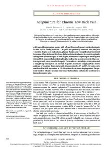
Composite Referred Pain Patterns Z Joint Injection of Hypertonic Saline Solution
Case 2 Differential Diagnosis • 45 year-old male presents with daily pain in the right sacroiliac, buttocks, abdominal and inguinal regions, lateral hip and right testicle since a lifting injury 5 years earlier. • Palpation reveals pain at the lower thoracic and upper lumbar spinous processes and paraspinal muscles, referred pain to the abdomen and right testicle. • Taut bands, painful nodules, localized pain in the multifidi muscles and referred pain to ipsilateral lower lumbar spine and abdomen . • Radiographic impression of lower lumbar DDD/DJD
Class Discussion
• What five differential diagnoses would you select? • How do you support them?
Did you consider? • Post-traumatic chronic pain syndrome G89.21 • Myofascial pain syndrome (T/L multifidi and/or QL) M54.6 • Maigne’s syndrome or Thoracolumbar Junction Syndrome M54.15 • Lumbar radiculopathy (L1-2) M54.16 • Degenerative joint and disc disease M51.36
Maigne’s Syndrome Thoracolumbar Junction Syndrome • Of 350 patients seen in a back pain clinic, 40% were found to have pain of thoracolumbar origin. Maigne R. - Low back pain of thoracolumbar origin. Arch. Phys. Med. Rehabil. 1980, 61, 389-395.
Maigne’s Syndrome Thoracolumbar Junction Syndrome • Neuropathic pain is found in three well described regions and serves as the principal clinical component in diagnosing “Lumbar Dorsal Ramus Syndrome” (LDRS).
Maigne’s Syndrome Thoracolumbar Junction Syndrome • The patient will not usually have spontaneous pain at the offending spinal level. • Pain can be provoked by palpation of the facet joints, or the level can remain veiled, with only the referred pain as evidence of the defect.
Maigne’s Syndrome Thoracolumbar Junction Syndrome • Usually unilateral, bilateral cases have been described... • Patients will not have pain radiating below the knee, which is more typical of anterior ramus involvement.
Maigne’s Syndrome Thoracolumbar Junction Syndrome • Radiographic evidence is non-contributory. • MRI, CT and myelography are all ineffective at localizing the at-fault level. • The typical degenerative changes seen on most images may lead to unnecessary surgery or false diagnosis. • The posterior ramus is far removed from herniating or bulging discs.
Maigne’s Syndrome Thoracolumbar Junction Syndrome • Pain relieved by injection of local anesthetic into the correct facet joint. • This diagnostic procedure can also be therapeutic; the injection of steroids or radiofrequency denervation of the medial branch can be added for refractory cases.
Maigne’s Syndrome Thoracolumbar Junction Syndrome • “Thoracolumbar junction syndrome is particularly responsive to spinal manipulative therapy and no further treatment is required in most cases as long as it is performed adequately.” •
Soo-Ryu Kim, et al. Thoracolumbar Junction Syndrome Causing Pain around Posterior Iliac Crest: A Case Report. Korean J Fam Med. 2013 Mar; 34(2): 152–155.
Thoracolumbar Syndrome A Report of Two Cases • Spinal manipulation of the thoracolumbar has been demonstrated effective with relief of a chronic thoracolumbar syndrome. •
Proctor D, Dupuis P, Cassidy D. Thoracolumbar syndrome as a cause of low-back pain: a report of two cases. The Journal of the CCA/Volume 29 No. 2/June 1985.
References • • • • • • • • • •
Scott-Charlton, W.and Roebuck, D.J. The Significance of Posterior Primary Divisions of Spinal Nerves in Pain Syndrome. The Medical Journal of Australia. 1972; 2:945–948. Maigne, R. Low back pain of thoracolumbar origin (T11-T12-L1). In: Maigne, R., Second Edition: Diagnosis and Treatment of Pain of Vertebral Origin. Taylor and Francis Group, 2006:289–98. McCall IW, Park WH, O’Brien JP. Induced pain referral from posterior lumbar elements in normal subjects. Spine 1979;4441–6. Marks R. Distribution of pain provoked from lumbar facet joints and related structures during diagnostic spinal infiltration. Pain 1989;39:37–40. Fukui, S. Distribution of Referred Pain from the Lumbar Zygapophyseal Joints and Dorsal Rami. The Clinical Journal of Pain 1997:13;303–307. Sherrington, C.S., Experiments in Examination of the Peripheral Distribution of the Fibres of the Posterior roots of some Spinal Nerves. Philosophical Transactions of the Royal Society of London, vol. clxxxiv. 1893. Maigne, R. Low Back Pain of Thoracolumbar Origin. Archives of Physical Medicine and Rehabilitation. 1980:61;389–395. Scott-Charlton, W.and Roebuck, D.J. The Significance of Posterior Primary Divisions of Spinal Nerves in Pain Syndrome. The Medical Journal of Australia. 1972; 2:945–948. Soo-Ryu Kim, et al. Thoracolumbar Junction Syndrome Causing Pain around Posterior Iliac Crest: A Case Report. Korean J Fam Med. 2013 Mar; 34(2): 152–155. Proctor D, Dupuis P, Cassidy D. Thoracolumbar syndrome as a cause of low-back pain: a report of two cases. The Journal of the CCA/Volume 29 No. 2/June 1985.
Recommended Text
Closing Statement …
• Diagnosis is the key to successful treatment!