International Journal of Clinical Medicine, 2014, 5, 1087-1098 Published Online September 2014 in SciRes. http://www.scirp.org/journal/ijcm http://dx.doi.org/10.4236/ijcm.2014.517139
A Simple Approach of Low Back Pain Hani Almoallim1,2,3*, Samar Alwafi1,2, Khaled Albazli1,2, Manal Alotaibi1,2, Tuqa Bazuhair1,2 1
Department of Medicine, Medical College, Umm Alqura University, Makkah, KSA Alzaidi Chair of Research in Rheumatic Diseases, Medical College, Umm Alqura University, Makkah, KSA 3 Department of Medicine, Dr. Soliman Fakeeh Hospital, Jeddah, KSA * Email:
[email protected] 2
Received 1 July 2014; revised 2 August 2014; accepted 3 September 2014 Copyright © 2014 by authors and Scientific Research Publishing Inc. This work is licensed under the Creative Commons Attribution International License (CC BY). http://creativecommons.org/licenses/by/4.0/
Abstract Low back pain (LBP) is primarily managed in general practice and commonly underestimated or misdiagnosed by physicians. This article presents comprehensive review for diagnosis and evaluation of LBP according to current clinical studies guidelines. Our objectives are to define LBP, to establish how to take a detailed history and how to physically examine it in order to enable physicians to make an appropriate differential diagnosis for LBP, and to identify relevant investigations and referrals of patients with LBP. The article first offers a quick description of inflammatory back pain then discusses the importance of screening red flag patients with LBP and the importance of its early detection. Finally, we summarize how to outline a primary plan for managing and treating LBP. The article is prepared in the format of question and answer to make it targeted and accessible.
Keywords Low Back Pain, Rheumatology, History, Examination, Management, Inflammatory Back Pain
1. What Is Back Pain? Low back pain (LBP) is defined as pain, muscle tension or stiffness localized below the costal margin and above the inferior gluteal folds, with or without leg pain [1]. It can be classified as a non-specific LBP, a serious condition, or as a radicular syndrome [2]. Classification of LBP as acute or chronic can be a useful aid for prognosis to guide management. It is often classified as acute (less than 6 weeks), sub-acute (6 - 12 weeks), and chronic (more than 12 weeks). There is a wide acceptance that the management of LBP should begin in primary care. Therefore, all physicians should know how to make a complete assessment for patients having LBP to provide them with the most *
Corresponding author.
How to cite this paper: Almoallim, H., Alwafi, S., Albazli, K., Alotaibi, M. and Bazuhair, T. (2014) A Simple Approach of Low Back Pain. International Journal of Clinical Medicine, 5, 1087-1098. http://dx.doi.org/10.4236/ijcm.2014.517139
H. Almoallim et al.
appropriate management and referrals. Clinical assessment by history taking and clinical examination is the cornerstone in diagnosis of LBP with a restricted use of clinical imaging. Anatomical or physiological abnormalities cannot explain most of LBP cases. However, imaging studies are important to exclude serious causes of LBP such as malignancies and infections and other anatomical degenerative changes.
2. Significance of Back Pain Non-specific LBP is now clearly recognized as a major public health problem. The symptom of LBP is the second most complaint after common cold. In 70% of cases, LBP has no obvious etiology or a well-known pathogenesis [1].
3. How Common Is It? Our review of the global prevalence of LBP shows that it is a major problem throughout the world and that it is most prevalent among females and persons ages 40 - 80 years [2]. Literature shows that there has been an increase in the reported lifetime prevalence of LBP to become as high as 84%, and the in prevalence of chronic LBP which reached 23%, with 11% - 12% of the population progressing to a disability [1] [3] [4].
4. Physician’s Awareness of Back Pain The impact of back pain on society and economy is substantial since it is the most frequent cause of disability for people under the age of 45 [5]. In 2009, the National Institute of Health and Care Excellence in United Kingdome published guidelines for management of non-specific LBP. In 2010, the Institute made a study to assess the impact of these guidelines on the management of back pain within primary care. It was concluded that these guidelines have not been well applied in management of LBP in primary care, which could be due to the lack of awareness of these guidelines or to adherence to other ones [6].
5. What Is the Common Prognosis of LBP? The long-term prognosis of LBP is generally good. In 2008, in Australian primary health care centers a cohort study was done on 973 patients with recent onset LBP to estimate the one-year prognosis and to identify the prognostic factors. This study found that 83% had mild or no pain, 86% had minimal or no disability at one year follow-up; however, only 72% had completely recovered [6]. In 2010, a survey was done on Australian general practice physicians (GP) about the application of LBP management guidelines showed that although the guidelines discourage the use of imaging, over one-quarter of patients were referred for imaging [6], and while the guidelines recommend that initial care should focus on advice and simple analgesics, only 20.5% received advice and 17.7% of patients received analgesics. The analgesics provided were typically non-steroidal anti-inflammatory drugs (37.4%) and opioids (19.6%). This indicates that the usual care provided by GPs for LBP does not match the care endorsed in international evidence-based guidelines and may not provide the best outcomes for patients. The unendorsed care may contribute to the high costs of managing LBP, and possibly to the fact that some aspects of the care provided carry a high risk of adverse effects [7].
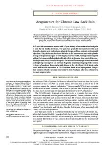
6. How to Approach LBP? The specific etiology of LBP is still unidentified in most of the patients presenting to healthcare but listening to the patient gives the physician the best opportunity to identify the causes of LBP [8]. A full history and physical examination is essential in developing a diagnostic plan to identify the cause(s) of symptoms and administering a therapeutic regimen to relieve the pain. A focused history and physical examination are essential in evaluating patients presenting with LBP to assess them for serious symptoms of neurologic compromise, inflammatory, or medical conditions. Most of the patients can be evaluated by history and physical examination alone if the duration of back pain is less than one month [8] but a through history and physical exam can guide clinicians for further indicated diagnostic studies in serious underlying conditions (see Figure 1). In the majority of cases, the pain is self-limited so no specific treatment is required.
7. How to Take the History? The history typically starts with a full analysis of pain by assessing the type of onset, site of pain and radiation,
1088
H. Almoallim et al.
Figure 1. Approach to the patient with low back pain.
character and continuity of pain, progression and intensity of pain at rest and movement, factors altering pain, severity of pain, and associated symptoms. Type of pain may help the physician recognize the structure possibly injured, deep, nagging, and dull pain usually indicates the bones. A dull ache indicates muscles. A sharp and shooting pain indicates nerve root. A sharp, bright, lightning-like pain might be nerve. A burning, pressure-like,
1089
H. Almoallim et al.
stinging, aching pain indicates sympathetic nerve. A throbbing diffused pain is mainly vascular. It is recommended in history taking to inquire about suggestive features of specific serious diagnosis associated with LBP like: cancer, infection, cauda equina syndrome, compression fractures, spinal stenosis, ankylosing spondylitis (AS), herniated disc or radiculopathy.
8. What Are the Alarming Keys to Systemic Symptoms? Systemic symptoms in history taking may raise the suspicion of cancer or infection. Hence, it is important to pay attention to clues suggestive of underlying systemic diagnosis by asking about red flags such as any previous history of malignancy, unexplained weight loss, pain at night, and pain over four weeks with no response to treatment. These factors should be investigated properly for an age over 50 and the duration of pain over four to six weeks. The American College of Radiology has identified ten “red flags” in the LBP investigation that should be obtained to guide further recommended diagnostic imaging [9] (Table 1).
9. What about Neurological Evidence? Symptoms of significant lumbar spinal stenosis include back pain, transient tingling in the legs, and ambulation-induced pain localized to the calf and distal lower extremity resolving with rest. Bowel or bladder dysfunction may be a symptom of severe compression, which calls for cauda equina syndrome. Urinary retention with overflowing incontinence is typically present with associated saddle anesthesia, bilateral sciatica, and leg weakness.
10. How to Do Physical Examination? The paradigm “look, move, feel” should be utilized in the examination of LBP [10]. However, we have modified the terminologies used to describe the steps involved in the examination of LBP (see Figure 2). It starts typically by general inspection to observe the gait and posture looking for any asymmetry or abnormal curvature. Following this, a screening examination including the active range of motion (ROM) is performed. Detection of gross limitation of movement in ROM is less reliable in diagnosis. However, it can be recorded as a baseline to observe further progressions or improvement. A systematic approach to check palpation further is then undertaken by checking the palpation over each of the spinous processes, then unilaterally on each side of the process looking for underlying intervertebral segmental stiffness, tenderness and pain reproduction. This should be performed on each segment from the thoracolumbar junction to the lumbosacral junction. The examination is completed with checking palpation around the sacrum and buttock. The physical examination should finish with a detailed neurological evaluation and special tests like the straight leg raise test and/or slump test (see Figure 2 and Table 2). In young adults, it is recommended to perform straight leg raise test as it assesses possible sciatica. Elderly with spinal stenosis may have a normal straight leg raise test. A focused neurologic examination should be obtained particularly for ankle and knee reflexes, ankle and great toe to assess dorsiflexion strength, and distribution of sensory complaints to document the presence of neurologic deficits.
11. When Should a Radiograph Be Used? Acute uncomplicated LBP without red flags is a benign self-limited condition that does not require imaging evaluation. In patients with red flags, MR has displaced CT and myelography as the initial imaging modality of choice in complicated LBP, with contrast useful for neoplasia, infection, and postoperative evaluation. However, CT is useful in patients with surgical fusion/instrumentation or bone structural abnormalities, and in patients with MRI contraindications. Myelography/CT, discography/CT, and radioisotope bone scans are useful in selected patients for problem solving.
12. What Makes the Back Hurt? As back pain is a very common symptom it could be due to many causes, both specific and non-specific [1] as Table 3 shows.
13. What Indicates an Inflammatory Back Pain? The diagnosis of inflammatory back pain (IBP) is often delayed in primary care. This may be due to inability to
1090
H. Almoallim et al.
differentiate IBP from mechanical back pain. IBP can be a lifelong problem and it can impair function significantly. Its diagnosis is still a challenge within rheumatologic diseases where the diagnosis is delayed by 8 - 11 years from the onset of symptoms [11]. The longer the diagnosis is delayed, the worse the functional outcome
History
History of presenting illness • Onset: any significant or mild trauma preceding onset or insidious onset (AS). • Duration: affects imaging and management decisions. • Alleviating/exacerbating factors: positions, timing, past treatments, exercise and/or rest. • Associated symptoms: sciatica, paresthesias, pseudoclaudication, hip/knee pain (inflammatory arthritis), bowel/bladder dysfunction. Review of Systems: • Visceral causes (Renal, GI, Pelvic). • Systemic symptoms of cancer or infection: (Fever, weight loss, night sweat or loss of appetite). Past medical and surgical history: • Previous cancer history. • Medications. • Osteoporosis/pathologic fractures. • Anxiety or depression. Social History: • Smoking, obesity, older age, intravenous (IV) drug use and work ergonomics.
Physical Exam
Table 1. How to diagnose back pain by history and physical examination.
• • • • • •
Inspect back and posture for any anatomical abnormalities. Palpate the back to assess vertebral or soft tissue tenderness (sensitive for spinal infection). Straight leg raise test to confirm radiculopathy. Neurologic assessment of L5 and S1 roots for patient suspected to have disc herniation. Evaluation for malignancy if history compatible with systemic diseases. Detect the baseline range of motion for the patient.
Red Flags appropriate for imaging: • Recent significant trauma, or milder trauma age >50. • Unexplained weight loss. • Unexplained fever. • Immunosuppression. • History of cancer. • IV drug use. • Osteoporosis, prolonged use of glucocorticoids. • Age >70. • Focal neurologic deficit progressive or disabling symptoms. • Duration greater than 6 weeks. • Prior surgery. Diagnostic Studies
• Plain x-rays
•
• CT • • MRI • •
Demonstrating bony abnormalities such as sacroiliac joint disease, fractures, spondylolisthesis, unstable fusions, abnormal facet joints, degenerative changes, and congenital abnormalities. Abnormal radiographs of the spine or non-diagnostic following trauma. Best for viewing soft tissues—indicated with neurologic signs/symptoms; most useful when there is concern for disk herniation, spinal stenosis, osteomyelitis, discitis, spinal epidural abscess, bone metastasis, arachnoiditis, neural tube defects. Detect the sacroiliac changes of AS before these are apparent on plain radiographs.
•
For patients with radiculopathy who may be surgical candidates and who have poor correlation between their radicular symptoms and neuroimaging. For patients with multilevel disease evident on neuroimaging.
• •
Sensitive for detecting occult infection or a neoplasm than are plain radiographs. Limited use in patients who have both normal plain films and a normal ESR.
Electromyography (EMG)
Radionuclide bone scans
Can detect infection, fracture, malignancy, spondylolisthesis, degenerative changes, disc space narrowing, and prior surgery. ESR can be used as a screening test if malignancy and infection are concerned, where are very unlikely in patients with an ESR