Neurologia medico-chirurgica Advance Publication Date: December 20, 2014
O RIGINAL A RTICLE
doi: 10.2176/nmc.oa.2014-0067 Online December 20, 2014
Neurol Med Chir (Tokyo) 55, 60–70, 2015
Elevated Cell Invasion in a Tumor Sphere Culture of RSV-M Mouse Glioma Cells Motonobu Nonaka,1 Toshio Yawata,1 Mitsuhiro Takemura,1 Youichirou Higashi,1 Eiichi Nakai,1 Keiji Shimizu,1 and Tetsuya Ueba1 1
Department of Neurosurgery, Kochi Medical School, Nankoku, Kochi
Abstract Cancer stem cells (CSCs) are the sole population possessing high self-renewal activity in tumors, with their existence affecting tumor recurrence. However, the invasive activity of CSCs has yet to be fully understood. In this article, we established a tumor sphere culture of RSV-M mouse glioma cells (RSVM-TS) and evaluated their migration and invasion activities. Histological analysis of a tumor formed by cranial injection of the RSV-M-TS cells showed highly invasive properties and similarities with human malignant glioma tissues. When the migration activity of both RSV-M and RSV-M-TS cells were compared by intracranial injection, rapid migration of RSV-M-TS cells was observed. To confirm the invasive capabilities of RSV-M-TS cells, a three-dimensional collagen invasion assay was performed in vitro using RSV-M, RSV-M-TS, and RSV-M-TS cells cultured with medium containing serum. RSV-M and RSV-MTS cultured with medium containing serum for 8 days indicated low migration activity, while moderate invasion activity was observed in RSV-M-TS cells. This activity was further enhanced by incubation with medium containing serum overnight. To identify the genes involved in this invasion activity, we performed quantitative polymerase chain reaction (PCR) array analysis of RSV-M and RSV-M-TS cells. Of 84 cancer metastasis-related genes, up-regulation was observed in 24 genes, while 4 genes appeared to be down-regulated in RSV-M-TS cells. These results suggest that the enhanced invasive activity of glioma sphere cells correlates with a number of tumor metastasis-related genes and plays a role in the dissemination and invasion of glioma cells. Key words: tumor sphere, glioma, invasion, metastatic gene expression, stem-like cells
Introduction
the immunoreactions, migration, and responses to various treatments under the complex circumstances in vivo. Neural stem-like cells isolated from the mouse glioma cell line GL261 were previously used for experimental immunotherapy12) demonstrating the significance of stem-like cells as a target of therapy. Studies on the dynamics of CSCs in tumor tissues remain limited, although putative stem cell fractions from CD133-positive cells in a glioma cell line have been shown to possess high migratory activity in vitro.13) Increased invasiveness in CD133-positive medulloblastoma cells was also shown to involve the expression of a membrane-type-1 matrix metalloproteinase.14) However, because human cell lines were used to bear the intracranial tumors in these studies, the immuno-reactions and differences between amino acid sequences in extracellular matrix and cytoskeleton molecules among different species have yet to be considered. Mouse tumor sphere models should therefore be useful in determining the precise migratory activity of CSCs in vivo.
Glioblastoma multiforme is the most common type of brain tumor with an average survival rate of 12–15 months, as well as chemo- and radio-resistance and recurrence after surgical resection.1–4) One of the main difficulties preventing a complete cure results from its ability to actively invade normal tissues, causing a small population of invading cells distant from the main tumor mass. Control of this subpopulation of invading tumor cells may therefore be effective in inhibiting local tumor recurrence after resection. Recently, a cancer stem cell (CSC) hypothesis was suggested whereby a small population of tumor cells is capable of forming a tumor mass whilst possessing resistance against modern therapy.5–10) In fact, strong resistance against chemo- and radiotherapy was previously observed in CSCs.5,11) Establishment of a mouse glioma stem model is therefore needed to analyze Received February 20, 2014; Accepted August 12, 2014
1
Neurologia medico-chirurgica Advance Publication Date: December 20, 2014
2
M. Nonaka et al.
Spheroid culture of glioma cell lines has been used to characterize their potential for migration, invasion, chemo-resistance and angiogenesis,15–23) as well as for indicating the malignant properties of high-grade gliomas including aggressive invasion and chemo-resistance. The properties of spheroid culture are similar to those of CSCs, but the similarities between these cells have yet to be documented. CSCs were described as having a highly invasive phenotype through histological studies of secondary tumor grown in immuno-competent mice. 8,24,25) However, most of these studies did not include quantitatively-assessed data of migratory and invasive activity. The identification of molecules involved in the invasion of CSCs is therefore important in understanding the dissemination of glioma cells. In this article, we demonstrated that tumor sphere cells of RSV-M glioma are possibly responsible for tumor dissemination. The tumor sphere cells showed high-self renewal activity and tumorigenicity, while their invasion was higher than that of the parent RSV-M cells in vivo and in vitro. Compared to parent RSV-M cells, many tumor metastasis-associated genes were up-regulated in the tumor sphere cells. Moreover, similar to fibronectin, which was up-regulated in this study, tenascin-C, which interacts with fibronectin showed enhanced expressed in the tumor sphere and invading tumor cells distant from the tumor mass.
Materials and Methods I. Culture For culture, we used RSV-M glioma cells, a cell line established from brain tumors induced in mouse strain C3H/HeN by intracerebral inoculation with chicken sarcoma cells producing the Schmidt-Ruppin strain of Rous sarcoma virus.26) RSV-M glioma cells were provided by Dr. T Kumanishi (Niigata University) and grown in Dulbecco’s Modified Eagle Medium (DMEM) supplemented with 10% fetal bovine serum, 100 unit/ml penicillin, and 50 µg/ml streptomycin in a humidified incubator at 37˚C with 5% CO2. Stem cells were isolated by neurosphere culture methods in DMEM/F12 containing N2 supplement, 20 ng/ml basic fibroblast growth factor (bFGF), 20 ng/ml epidermal growth factor (EGF), and 10 ng/ ml leukemia inhibitory factor (LIF) as described previously.8) For cell differentiation, the stem cells were washed and cultured in DMEM containing 10% Fetal Calf Serum (FCS). II. RT-PCR RNA was isolated from the cultured cells using an RNA isolation kit (QIAGEN, Santa Clarita,
California, USA) and reverse-transcribed with an oligo dT primer using the Superscript II First Strand cDNA Synthesis kit (Invitrogen, Carlsbad, California, USA). cDNA was amplified using AmpliTaq Gold (Applied Biosystems, Foster City, California, USA) according to the manufacturer’s instructions and the products were visualized by agarose gel electrophoresis. Primer sequences, the number of cycles, and annealing temperatures are listed in Table 1. For quantification of tenascin-C mRNA, real-time polymerase chain reaction (RT-PCR) was performed with initial denaturation at 94˚C for 10 min followed by 45 cycles of 20 sec at 94˚C, 20 sec at 57˚C, 20 sec at 72˚C, and 84˚C at 20 sec using the LightCycler system (Roche, Mannheim, Germany) and Quantitect SYBR Green PCR kit (QIAGEN).
III. Subcutaneous and intracranial transplantation of mouse glioma cells Eight-week-old female C3H/HeN mice were anesthetized by intraperitoneal administration of pentobarbital (50 mg/kg of body weight) then subcutaneously injected with 100 µl of glioma cells suspended in phosphate buffered saline (PBS) at a concentration of 1 × 10⁶ cells/ml. For intracranial injection of tumor cells, the mice were placed in a stereotactic apparatus (David Kopf Instruments, Tujunga, California, USA) after anesthetization. A head burr hole (0.5 mm in diameter) was made 3 mm to the right and 1 mm anterior to the bregma with an electric drill after performing a mid-line skin incision. A glioma cell suspension (1 × 10² or 1 × 10³ or 105 cells in 2 µl of PBS) was inoculated using a 10-µl Hamilton syringe with a 26-gauge needle connected to a stereotactic arm through the burr hole. The cell suspension was inoculated at a depth of 3 mm from the dura (corresponding to the lateral area of the right striatum) over a period of more than 2 min. The needle was left in place for 3 min and then withdrawn slowly over 1 min. For observation of the migratory activity of transplanted mouse glioma cells, the cells were labeled with VybrantTM DiI (Molecular Probes, Eugene, Oregon, USA ) according to the manufacturer’s instructions. Briefly, RSV-M and RSV-M-TS cells were incubated with 5 µl/ ml Vybrant DiI cell solution for 20 min before transplantation at 37˚C in a 5% CO2 humidified atmosphere whilst being protected from the light. This was followed by three washes with PBS. IV. Three-dimensional collagen invasion assay Twenty-four well transwell polycarbonate membrane inserts with 8.0-µm pores (KURABO, Osaka) were coated from the bottom with 20 µg/ml fibronectin (BD Biosciences, San Jose, California, USA). Collagen
Neurol Med Chir (Tokyo) 55, January, 2015
Neurologia medico-chirurgica Advance Publication Date: December 20, 2014
3
Up-regulation of Invasive Activity in Glioma Spheres Table 1 Primer sequences and polymerase chain reaction conditions Primer
Sequence
Annealing (˚C)
Products (bp)
Cycles
CD133F
aagcccgtctcgatactgacaac
60
167
35
CD133R
cactgatgccatgttcctagtgc
NestinF
gctacatacaggactctgctg
60
550
32
NestinR
aaactctagactcactggattct 54
732
35
60
105
35
54
361
35
60
432
35
56
450
35
56
136
35
54
340
35
60
114
30
60
152
30
60
134
32
60
112
23
Bmi1F
atgcatcgaacaaccagaat
Bmi1R
tcactttccagctctcca
SOX2F
tgggctctgtggtcaagtc
SOX2R
tgatcatgtcccggaggt
Notch1F
ttacagccaccatcacagccacacc
Notch1R
atgccctcggaccaatcaga
Notch2F
aaccgtgtaaccgatctggatg
Notch2R
ggtgagagcgcagaagtcaag
Notch3F
acactgggagttctctgt
Notch3R
gtctgctggcatgggata
Notch4F
tgcctctgccttcctggattc
Notch4R
ttctcgcagtgtggcccttc
Delta1F
tgtgacgagcactactacggagaag
Delta1R
agtagttcaggtcttggttgcagaa
MBPF
actcacacacgagaactacccatta
MBPR
ttcgaggtgtcacaatgttcttg
GFAPF
aaggccacctcaagaggaacatc
GFAPR
ctaagctttgggccctcacact
S100betaF
agagggtgacaagcacaagctg
S100betaR
ccatgaactcctggaagtcacac
ACTBF
ctaaggccaaccgtgaaaagatg
ACTBR
gtggtacgaccagaggcatacag
I (BD Biosciences, San Jose, California, USA) was added to the tubes then 10× concentrated DMEM and NaOH were added in a mixture at 0°C. After thorough mixing, the cells were added at 2.5 × 10⁶/ml with a final collagen concentration of 2.5 mg/ml. The cell–collagen mix was then added at 100 µl per well in transwells. The collagen was allowed to gel and then equilibrated for 30 min at 37°C in a CO2 incubator prior to the addition of culture media. Following polymerization of the collagen I droplet in the top compartment, 100 µL of DMEM medium with N2 supplement was added to the upper chamber and 1 mL of 10% FBS-containing glioma medium was applied to the lower well. After incubation for 24 hours at 37°C in the presence of 5% CO2, non-migrated cells on the upper surface were removed with cotton swabs and the remaining migrated cells on the lower surface were fixed and stained in May-Grünwald
Neurol Med Chir (Tokyo) 55, January, 2015
stain solution. Migrated cells were quantitated by imaging 3 random fields/filters at 40× magnification.
V. RT2 profiler PCR arrays RNA was extracted from RSV-M cells and the tumor sphere using an RNeasy Mini kit (QIAGEN) and then treated with DNase I (Promega, Tokyo) to remove contaminating DNA. Complementary DNA was synthesized on 1 µg of total DNasetreated RNA using an RT2 Profiler PCR array first strand kit C-02 (SA Biosciences) according to the protocol. RT2 Profiler PCR arrays of mouse tumor metastasis (Catalog no. PAMM-028; SA Biosciences) were performed according to the manufacturer’s instructions using the ABI Prism 7700 instrument (Applied Biosystems, Foster City, California, USA) and ABI Prism 7900 SDS software 2.1. Acquired data were analyzed with the PCR array data analysis template downloaded from the SuperArray
Neurologia medico-chirurgica Advance Publication Date: December 20, 2014
4
M. Nonaka et al.
Website (www.sabioscience.com) and normalized to the expression level of housekeeping control genes. The specificity of the SYBR Green assay was confirmed by melting point analysis.
expression of NSC markers was examined by semiquantitative RT-PCR (Fig. 1D). The expression of the stem cells markers prominin/CD133 and nestin were not altered between RSV-M and RSV-M-TS,
VI. Immunohistochemistry Tissue specimens were postfixed in 4% paraformaldehyde overnight and stored at 4°C in 30% sucrose prior to cryosectioning. For immunohistochemistry, 5-μm sections were cut and examined for TenascinC expression using immunohistochemistry. Antigen retrieval was carried out using an autoclave for 10 min in 10 mmol/L sodium citrate buffer. Tenascin-C was stained with rabbit polyclonal anti-Tenascin-C antibody (dilution, 1:100; Santa Cruz Biotechnology Inc., Santa Cruz, California, USA). Bound antibodies were detected using Envision/AP (DAKO, Carpinteria, California, USA) followed by counterstaining with hematoxylin.
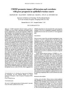
Results I. Establishment of a tumor sphere culture from mouse glioma RSV-M cells To isolate the mouse glioma tumor sphere, RSV-M cells were cultured with neurosphere culture medium containing growth factors EGF, bFGF, and LIF for 3 weeks. A small tumor sphere was observed several days after initiation of culture and started to proliferate steadily after approximately 2 weeks (Fig. 1A). The RSV-M tumor spheres (RSV-M-TS) continued to proliferate and were able to be maintained over 1 year under the same culture conditions. The EGF- and bFGF-dependency of neurosphere culture including neural stem cell (NSC) growth is well documented;27,28) therefore, we also examined the growth factor dependency of RSV-M-TS cell growth. Contrary to our expectation, RSV-M-TS cells grew in both the absence and presence of EGF and bFGF (Fig. 1B). That is, the growth factor requirements of RSV-M-TS cells were different from those of neurosphere culture. To determine the self-renewal activity of RSV-M and RSV-M-TS cells, clonogenicity was assayed by plating cells at serial dilutions down to 1 cell/well. When clonogenicity was compared between cells at a plating density of 100 cells/ well, both cell types were found to proliferate until confluent. However, RSV-M-TS cells proliferated at a significantly higher rate, increasing more than 2-fold and 7-fold at a plating density of 10 cells/ well and 1 cell/well, respectively (Fig. 1C). This data indicates that cultures of RSV-M-TS contain more clonogenic cells than RSV-M. To determine whether the RSV-M-TS cells were enriched with populations of neural stem-like cells,
A
B
C
D
E
Fig. 1 Isolation and characterization of a tumor sphere culture of the mouse glioma cell line RSV-M. A: Tumor spheres (right) were isolated from RSV-M (left) according to the neurosphere culture method. B: Effects of EGF and bFGF on the growth of tumor sphere cells were measured by cell counting. C: The clonogenic self-renewal capacity of parent and tumor sphere cells was measured by limiting dilution into 96-well plates. Cells were seeded at concentrations of 1 cell/well to 100 cells/well. The number of wells showing cell growth was counted after 1 month. D: Expression levels of stem glial and oligodendrocytic cell markers in neurosphere isolated from mouse brain, parent, and tumor sphere cells of RSV-M were examined by semiquantitative RT-PCR. E: Tumorigenicity of RSV-M and tumor sphere cells was investigated in syngeneic C3H/ HeN mice (left). One hundred thousand parent RSV-M (left) and dissociated tumor sphere (right) cells were injected subcutaneously into the flanks. A photograph was taken of a representative mouse 3 weeks after injection. H&E staining of the tumor is formed by intracranial injection of 100 RSV-M-TS cells (right). bFGF: basic fibroblast growth factor, EGF: epidermal growth factor, H&E: hematoxylin and eosin, RT-PCR: real-time polymerase chain reaction.
Neurol Med Chir (Tokyo) 55, January, 2015
Neurologia medico-chirurgica Advance Publication Date: December 20, 2014
5
Up-regulation of Invasive Activity in Glioma Spheres
while only SRY-related HMG-box gene 2 (SOX2), a NSC marker, was up-regulated in RSV-M-TS cells. Furthermore, there were no obvious differences in the expression of astrocytic markers GFAP and S100beta between both cultured cells. This result indicates the complexity of using neural stem and differentiated cell marker expression in the identification of CSCs in RSV-M cells.
II. Tumorigenic potential of RSV-M-TS cells Many reports have suggested that tumor sphere cultures contain a considerable percentage of tumorigenic cells. In order to determine the tumorigenicity of our tumor sphere culture, RSV-M and mechanically dissociated RSV-M-TS cells were transplanted into the subcutaneous (S.C.) or brain of syngeneic mice C3H/HeN. One month after S.C. transplantation of 1 × 10⁵ RSV-M-TS cells but not 1 × 10⁵ RSV-M cells, a tumor mass was observed (Fig. 1E). Moreover, the minimum number of transplanted RSV-M-TS cells required for tumor formation was only 100 cells in the brain, whereas transplantation of the same number of parent RSV-M cells did not result in a tumor mass. Histological analysis showed infiltration of tumor cells into the normal brain, resembling primary human glioblastoma tissues (Fig. 1E). These results suggest that RSV-M-TS cells contain a considerable percentage of tumorigenic cells compared to parental RSV-M cells, potentiating the invasive properties in vivo. III. Migratory activity of RSV-M and RSV-M-TS cells in mouse brain Mouse brain tumor models transplanted with
A
B
RSV-M-TS cells showed the infiltrative property of malignant gliomas (Fig. 1E). The migratory ability of CSCs has not been well studied in the past; thus, mouse syngeneic models may be suitable since immunorejection against allogeneic cells can be excluded. DiI-labeled RSV-M and RSV-M-TS cells were transplanted into the striatum of syngeneic mice and the migration distances from the transplantation sites observed (Fig. 2B). On the day of transplantation, transplanted cells were observed as a small mass with strong red fluorescence. Three days after transplantation, the tumor cells had migrated along the external capsule, while at 7 days, the red fluorescent signals were diffuse and weak and it became difficult to trace migration. Therefore, the number of cells that migrated to the most lateral (towards the side of the head) position of the external capsule were counted 3 days after transplantation. The number of RSV-M-TS cells that migrated from the transplant site to this most lateral position increased 2.86-fold compared to RSV-M cells (Fig. 2C, D). This result shows that the migratory activity of tumor spheres consisting of RSV-M cells was higher than that of parental RSV-M cells in vivo.
IV. Invasive activity of RSV-M and RSV-M-TS in three-dimensional transwell assay The above results suggest that RSV-M-TS cells possess highly invasive activity in vivo. To confirm this, we measured the invasive activity of RSV-M and RSV-M-TS cells in a three-dimensional collagen matrix (Fig. 3A, B). In this assay, the invasion of RSV-M-TS cells cultured with medium containing
C
D
Fig. 2 Comparison of the migratory activities of RSV-M and RSV-M-TS cells in mouse brain. DiI-labeled cells (1 × 10⁵) were injected into mouse striatum and their behavior traced by observation of red fluorescence. A: Diagram showing the injection site of tumor cells. The red horizontal bar indicates the point where the migrating cells were counted. B: The migration distance of DiI-labeled cells was compared at 0 day, 3 days, and 7 days after injection. C: RSV-M-TS cells showed enhanced migration along the external capsule compared to RSV-M cells 3 days after injection. D: Results represent the average number of cells that migrated over the red horizontal bar in (A) from five independent experiments.
Neurol Med Chir (Tokyo) 55, January, 2015
Neurologia medico-chirurgica Advance Publication Date: December 20, 2014
6
M. Nonaka et al.
A
B Fig. 3 Invasive activity of RSV-M and RSV-M derivatives in the 3-dimensional collagen I matrix. A: Photomicrographs of RSV-M (upper left), RSV-M-TS (upper right) and RSV-M-TS cells treated with serum for 12 hours (lower left) or 8 days (lower right) migrating onto lower surfaces of 8 µm-pore transwells. Magnification is 100×. B: The number of migrating cells was determined by counting 4 random fields (40-fold magnification) in each of three independent experiments.
serum for 24 hours or 8 days was also analyzed to determine the effect of serum during culture. The invasive activity was significantly enhanced (more than 11-fold) in RSV-M-TS cells compared to RSV-M cells. Furthermore, culture of RSV-MTS cells with 10% serum for 24 hours induced a three-fold increase in invasion. This enhanced invasive activity was decreased in RSV-M-TS cells after long-term culture (8 days) in medium containing serum. This result suggests that tumor sphere culture and short-term incubation of tumor sphere with serum increase invasive activity, whilst long-term exposure to serum decreases invasion.
V. Profiling the gene expression involved with cell migration/invasion in RSV-M-TS Since the tumor sphere culture showed highly invasive activity in vivo and in vitro (Figs. 2, 3), gene expression profiling was deemed the next important step to discover the molecules and pathways involved in the invasion of tumor sphere cells. Using a tumor metastasis PCR array, we examined
Fig. 4 Relative expression comparison of 84 metastasisrelated genes between the RSV-M-TS and parent RSV-M cells. The figure shows a log transformation plot of the relative expression level of each gene (2-DCt) in RSV-M (x-axis) and RSV-M-TS (y-axis). The middle diagonal line indicates equal expression levels, whereas genes outside the dotted line differed by 2-fold or more.
the expression profiles and compared the relative expression of tumor metastasis genes in the RSV-M and RSV-M-TS cells (Fig. 4, Table 2). A scatter plot of the results showed the positions of several noteworthy genes based on large-fold differences in expression between RSV-M and RSV-M-TS. Of 84 cancer metastasis-related genes, 28 genes showed at least a 4-fold increase or 0.25-fold reduction in expression in RSV-M-TS cells.
VI. Tenascin-C up-regulation in RSV-M-TS and invading tumor cells in mouse brain Of the genes up-regulated in RSV-M-TS cells, up-regulation of Fn1 (fibronectin) was previously reported in tumor spheroids of a glioma cell line. 29) Since enhanced migration of glioma cells on fibronectin through soluble tenascin-C has also been shown, 30) we also examined the expression of tenascin-C in normal neurosphere, RSV-M and RSV-M-TS cells (Fig. 5). Quantitative PCR analysis showed an approximately 8-fold increase in expression of tenascin-C in RSV-M-TS cells compared with RSV-M cells. To confirm the expression in vivo, we performed immunohistochemical analysis of tumor tissues generated by transplantation of RSV-M-TS cells into mouse brain. Tenascin-C expression was observed in invading
Neurol Med Chir (Tokyo) 55, January, 2015
Neurologia medico-chirurgica Advance Publication Date: December 20, 2014
7
Up-regulation of Invasive Activity in Glioma Spheres Table 2 Changes in relative expression of tumor metastasis genes between RSV-M and RSV-M-TS cells Gene Symbol Cd44 Cdh11 Cdh6 Cdh8 Chd4 Ctsk Ctsl Elane Ephb2 Fn1 Kiss1r Hgf Il18 Cxcr2 Itga7 Itgb3 Kiss1 Mcam Met Mmp2 Mmp7 Mtss1 Myc Pten Rb1 Sstr2 Syk Cdh1 Mycl1 Nme1 Plaur
Fold change RSV-M-TS /RSV-M 4.3651 15.4122 6.8971 18.5841 4.0727 19.7804 4.0446 22.2541 22.2541 4.1011 5.9628 19.3733 15.6273 6.0881 18.2017 6.8971 20.7638 4.8099 5.049 4.1296 17.9511 3.6452 3.023 3.0441 3.521 9.4873 23.8514 –8.6578 –3.8477 –4.3893 –6.56141
Putative function Cell adhesion Cell adhesion Cell adhesion Cell adhesion Transcription factor and regulator Lysosomal protease Lysosomal protease protease Receptor Cell adhesion Receptor Cell growth and proliferation Cytokine Receptor Cell adhesion Cell adhesion Cell adhesion Cell adhesion Cell growth and proliferation Extracellular matrix-protein Extracellular matrix-protein Cell adhesion Cell growth and proliferation Cell growth and proliferation Cell growth and proliferation Cell growth and proliferation Cell adhesion Cell adhesion Transcription factor and regulator Nucleic acid synthesis Cell adhesion
Average raw Ct RSV-M-TS 20.1 29.84 27.83 29.57 19.76 29.84 17.04 29.31 29.31 31.75 28.58 29.51 27.54 31.18 27.2 30.98 29.41 24.57 24.81 29.89 29.62 26.18 21.03 22.12 26.13 29.03 29.21 35 32.62 21.41 25.22
Average raw Ct RSV-M 23.44 35 31.83 35 23 35 20.27 35 35 35 32.37 35 32.72 35 32.6 34.98 35 28.05 28.36 33.15 35 29.62 23.84 24.94 29.16 33.49 35 33.1 31.89 20.49 23.72
The table lists genes that exhibited at least a 3-fold difference in expression in the RSV-M-TS cell sample compared with RSV-M. The raw threshold cycle (Ct) values seen in the two samples are also listed for comparison.
B
A Neurol Med Chir (Tokyo) 55, January, 2015
Fig. 5 Enhanced expression of tenascin-C in RSV-M-TS and migrating cells. A: Tenascin-C expression in normal neurosphere, RSV-M and RSV-M-TS cells was examined by quantitativepolymerase chain reaction. B: Tenascin-C expressing cells were frequently localized at the tumor front (left) and external capsule (right) in the mouse brain 3 weeks after transplantation of 1 × 10⁵ RSV-M-TS cells into the striatum. Arrows indicate the tenascin-C expressing cells.
Neurologia medico-chirurgica Advance Publication Date: December 20, 2014
8
M. Nonaka et al.
cells attached to the tumor mass. This result suggests a role of tenascin-C and fibronectin in migration of RSV-M glioma cells and that tumor sphere cells can produce extracellular matrix for migration and invasion.
Discussion In this study, we established a mouse glioma tumor sphere model, revealing that RSV-M-TS cells show high tumorigenicity and migratory activity. It is well known that excessive passage of tumor cell lines including RSV-M cells tends to result in a loss of tumorigenicity. The method of tumor sphere culture described here, however, appears to maintain tumorigenicity and partly recapture tumor phenotypes resembling glioma patient tissues.8,14,24,25) Tumor sphere culture of RSV-M cells may also enrich CSCs capable of recreating the tumor phenotype seen in vivo. CSCs derived from brain tumors were identified and isolated using a NSCs marker; however, we did not detect obvious up-regulation of CD133, nestin, or Bmi1 in RSV-M-TS cells. Only SOX2 was up-regulated in RSV-M-TS cells (Fig. 1). In a previous report, stem cell culture derived from a mouse glioma cell line showed the up-regulation of nestin, but not CD133.12) In addition, the existence of a CD133 negative tumor spheres derived from human glioma was also reported.31–33) It has further been suggested that CSCs could arise from various cells of neural lineage.34) If the expression pattern of stem cells markers is affected by unique genetic and epigenetic alterations in their lineage, an absolute marker for CSCs may not exist. The failure to detect a strong correlation between sphere formation and stem cell markers in RSV-M cells is consistent with other publications demonstrating the insufficiency of these markers in glioma stem cells.35,36) The difference between RSV-M-TS and neurosphere cells, with respect to their dependency on EGF and bFGF for growth, may be a key phenotype for identifying cell origin. In experimental glioma and oligodendroglioma models, tumor spheres showed efficient self-renewal and proliferation activity independent of either growth factor, suggesting that oligodendrocyte progenitor cells (OPCs) are candidates for tumor origin. RSV-M-TS cells may be more similar to OPCs than to NSCs.37,38) Because the lack of a reliable phenotype for identifying CSCs currently restricts tumorigenicity and sphere formation, further studies are needed to characterize CSCs derived from various tumor tissues. In this article, up-regulation of migratory activity in RSV-M-TS cells was observed both in vivo and in vitro compared to the parent RSV-M cells. The maximum
difference in migration activity was observed at Day 3, consistent with previous reports in vivo and in vitro.39,40) Most CSCs transplanted into the mouse brain differentiate and form a tumor mass displaying histological similarity with clinical specimens of gliomas.8) If differentiated cells tend to lose their migratory activity, robust migration of RSV-M-TS cells should be observed within several days after transplantation. To understand how cell differentiation affects migration, the identification of reliable stem and differentiated cell markers is necessary. Several studies have suggested that transplanted NSCs migrate toward glioma in vivo.41,42) Whether endogenous NSCs show similar behavior remains unknown; however, NSCs usually differentiate and then acquire migration capability.43) CSCs derived from brain tumors are thought to resemble NSC and may therefore need to differentiate prior to migration. This study demonstrated that RSV-M-TS cells possess high migratory activity compared to RSV-M. Furthermore, short-term incubation of RSV-M-TS cells with medium containing serum robustly up-regulated their migratory activity. A short incubation period is usually insufficient to induce cell differentiation, because differentiation of NSC is induced over 5–7 days in vitro. The differentiation level of RSV-M-TS cells might resemble that of progenitors of neural lineage cells, that is, not yet fully differentiated cells. Since the tumorigenicity and clonogenicity of RSV-M were obviously enriched by tumor sphere culture, characterization of the biological behavior of this population remains important. Tumor metastasis PCR array analysis of the RSV-M and RSV-M-TS cells demonstrated significant up-regulation rather than down-regulation of various metastatic factors in the RSV-M-TS cells (Table 2). The up-regulated genes included Cd44, Ctsl, Ephb2, Hgf, Mmp-2, Mmp-7, and c-Met, the expressions of which are reportedly correlated with invasion or migration of glioblastomas.44–51) Furthermore, Cd44 was identified as an invasive marker of gliomas and is reportedly up-regulated in CD133+ CSCs in glioblastoma.52) c-Met in particular is strongly overexpressed in malignant glioma and stimulates hepatocyte growth factor (HGF)-induced cell migration of glioma.47,51) Among the down-regulated genes in RSV-M-TS, Nme1 is reportedly a suppressor of invasion and migration.53) Our results are in agreement with other studies that previously reported that a number of adhesion molecules, extracellular-matrix molecules, and cytokines are related to the onset and progression of tumor metastasis.39–43) It has been reported that the radio-resistance of gliomas is involved with the existence of CSCs.54) Populations of CSCs can survive after radiotherapy
Neurol Med Chir (Tokyo) 55, January, 2015
Neurologia medico-chirurgica Advance Publication Date: December 20, 2014
Up-regulation of Invasive Activity in Glioma Spheres
through up-regulation of DNA repair and anti-apoptotic pathways.9) At the same time, promotion of migration and invasion of glioma cells as a result of sublethal doses of irradiation, partly due to enhanced expression of Mmp-2, has also been suggested.54) The expression level of Mmp-2 in RSV-M-TS cells was increased to more than 4-fold compared to RSV-M cells. If migratory phenotype of CSCs is characterized by enhanced Mmp-2 expression, our model may be useful for investigating mechanisms of radioresistance and dissemination in malignant glioma. Further studies to investigate migratory activity and metastatic gene expression in human glioma spheres are required to broaden the significance of the data reported here.
Acknowledgment This research was partially supported by a Grantin-Aid for Scientific Research (C), 2010–2013 from the Ministry of Education, Science, Sports and Culture to Toshio Yawata (22591614).
Conflicts of Interest Disclosure The authors have no personal, financial, or institutional interests in any of the drugs, materials, or devices cited in this article. All authors who are members of The Japan Neurosurgical Society (JNS) have registered online their self-reported COI disclosure statements (available from the JNS website).
References 1) Central Brain Tumor Registry of the United States (CBTRUS): Statistical Report: Primary Brain Tumors in the United States, 1995–1999. CBTRUS, 2002 2) Stupp R, Mason WP, van den Bent MJ, Weller M, Fisher B, Taphoorn MJ, Belanger K, Brandes AA, Marosi C, Bogdahn U, Curschmann J, Janzer RC, Ludwin SK, Gorlia T, Allgeier A, Lacombe D, Cairncross JG, Eisenhauer E, Mirimanoff RO; European Organisation for Research and Treatment of Cancer Brain Tumor and Radiotherapy Groups; National Cancer Institute of Canada Clinical Trials Group: Radiotherapy plus concomitant and adjuvant temozolomide for glioblastoma. N Engl J Med 352: 987–996, 2005 3) Prados MD, Berger MS, Wilson CB: Primary central nervous system tumors: advances in knowledge and treatment. CA Cancer J Clin 48: 331–360, 321, 1998 4) Laws ER, Thapar K: Brain tumors. CA Cancer J Clin 43: 263–271, 1993 5) Hemmati HD, Nakano I, Lazareff JA, Masterman-Smith M, Geschwind DH, Bronner-Fraser M, Kornblum HI: Cancerous stem cells can arise from pediatric brain
Neurol Med Chir (Tokyo) 55, January, 2015
9
tumors. Proc Natl Acad Sci USA 100: 15178–15183, 2003 6) Singh SK, Clarke ID, Terasaki M, Bonn VE, Hawkins C, Squire J, Dirks PB: Identification of a cancer stem cell in human brain tumors. Cancer Res 63: 5821–5828, 2003 7) Galli R, Binda E, Orfanelli U, Cipelletti B, Gritti A, De Vitis S, Fiocco R, Foroni C, Dimeco F, Vescovi A: Isolation and characterization of tumorigenic, stem-like neural precursors from human glioblastoma. Cancer Res 64: 7011–7021, 2004 8) Singh SK, Hawkins C, Clarke ID, Squire JA, Bayani J, Hide T, Henkelman RM, Cusimano MD, Dirks PB: Identification of human brain tumour initiating cells. Nature 432: 396–401, 2004 9) Bao S, Wu Q, McLendon RE, Hao Y, Shi Q, Hjelmeland AB, Dewhirst MW, Bigner DD, Rich JN: Glioma stem cells promote radioresistance by preferential activation of the DNA damage response. Nature 444: 756–760, 2006 10) Bao S, Wu Q, Sathornsumetee S, Hao Y, Li Z, Hjelmeland AB, Shi Q, McLendon RE, Bigner DD, Rich JN: Stem cell-like glioma cells promote tumor angiogenesis through vascular endothelial growth factor. Cancer Res 66: 7843–7848, 2006 11) Eramo A, Ricci-Vitiani L, Zeuner A, Pallini R, Lotti F, Sette G, Pilozzi E, Larocca LM, Peschle C, De Maria R: Chemotherapy resistance of glioblastoma stem cells. Cell Death Differ 13: 1238–1241, 2006 12) Pellegatta S, Poliani PL, Corno D, Menghi F, Ghielmetti F, Suarez-Merino B, Caldera V, Nava S, Ravanini M, Facchetti F, Bruzzone MG, Finocchiaro G: Neurospheres enriched in cancer stem-like cells are highly effective in eliciting a dendritic cell-mediated immune response against malignant gliomas. Cancer Res 66: 10247–10252, 2006 13) Annabi B, Lachambre MP, Plouffe K, Sartelet H, Béliveau R: Modulation of invasive properties of CD133+ glioblastoma stem cells: a role for MT1-MMP in bioactive lysophospholipid signaling. Mol Carcinog 48: 910–919, 2009 14) Annabi B, Rojas-Sutterlin S, Laflamme C, Lachambre MP, Rolland Y, Sartelet H, Béliveau R: Tumor environment dictates medulloblastoma cancer stem cell expression and invasive phenotype. Mol Cancer Res 6: 907–916, 2008 15) Janik P, Szaniawska B, Miloszewska J, Pietruszewska E, Kowalczyk D: The role of protein kinase C in migration of rat glioma cells from spheroid cultures. Cancer Lett 63: 167–170, 1992 16) Engebraaten O, Schwachenwald R, Valen H, Bjerkvig R, Laerum OD, Backlund EO: Effects of high and low single dose irradiation on glioma spheroid invasion into normal rat brain tissue in vitro. Anticancer Res 12: 1501–1506, 1992 17) Pedersen PH, Ness GO, Engebraaten O, Bjerkvig R, Lillehaug JR, Laerum OD: Heterogeneous response to the growth factors [EGF, PDGF (bb), TGF-alpha, bFGF, IL-2] on glioma spheroid growth, migration and invasion. Int J Cancer 56: 255–261, 1994
Neurologia medico-chirurgica Advance Publication Date: December 20, 2014
10
M. Nonaka et al.
18) Kitahara M, Katakura R, Suzuki J, Sasaki T: Experimental combination chemotherapy of ACNU and 5-FU against cultured glioma model (spheroid) and subcutaneous rat glioma. Int J Cancer 40: 557–563, 1987 19) Kolchinsky A, Roninson IB: Drug resistance conferred by MDR1 expression in spheroids formed by glioblastoma cell lines. Anticancer Res 17: 3321–3327, 1997 20) Fehlauer F, Muench M, Richter E, Rades D: The inhibition of proliferation and migration of glioma spheroids exposed to temozolomide is less than additive if combined with irradiation. Oncol Rep 17: 941–945, 2007 21) Shweiki D, Neeman M, Itin A, Keshet E: Induction of vascular endothelial growth factor expression by hypoxia and by glucose deficiency in multicell spheroids: implications for tumor angiogenesis. Proc Natl Acad Sci USA 92: 768–772, 1995 22) Goldbrunner RH, Wagner S, Roosen K, Tonn JC: Models for assessment of angiogenesis in gliomas. J Neurooncol 50: 53–62, 2000 23) Tunici P, Bissola L, Lualdi E, Pollo B, Cajola L, Broggi G, Sozzi G, Finocchiaro G: Genetic alterations and in vivo tumorigenicity of neurospheres derived from an adult glioblastoma. Mol Cancer 3: 25, 2004 24) Chong YK, Toh TB, Zaiden N, Poonepalli A, Leong SH, Ong CE, Yu Y, Tan PB, See SJ, Ng WH, Ng I, Hande MP, Kon OL, Ang BT, Tang C: Cryopreservation of neurospheres derived from human glioblastoma multiforme. Stem Cells 27: 29–39, 2009 25) Yu SC, Ping YF, Yi L, Zhou ZH, Chen JH, Yao XH, Gao L, Wang JM, Bian XW: Isolation and characterization of cancer stem cells from a human glioblastoma cell line U87. Cancer Lett 265: 124–134, 2008 26) Kumanishi T, Ikuta F, Yamamoto T: Brain tumors induced by Rous sarcoma virus, Schmidt-Ruppin strain. 3. Morphology of brain tumors induced in adult mice. J Natl Cancer Inst 50: 95–109, 1973 27) Gritti A, Frölichsthal-Schoeller P, Galli R, Parati EA, Cova L, Pagano SF, Bjornson CR, Vescovi AL: Epidermal and fibroblast growth factors behave as mitogenic regulators for a single multipotent stem cell-like population from the subventricular region of the adult mouse forebrain. J Neurosci 19: 3287–3297, 1999 28) Svendsen CN, Caldwell MA, Ostenfeld T: Human neural stem cells: isolation, expansion and transplantation. Brain Pathol 9: 499–513, 1999 29) Glimelius B, Norling B, Nederman T, Carlsson J: Extracellular matrices in multicellular spheroids of human glioma origin: increased incorporation of proteoglycans and fibronectin as compared to monolayer cultures. APMIS 96: 433–444, 1988 30) Deryugina EI, Bourdon MA: Tenascin mediates human glioma cell migration and modulates cell migration on fibronectin. J Cell Sci 109 (Pt 3): 643–652, 1996
31) Joo KM, Kim SY, Jin X, Song SY, Kong DS, Lee JI, Jeon JW, Kim MH, Kang BG, Jung Y, Jin J, Hong SC, Park WY, Lee DS, Kim H, Nam DH: Clinical and biological implications of CD133-positive and CD133-negative cells in glioblastomas. Lab Invest 88: 808–815, 2008 32) Nishide K, Nakatani Y, Kiyonari H, Kondo T: Glioblastoma formation from cell population depleted of Prominin1-expressing cells. PLoS One 4: e6869, 2009 33) Clément V, Dutoit V, Marino D, Dietrich PY, Radovanovic I: Limits of CD133 as a marker of glioma self-renewing cells. Int J Cancer 125: 244–248, 2009 34) Fine HA: Glioma stem cells: not all created equal. Cancer Cell 15: 247–249, 2009 35) Ogden AT, Waziri AE, Lochhead RA, Fusco D, Lopez K, Ellis JA, Kang J, Assanah M, McKhann GM, Sisti MB, McCormick PC, Canoll P, Bruce JN: Identification of A2B5+CD133- tumor-initiating cells in adult human gliomas. Neurosurgery 62: 505–514; discussion 514–515, 2008 36) Sun Y, Kong W, Falk A, Hu J, Zhou L, Pollard S, Smith A: CD133 (Prominin) negative human neural stem cells are clonogenic and tripotent. PLoS ONE 4: e5498, 2009 37) Jiang Y, Boije M, Westermark B, Uhrbom L: PDGF-B Can sustain self-renewal and tumorigenicity of experimental glioma-derived cancer-initiating cells by preventing oligodendrocyte differentiation. Neoplasia 13: 492–503, 2011 38) Persson AI, Petritsch C, Swartling FJ, Itsara M, Sim FJ, Auvergne R, Goldenberg DD, Vandenberg SR, Nguyen KN, Yakovenko S, Ayers-Ringler J, Nishiyama A, Stallcup WB, Berger MS, Bergers G, McKnight TR, Goldman SA, Weiss WA: Non-stem cell origin for oligodendroglioma. Cancer Cell 18: 669–682, 2010 39) Tonn JC, Haugland HK, Saraste J, Roosen K, Laerum OD: Differential effects of vincristine and phenytoin on the proliferation, migration, and invasion of human glioma cell lines. J Neurosurg 82: 1035–1043, 1995 40) Vajkoczy P, Goldbrunner R, Farhadi M, Vince G, Schilling L, Tonn JC, Schmiedek P, Menger MD: Glioma cell migration is associated with gliomainduced angiogenesis in vivo. Int J Dev Neurosci 17: 557–563, 1999 41) Aboody KS, Brown A, Rainov NG, Bower KA, Liu S, Yang W, Small JE, Herrlinger U, Ourednik V, Black PM, Breakefield XO, Snyder EY: Neural stem cells display extensive tropism for pathology in adult brain: evidence from intracranial gliomas. Proc Natl Acad Sci USA 97: 12846–12851, 2000 42) Tang Y, Shah K, Messerli SM, Snyder E, Breakefield X, Weissleder R: In vivo tracking of neural progenitor cell migration to glioblastomas. Hum Gene Ther 14: 1247–1254, 2003 43) Goldman SA: Adult neurogenesis: from canaries to the clinic. J Neurobiol 36: 267–286, 1998
Neurol Med Chir (Tokyo) 55, January, 2015
Neurologia medico-chirurgica Advance Publication Date: December 20, 2014
Up-regulation of Invasive Activity in Glioma Spheres 44) Ariza A, López D, Mate JL, Isamat M, Musulén E, Pujol M, Ley A, Navas-Palacios JJ: Role of CD44 in the invasiveness of glioblastoma multiforme and the noninvasiveness of meningioma: an immunohistochemistry study. Hum Pathol 26: 1144–1147, 1995 45) Levicar N, Dewey RA, Daley E, Bates TE, Davies D, Kos J, Pilkington GJ, Lah TT: Selective suppression of cathepsin L by antisense cDNA impairs human brain tumor cell invasion in vitro and promotes apoptosis. Cancer Gene Ther 10: 141–151, 2003 46) Nakada M, Niska JA, Tran NL, McDonough WS, Berens ME: EphB2/R-Ras signaling regulates glioma cell adhesion, growth, and invasion. Am J Pathol 167: 565–576, 2005 47) Yamamoto S, Wakimoto H, Aoyagi M, Hirakawa K, Hamada H: Modulation of motility and proliferation of glioma cells by hepatocyte growth factor. Jpn J Cancer Res 88: 564–577, 1997 48) Xie Q, Gao CF, Shinomiya N, Sausville E, Hay R, Gustafson M, Shen Y, Wenkert D, Vande Woude GF: Geldanamycins exquisitely inhibit HGF/SF-mediated tumor cell invasion. Oncogene 224: 3697–3707, 2005 49) Chintala SK, Sawaya R, Gokaslan ZL, Rao JS: Modulation of matrix metalloprotease-2 and invasion in human glioma cells by alpha 3 beta 1 integrin. Cancer Lett 103: 201–208, 1996 50) Thorns V, Walter GF, Thorns C: Expression of
Neurol Med Chir (Tokyo) 55, January, 2015
11
MMP-2, MMP-7, MMP-9, MMP-10 and MMP-11 in human astrocytic and oligodendroglial gliomas. Anticancer Res 23: 3937–3944, 2003 51) Eckerich C, Zapf S, Fillbrandt R, Loges S, Westphal M, Lamszus K: Hypoxia can induce c-Met expression in glioma cells and enhance SF/HGF-induced cell migration. Int J Cancer 121: 276–283, 2007 52) Liu G, Yuan X, Zeng Z, Tunici P, Ng H, Abdulkadir IR, Lu L, Irvin D, Black KL, Yu JS: Analysis of gene expression and chemoresistance of CD133+ cancer stem cells in glioblastoma. Mol Cancer 5: 67, 2006 53) Jung S, Paek YW, Moon KS, Wee SC, Ryu HH, Jeong YI, Sun HS, Jin YH, Kim KK, Ahn KY: Expression of Nm23 in gliomas and its effect on migration and invasion in vitro. Anticancer Res 26: 249–258, 2006 54) Wild-Bode C, Weller M, Rimner A, Dichgans J, Wick W: Sublethal irradiation promotes migration and invasiveness of glioma cells: implications for radiotherapy of human glioblastoma. Cancer Res 61: 2744–2750, 2001
Address reprint requests to: Toshio Yawata, PhD, Department of Neurosurgery, Kochi Medical School, Kohasu, Okoh-cho, Nankoku, Kochi 783-8505, Japan. e-mail:
[email protected]